. 2007 Apr 1;178(7):4528-37.
doi: 10.4049/jimmunol.178.7.4528.

MyD88-dependent signals are essential for the host immune response in experimental brain abscess

Affiliations

MyD88-dependent signals are essential for the host immune response in experimental brain abscess

Tammy Kielian et al. J Immunol. .

Abstract

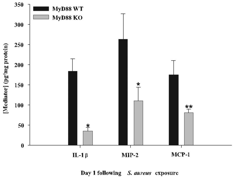
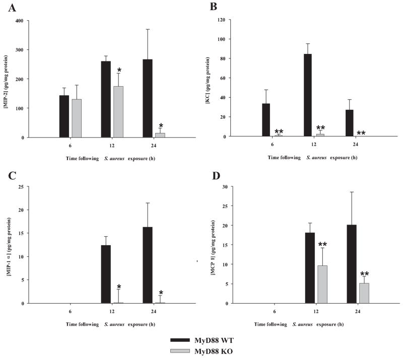
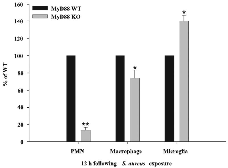
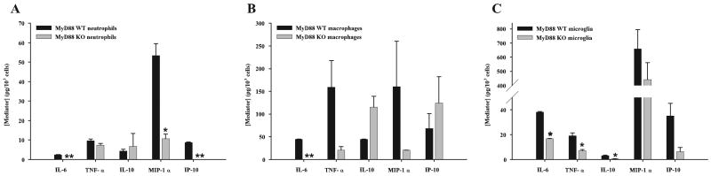
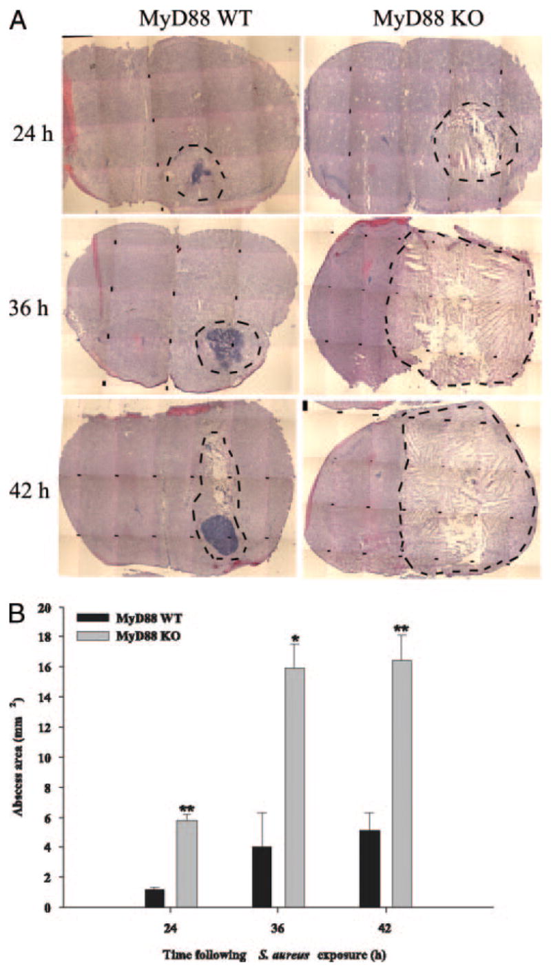
Brain abscesses form in response to a parenchymal infection by pyogenic bacteria, with Staphylococcus aureus representing a common etiologic agent of human disease. Numerous receptors that participate in immune responses to bacteria, including the majority of TLRs, the IL-1R, and the IL-18R, use a common adaptor molecule, MyD88, for transducing activation signals leading to proinflammatory mediator expression and immune effector functions. To delineate the importance of MyD88-dependent signals in brain abscesses, we compared disease pathogenesis using MyD88 knockout (KO) and wild-type (WT) mice. Mortality rates were significantly higher in MyD88 KO mice, which correlated with a significant reduction in the expression of several proinflammatory mediators, including but not limited to IL-1beta, TNF-alpha, and MIP-2/CXCL2. These changes were associated with a significant reduction in neutrophil and macrophage recruitment into brain abscesses of MyD88 KO animals. In addition, microglia, macrophages, and neutrophils isolated from the brain abscesses of MyD88 KO mice produced significantly less TNF-alpha, IL-6, MIP-1alpha/CCL3, and IFN-gamma-induced protein 10/CXCL10 compared with WT cells. The lack of MyD88-dependent signals had a dramatic effect on the extent of tissue injury, with significantly larger brain abscesses typified by exaggerated edema and necrosis in MyD88 KO animals. Interestingly, despite these striking changes in MyD88 KO mice, bacterial burdens did not significantly differ between the two strains at the early time points examined. Collectively, these findings indicate that MyD88 plays an essential role in establishing a protective CNS host response during the early stages of brain abscess development, whereas MyD88-independent pathway(s) are responsible for pathogen containment.


Conflict of interest statement

Disclosures

The authors have no financial conflict of interest.

Figures

FIGURE 1
FIGURE 1
MyD88 KO mice exhibit defects in the induction of several proinflammatory mediators. MyD88 KO and WT mice (n = 4 or 5 per group) were sacrificed 1 day following S. aureus infection, whereupon IL-1β, MIP-2, and MCP-1 protein levels were quantitated by ELISA (mean ± SD). Results were normalized to the amount of total protein recovered to correct for differences in abscess sampling size. Significant differences are denoted by asterisks (*, p < 0.05; **, p < 0.001). Results are representative of three independent experiments.
FIGURE 2
FIGURE 2
MyD88-dependent signals control the expression of select cytokines during the early stage of brain abscess development. MyD88 KO and WT mice (n = 4 to 5 per group) were infected with S. aureus intracerebrally as described in Materials and Methods. Animals were sacrificed at 6, 12, or 24 h following bacterial exposure, whereupon multiplex microbead array analysis was performed on abscess homogenates to allow for the simultaneous quantitation of 20 distinct mediators at the protein level. Results are shown for abscess-associated IL-1β (A), IL-1α (B), IL-12 p70 (C), IL-6 (D), and IL-10 (E) (mean ± SD) and were normalized based on the amount of total protein recovered from abscesses to correct for differences in tissue sampling size. Significant differences in cytokine expression between brain abscesses in MyD88 KO and WT mice are indicated by asterisks (*, p < 0.05; **, p < 0.001). Results are representative of two independent experiments.
FIGURE 3
FIGURE 3
Chemokine induction during acute brain abscess development is highly dependent on MyD88. MyD88 KO and WT mice (n = 4 or 5 per group) were infected with S. aureus intracerebrally as described in Materials and Methods. Animals were sacrificed at 6, 12, or 24 h following bacterial exposure, whereupon multiplex microbead array analysis was performed on abscess homogenates to allow the simultaneous quantitation of 20 distinct mediators at the protein level. Results are shown for abscess-associated MIP-2 (A), KC (B), MIP-1α (C), and MCP-1 (D) (mean ± SD) and were normalized based on the amount of total protein recovered from abscesses to correct for differences in tissue sampling size. MIP-2 levels were determined using a standard sandwich ELISA approach because this chemokine was not included in the multiplex array. Significant differences in chemokine expression between brain abscesses of MyD88 KO and WT mice are indicated by asterisks (*, p < 0.05; **, p < 0.001). Results are representative of two independent experiments.
FIGURE 4
FIGURE 4
Neutrophil and macrophage influx into brain abscesses is significantly impaired in MyD88 KO mice. MyD88 KO and WT mice (n = 10 per group) were infected with S. aureus intracerebrally and sacrificed at 12 h following bacterial exposure, whereupon abscess-associated neutrophils (Gr-1+, CD11b+, and CD45high), macrophages (Gr-1, CD11b+, and CD45high), and microglia (Gr-1, CD11b+, and CD45low) were quantitated by FACS analysis. Results are expressed as the percentage of cells normalized to WT values (set at 100%) and represent the mean ± SEM from three independent experiments. Significant differences in abscess-associated cells recovered from MyD88 KO vs WT animals are denoted by asterisks (*, p < 0.05; **, p < 0.001).
FIGURE 5
FIGURE 5
The expression of numerous cytokines and chemokines is attenuated in abscess-associated cells from MyD88 KO mice. Abscess-associated neutrophils (A), macrophages (B), and microglia (C) were recovered from the lesions of MyD88 KO and WT mice at 12 h following S. aureus infection as described in Materials and Methods. Cells were cultured overnight without additional stimulation and conditioned supernatants were analyzed for mediator expression by multiplex microbead array assays. The amount of each cytokine/chemokine was normalized based on total cell numbers and represents the mean ± SEM from three independent experiments. Significant differences in mediator expression between the various cell types recovered from MyD88 KO vs WT mice are denoted with asterisks (*, p < 0.05; **, p < 0.001).
FIGURE 6
FIGURE 6
Brain abscess size is significantly greater in MyD88 KO mice. MyD88 KO and WT mice (n = 3 or 4 per group per time point) were injected intracerebrally with S. aureus and sacrificed at 24, 36, and 42 h following bacterial exposure, whereupon brain tissues were flash frozen on dry ice for subsequent cryostat sectioning. Serial sections were prepared throughout the entire abscess to ensure that the maximal cross-sectional area was identified and stained with H&E to demarcate the extent of tissue damage. A, A series of microscopic images (original magnification, ×4) were assembled for each individual MyD88 KO or WT animal to demonstrate the extent of abscess formation and edema/necrosis. Abscess margins (including cerebritis) are demarcated with dotted lines. B, Abscess area (mm2; mean ± SD) was quantitated using the MetaMorph image analysis program by measuring the largest lesion size for each tissue specimen. Significant differences in brain abscess size between MyD88 KO vs WT mice are denoted with asterisks (*, p < 0.05; **, p < 0.001).

Similar articles

Cited by

References

    1. Kielian T. Immunopathogenesis of brain abscess. J Neuroinflammation. 2004;1:16. - PMC - PubMed
    1. Bloch O, Papadopoulos MC, Manley GT, Verkman AS. Aquaporin-4 gene deletion in mice increases focal edema associated with staphylococcal brain abscess. J Neurochem. 2005;95:254–262. - PubMed
    1. Lo WD, Wolny A, Boesel C. Blood-brain barrier permeability in staphylococcal cerebritis and early brain abscess. J Neurosurg. 1994;80:897–905. - PubMed
    1. Kielian T, Hickey WF. Proinflammatory cytokine, chemokine, and cellular adhesion molecule expression during the acute phase of experimental brain abscess development. Am J Pathol. 2000;157:647–658. - PMC - PubMed
    1. Baldwin AC, Kielian T. Persistent immune activation associated with a mouse model of Staphylococcus aureus-induced experimental brain abscess. J Neuroimmunol. 2004;151:24–32. - PubMed

Publication types

Substances