. 2007 Apr 1;178(7):4641-9.
doi: 10.4049/jimmunol.178.7.4641.

IL-1beta causes an increase in intestinal epithelial tight junction permeability

Affiliations

IL-1beta causes an increase in intestinal epithelial tight junction permeability

Rana M Al-Sadi et al. J Immunol. .

Abstract

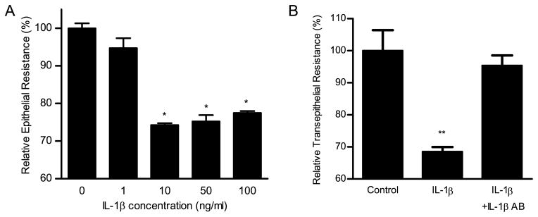
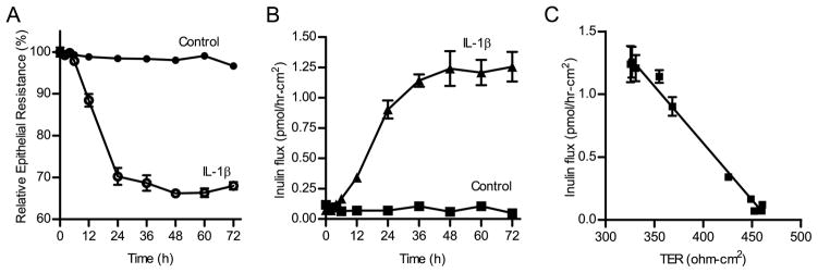
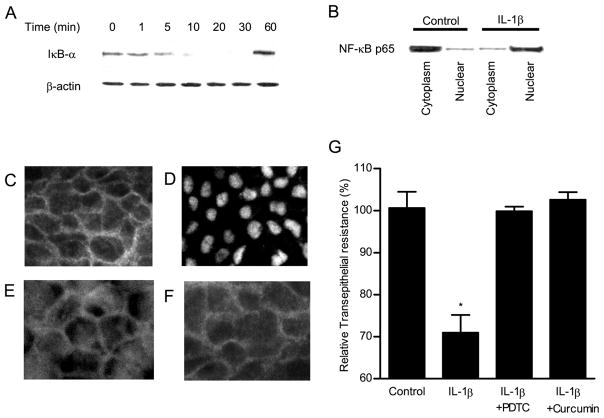
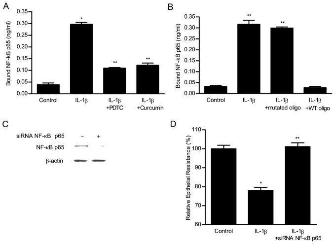
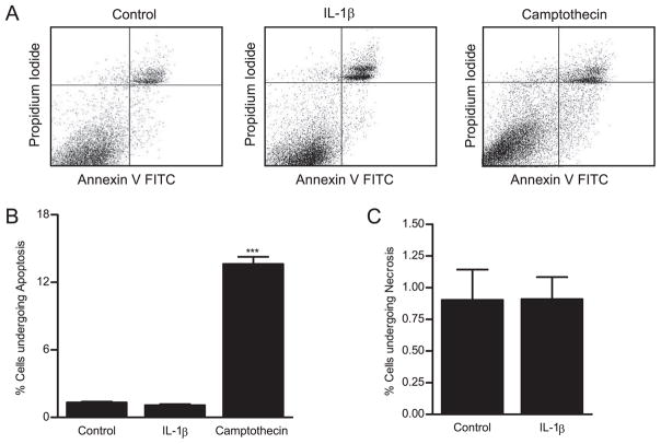
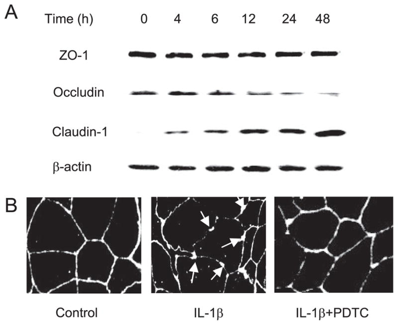
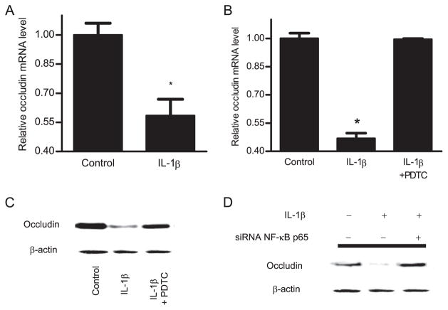
IL-1beta is a prototypical proinflammatory cytokine that plays a central role in the intestinal inflammation amplification cascade. Recent studies have indicated that a TNF-alpha- and IFN-gamma-induced increase in intestinal epithelial paracellular permeability may be an important mechanism contributing to intestinal inflammation. Despite its central role in promoting intestinal inflammation, the role of IL-1beta on intestinal epithelial tight junction (TJ) barrier function remains unclear. The major aims of this study were to determine the effect of IL-1beta on intestinal epithelial TJ permeability and to elucidate the mechanisms involved in this process, using a well-established in vitro intestinal epithelial model system consisting of filter-grown Caco-2 intestinal epithelial monolayers. IL-1beta (0-100 ng/ml) produced a concentration- and time-dependent decrease in Caco-2 transepithelial resistance. Conversely, IL-1beta caused a progressive time-dependent increase in transepithelial permeability to paracellular marker inulin. IL-1beta-induced increase in Caco-2 TJ permeability was accompanied by a rapid activation of NF-kappaB. NF-kappaB inhibitors, pyrrolidine dithiocarbamate and curcumin, prevented the IL-1beta-induced increase in Caco-2 TJ permeability. To further confirm the role of NF-kappaB in the IL-1beta-induced increase in Caco-2 TJ permeability, NF-kappaB p65 expression was silenced by small interfering RNA transfection. NF-kappaB p65 depletion completely inhibited the IL-1beta-induced increase in Caco-2 TJ permeability. IL-1beta did not induce apoptosis in the Caco-2 cell. In conclusion, our findings show for the first time that IL-1beta at physiologically relevant concentrations causes an increase in intestinal epithelial TJ permeability. The IL-1beta-induced increase in Caco-2 TJ permeability was mediated in part by the activation of NF-kappaB pathways but not apoptosis.


Conflict of interest statement

Disclosures

The authors have no financial conflict of interest.

Figures

FIGURE 1
FIGURE 1
A, Effect of increasing concentration of IL-1β (0, 1, 10, 50, and 100 ng/ml) on Caco-2 TER. Filter-grown Caco-2 monolayers were treated with IL-1β for a 48-h experimental period. IL-1β produced a concentration-dependent decrease in Caco-2 TER. Data represent means ± SE of TER (n = 6). *, p < 0.0001 vs control. B, The effect of IL-1β mAb (100 ng/ml) on an IL-1β-induced drop in Caco-2 TER. IL-1β mAb pre-treatment prevented the IL-1β-induced drop in Caco-2 TER. Data represent means ± SE of TER (n = 6).**, p < 0.0001 vs control.
FIGURE 2
FIGURE 2
Time-course effect of IL-1β on Caco-2 TER and paracellular permeability. The effect of IL-1β (10 ng/ml) on Caco-2 TER and mucosal-to-serosal flux of paracellular marker, inulin were measured over a 72-h experimental period. A, Time-course effect of IL-1β on Caco-2 TER (n = 4). B, Time-course effect of IL-1β on transepithelial inulin flux (n = 3). C, Graph of TER vs inulin flux (r = 0.97).
FIGURE 3
FIGURE 3
Time-course effect of IL-1β on NF-κB activation in Caco-2 monolayers. Filter-grown Caco-2 monolayers were treated with IL-1β (10 ng/ml) for increasing time periods (0–60 min). A, The IκB-α protein expression was determined by Western blot analysis as described in Materials and Methods. B, Filter-grown Caco-2 monolayers were treated with IL-1β (10 ng/ml) for 30 min, and changes in the NF-κB p65 expression in the cytoplasmic and nuclear fractions were assayed by Western blot analysis. The effect of IL-1β (10 ng/ml) on NF-κB p65 cytoplasmic-to-nuclear translocation as determined by immunofluorescent Ab labeling as described in Materials and Methods. C, Control or untreated Caco-2 monolayers. D, Caco-2 monolayers treated for 30 min with IL-1β (10 ng/ml). Caco-2 monolayers pretreated with (E) curcumin (5 μM) or (F) PDTC (100 μM) before the 30-min treatment with IL-1β. IL-1β caused a rapid cytoplasmic-to-nuclear translocation of NF-κB p65 subunit. Original magnification, ×400. G, The effect of NF-κB inhibitors PDTC and curcumin on IL-1β-induced drop in Caco-2 TER. PDTC (100 μM) and curcumin (5 μM) significantly prevented the IL-1β-induced drop in Caco-2 TER (n = 6). *, p < 0.01 vs control. **, p < 0.01 vs IL-1β-treated monolayers.
FIGURE 4
FIGURE 4
ELISA-binding assay of IL-1β-activated NF-κB p65 binding to the oligonucleotide probe containing the κB-binding site. The binding of p65 to binding site was expressed as a concentration of NF-κB p65 (nanograms per milliliter) bound to the binding site. A, IL-1β caused a significant increase in NF-κB binding. NF-κB inhibitors PDTC (100 μM) and curcumin (5 μM) inhibited the IL-1β-induced NF-κB binding. B, The addition of wild-type (WT) oligonucleotide containing the consensus NF-κB-binding site in excess (100:1 ratio) as a competitive inhibitor prevented the binding of NF-κB to the DNA probe. In contrast, the addition of excess oligonucleotide containing a mutated NF-κB-binding motif did not inhibit the NF-κB binding. C, Effect of NF-κB p65 depletion by siRNA on the IL-1β-induced drop on Caco-2 TER. Caco-2 monolayers were transfected with NF-κB p65 siRNA for a 96-h time period as described in Materials and Methods. D, NF-κB p65 siRNA resulted in a marked depletion in NF-κB p65 protein expression. B, NF-κB p65 siRNA transfection prevented the IL-1β-induced drop in Caco-2 TER. *, p < 0.001 vs control; **, p < 0.001vs IL-1β treatment.
FIGURE 5
FIGURE 5
The effect of IL-1β on Caco-2 cell apoptosis. The IL-1β effect on Caco-2 cell apoptosis and necrosis was assessed by Annexin VFITC and propidium iodide labeling as described in Materials and Methods. Flow cytometric analysis of Annexin VFITC and propidium iodide staining was performed. A, The dot plots represent a total of 20,000 events for each sample. Upper left panel, Necrotic cells; lower right panel, apoptotic cells. Caco-2 monolayers were treated with IL-1β (10 ng/ml) for 48 h. Subsequently, Caco-2 cells were trypsinized and labeled with Annexin VFITC (apoptosis) or propidium iodide (necrosis). Camptothecin was used as a positive control for apo-ptosis. IL-1β did not induce (B) apoptosis or (C) cell necrosis in IL-1β-treated Caco-2 cells.
FIGURE 6
FIGURE 6
A, Time-course effect of IL-1β on TJ protein expression in Caco-2 monolayers. Filter-grown Caco-2 monolayers were treated with IL-1β (10 ng/ml) for increasing time periods (0–48 h). The protein expression was determined by Western blot analysis as described in Materials and Methods. IL-1β did not cause any change in the ZO-1 protein expression. IL-1β caused a progressive down-regulation in occludin protein expression and up-regulation in claudin-1 expression. B, Effect of IL-1β on occludin junctional localization. The junctional localization of occludin protein in filter-grown Caco-2 monolayers was assessed by immunofluorescent Ab labeling. IL-1β produced a significant disturbance in occludin junctional localization (arrows). PDTC prevented the IL-1β-induced alteration in junctional localization of occludin protein.
FIGURE 7
FIGURE 7
A, Effect of IL-1β on occludin mRNA levels was assessed by real-time PCR. Filter-grown Caco-2 cells were treated with 10 ng/ml IL-1β for 16 h. B, Pretreatment with PDTC (100 μM) prevented the IL-1β-induced decrease in occludin mRNA levels. Data are represented as means of four replicates ± SE; *, p < 0.001 vs control. C, Effect of NF-κB inhibitor, PDTC (100 μM), and (D) NF-κB p65 depletion by siRNA on IL-1β-induced down-regulation of occludin protein expression. Caco-2 monolayers were treated with IL-1β for the 48-h experimental period. PDTC (100 μM) and NF-κB p65 siRNA significantly prevented the IL-1β-induced down-regulation in occludin protein expression.

Similar articles

Cited by

References

    1. Dinarello CA. Biologic basis for interleukin-1 in disease. Blood. 1996;87:2095–2147. - PubMed
    1. Ligumsky M, Simon PL, Karmeli F, Rachmilewitz D. Role of interleukin 1 in inflammatory bowel disease—enhanced production during active disease. Gut. 1990;31:686–689. - PMC - PubMed
    1. Spiller RC, Jenkins D, Thornley JP, Hebden JM, Wright T, Skinner M, Neal KR. Increased rectal mucosal enteroendocrine cells, T lymphocytes, and increased gut permeability following acute Campylobacter enteritis and in post-dysenteric irritable bowel syndrome. Gut. 2000;47:804–811. - PMC - PubMed
    1. Casini-Raggi V, Kam L, Chong YJ, Fiocchi C, Pizarro TT, Cominelli F. Mucosal imbalance of IL-1 and IL-1 receptor antagonist in inflammatory bowel disease: a novel mechanism of chronic intestinal inflammation. J Immunol. 1995;154:2434–2440. - PubMed
    1. Isaacs KL, Sartor RB, Haskill S. Cytokine messenger RNA profiles in inflammatory bowel disease mucosa detected by polymerase chain reaction amplification. Gastroenterology. 1992;103:1587–1595. - PubMed

Publication types

MeSH terms