. 2007 Mar 27;104(13):5668-73.
doi: 10.1073/pnas.0701331104. Epub 2007 Mar 19.

Wnt/beta-catenin/CBP signaling maintains long-term murine embryonic stem cell pluripotency

Affiliations

Wnt/beta-catenin/CBP signaling maintains long-term murine embryonic stem cell pluripotency

Tomoyuki Miyabayashi et al. Proc Natl Acad Sci U S A. .

Abstract

Embryonic stem cells (ESCs) represent an important research tool and a potential resource for regenerative medicine. Generally, ESCs are cocultured with a supportive feeder cell layer of murine embryonic fibroblasts, which maintain the ESCs' capacity for self-renewal and block spontaneous differentiation. These cumbersome conditions, as well as the risk of xenobiotic contamination of human ESCs grown on murine embryonic fibroblasts, make it a priority to develop chemically defined methods that can be safely used for the expansion of ESCs. Using a high-throughput, cell-based assay, we identified the small molecule IQ-1 that allows for the Wnt/beta-catenin-driven long-term expansion of mouse ESCs and prevents spontaneous differentiation. We demonstrate that IQ-1, by targeting the PR72/130 subunit of the serine/threonine phosphatase PP2A, prevents beta-catenin from switching coactivator usage from CBP to p300. The increase in beta-catenin/CBP-mediated transcription at the expense of beta-catenin/p300-mediated transcription is critical for the maintenance of murine stem cell pluripotency.


Conflict of interest statement

The authors declare no conflict of interest.

Figures
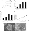
Fig. 1.
Fig. 1.
IQ-1 maintains undifferentiated state of ESCs. (a) Structure of IQ-1. (b) IQ-1 dose-dependently maintains alkaline phosphatase activity. Quantification of alkaline phosphatase activity in ESCs in a feeder-free system was performed as described in Experimental Procedures. (c) IQ-1 maintains SSEA-1 expression dose-dependently. Quantification of SSEA-1 expression in ESCs, in a feeder-free system and supplemented with either IQ-1 or LIF, was assayed as described in Experimental Procedures. SSEA-1 expression was analyzed 7 days after addition of IQ-1. (d) IQ-1 enabled ESCs to proliferate in the undifferentiated state for at least 65 days, without MEF feeders or LIF. ESCs, in media supplemented with 4 μg/ml IQ-1, are passaged 2–3 times every week at 1 × 10⁵ to 1 × 10⁶ cells per 6-cm dish and counted. All error bars represent ±SD. (e) Alkaline phosphatase staining of ESCs grown in a feeder-free system in the presence or absence of IQ-1 (4 μg/ml) for 65 days.
Fig. 2.
Fig. 2.
IQ-1 maintains ESCs' self-renewal independently of LIF. (a) IQ-1 increases Nanog gene expression significantly compared with LIF. mRNA was isolated from ESCs cultured under a feeder-free system in the presence of either IQ-1 (4 μg/ml) or LIF (1,000 units/ml) for 21 h. Real-time RT-PCR for Nanog was performed. The control expression level of Nanog at day 0 was set at 1. (b) Removal of IQ-1 decreases Nanog gene expression in ESCs. IQ-1 was removed for 3 days from ESCs previously cultured under a feeder-free system in the presence of IQ-1. mRNA was isolated at day 0 or day 3, and real-time RT-PCR was performed to assay for Nanog gene expression. (c) Effects of IQ-1 were not mediated through Stat3 signaling pathway as judged by luciferase reporter assay. Feeder-free ESCs, transfected with the pSTAT3-TA-Luc reporter, were exposed to either IQ-1 at the indicated doses or LIF. R.L.U., relative light unit. All error bars represent mean ± SD.
Fig. 3.
Fig. 3.
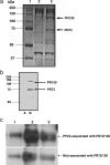
IQ-1 modulates Wnt signaling via interaction with PR72/130. (a) Coomassie stain of affinity chromatography isolation of IQ-1's molecular target(s), which was performed as described in Experimental Procedures. The two bands at 72 kDa and 130 kDa (labeled) were identified by mass spectral sequencing as the differentially spliced regulatory subunits PR72/130 of the serine/threonine protein phosphatase, PP2A. Lane 1, marker; lane 2, negative control; lane 3, pull-down. (b) Immunoblotting, using PR72/130 antisera, was performed to confirm the identity of the two bands. A, inactive biotinylated substrate pull-down; B, biotinylated IQ-1 pull-down. (c) Nuclear lysates from P19 cells treated with DMSO (control) or 10 μM IQ-1 were coimmunoprecipitated with antisera to PR72/130 and immunoblotted for PP2A and Nkd (described in Experimental Procedures). Lane 1 (control) was immunoblotted by using rabbit IgG. Lane 2 shows PP2A (Upper) and Nkd (Lower) from DMSO control-treated P19 cells. Lane 3 demonstrates the disruption of the PR72/130 complex, as judged by the diminished signal for PP2A (Upper) and Nkd (Lower) from P19 cells exposed to 10 μM IQ-1.
Fig. 4.
Fig. 4.
IQ-1 Maintenance of ESCs is Wnt/β-catenin/CBP-dependent. (a) Wnt/β-catenin coactivator switching model. A key feature of this model is that β-catenin/CBP-mediated transcription is critical for “stem/progenitor” cell proliferation, whereas coactivator switching to β-catenin/p300-mediated transcription is critical to initiate a differentiative program (25, 26). TCF, T cell factor. (b) IQ-1 increases the β-catenin/CBP complex at the expense of the β-catenin/p300 complex. P19 cells were treated with Wnt3A supplemented with IQ-1, the β-catenin/CBP antagonist ICG-001, DMSO, or DMSO-only control. Nuclear lysates were coimmunoprecipitated with anti-CBP or anti-p300 antibody and immunoblotted for β-catenin. (c) Phosphorylation of p300 Ser-89, in a PKC-dependent manner, increases the β-catenin/p300 interaction. After in vitro phosphorylation with PKCα, wild-type p300 (1–110 aa) and the mutant p300 (p300 S89A) were mixed with P19 lysates and coimmunoprecipitated by using a β-catenin-specific antibody. Western blot analysis for p300 (Upper) or β-catenin loading control (Lower) was performed. Lane 1, β-catenin/p300 binding; lane 2, PKCα phosphorylated p300/β-catenin binding; lane 3, S89A p300/β-catenin binding; lane 4, PKCα phosphorylated S89A p300/β-catenin binding. (d) IQ-1 decreases the phosphorylation of p300 Ser-89. P19 cells were treated with IQ-1 or DMSO (control) and exposed to purified Wnt3A for 24 h. Cell lysates were immunoblotted by using antibodies specific for p300 or p300 phosphorylated at position Ser 89. Lane 1, negative control [vector control (VC) and DMSO]; lane 2, Wnt3a plus DMSO control: phospho Ser-89 p300 immunoblot (Top) and p300 immunoblot (Middle); lane 3, Wnt3a plus 10 μM IQ-1: phospho Ser-89 p300 immunoblot (Top) and p300 immunoblot (Middle). α-Tubulin is the loading control (Bottom).
Fig. 5.
Fig. 5.
Pluripotency of long-term-cultured ESCs. (a) ESCs cultured in media in the presence of Wnt3A and IQ-1 (4 μg/ml) for 48 days were able to form embryoid bodies (Left). ESCs lose their ability to form embryoid bodies after 3 days of culturing in the absence of IQ-1 (Right). (b) Embryoid bodies, derived from IQ-1-treated ESCs, were cultured (adherence culture) for 7–14 days to induce further differentiation. Immunofluorescence staining for α-fetoprotein, smooth muscle actin, GATA4, MAP2, β-III tubulin, and oligodendrocytes demonstrated that long-term culture of ESCs in the presence of IQ-1 preserves pluripotency. Shown are the endoderm (Upper Left), mesoderm (Lower Left), and ectoderm (Right). (c) Model depicting the mechanism of action of IQ-1.

References

    1. Dravid G, Ye Z, Hammond H, Chen G, Pyle A, Donovan P, Yu X, Cheng L. Stem Cells. 2005;23:1489–1501. - PubMed
    1. Willert K, Brown JD, Danenberg E, Duncan AW, Weissman IL, Reya T, Yates JR, III, Nusse R. Nature. 2003;423:448–452. - PubMed
    1. Haegele L, Ingold B, Naumann H, Tabatabai G, Ledermann B, Brandner S. Mol Cell Neurosci. 2003;24:696–708. - PubMed
    1. Sato N, Meijer L, Skaltsounis L, Greengard P, Brivanlou A. Nat Med. 2004;10:55–63. - PubMed
    1. Chenn A, Walsh CA. Science. 2002;297:365–369. - PubMed