

Alteration in the gene expression pattern of primary monocytes after adhesion to endothelial cells
- PMID: 17372200
- PMCID: PMC1838499
- DOI: 10.1073/pnas.0700732104
Alteration in the gene expression pattern of primary monocytes after adhesion to endothelial cells
Abstract
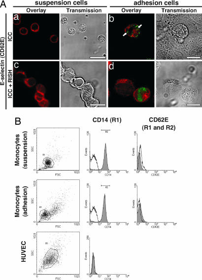
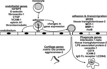
Monocytes originate from precursors made in the bone and remain in the circulation for nearly 24 h. Much effort has been done to identify the molecules regulating transendothelial migration of monocytes during inflammatory conditions. In contrast, considerably less is known about the process of constitutive monocyte emigration although nearly 340 million monocytes leave the circulation each day in healthy individuals. Previous studies indicated that chemokines were up-regulated in monocytes cocultured with endothelial cells that induce the retraction of the latter cell type, thereby increasing vascular permeability. Thus, we hypothesized that the utilities required for efficient constitutive monocyte extravasation are generated by monocytes themselves because of adhesion to naïve endothelial cells. To test this hypothesis, cDNA microarray analysis was performed to determine the changes in the gene expression pattern of primary monocytes that have been attached to endothelial cells compared with monocytes that were held in suspension, and we were able to identify three major groups of genes. The first group includes genes such as matrix metalloproteinase 1, monocyte chemoattractant protein 1, and tissue transglutaminase 2, which are likely required for monocyte extravasation. The second group consists of genes that are expressed in phagocytes such as caveolin-1 and CD74. Finally, the third group comprises genes that are expressed in cells of endothelial tissue and cartilage including E-selectin, fibronectin-1, matrix Gla protein, and aggrecanase-2. In summary, we conclude that adhesion of peripheral blood monocytes to naïve endothelial cells has two effects: mandatory extravasation-specific genes are regulated, and the differentiation program of monocytes is initiated.
Conflict of interest statement
The authors declare no conflict of interest.
Figures


Similar articles
-
Monocytes induce E-selectin gene expression in endothelial cells: role of CD11/CD18 and extracellular matrix proteins.Eur J Immunol. 1996 Dec;26(12):2944-51. doi: 10.1002/eji.1830261221. Eur J Immunol. 1996. PMID: 8977290
-
An endothelial cell adhesion protein for monocytes recognized by monoclonal antibody IG9. Expression in vivo in inflamed human vessels and atherosclerotic human and Watanabe rabbit vessels.Lab Invest. 1994 Jun;70(6):836-49. Lab Invest. 1994. PMID: 8015288
-
C-reactive protein promotes adhesion of monocytes to endothelial cells via NADPH oxidase-mediated oxidative stress.J Cell Biochem. 2012 Mar;113(3):857-67. doi: 10.1002/jcb.23415. J Cell Biochem. 2012. PMID: 22020763
-
Molecular mechanism of blood monocyte adhesion to vascular endothelial cells.Nouv Rev Fr Hematol (1978). 1992;34 Suppl:S55-9. Nouv Rev Fr Hematol (1978). 1992. PMID: 1340530 Review.
-
Human circulating monocytes as multipotential progenitors.Keio J Med. 2007 Jun;56(2):41-7. doi: 10.2302/kjm.56.41. Keio J Med. 2007. PMID: 17609587 Review.
Cited by
-
Central Nervous System Barriers Impact Distribution and Expression of iNOS and Arginase-1 in Infiltrating Macrophages During Neuroinflammation.Front Immunol. 2021 Apr 15;12:666961. doi: 10.3389/fimmu.2021.666961. eCollection 2021. Front Immunol. 2021. PMID: 33936108 Free PMC article.
-
Phenotypic and functional changes in blood monocytes following adherence to endothelium.PLoS One. 2012;7(5):e37091. doi: 10.1371/journal.pone.0037091. Epub 2012 May 15. PLoS One. 2012. PMID: 22615904 Free PMC article.
-
Endothelial Basement Membrane Laminins as an Environmental Cue in Monocyte Differentiation to Macrophages.Front Immunol. 2020 Oct 30;11:584229. doi: 10.3389/fimmu.2020.584229. eCollection 2020. Front Immunol. 2020. PMID: 33193400 Free PMC article.
-
D6 facilitates cellular migration and fluid flow to lymph nodes by suppressing lymphatic congestion.Blood. 2011 Dec 1;118(23):6220-9. doi: 10.1182/blood-2011-03-344044. Epub 2011 Oct 6. Blood. 2011. PMID: 21979941 Free PMC article.
-
Jagged1 Instructs Macrophage Differentiation in Leprosy.PLoS Pathog. 2016 Aug 17;12(8):e1005808. doi: 10.1371/journal.ppat.1005808. eCollection 2016 Aug. PLoS Pathog. 2016. PMID: 27532668 Free PMC article.
References
-
- Cruse JM, Lewis RE. Illustrated Dictionary of Immunology. Boca Raton, FL: CRC Press; 1995.
-
- Ley K. Cardiovasc Res. 1996;32:733–742. - PubMed
-
- Olson TS, Ley K. Am J Physiol. 2002;283:R7–R28. - PubMed
-
- Zhao B, Stavchansky SA, Bowden RA, Bowman PD. Am J Physiol. 2003;284:C1577–C1583. - PubMed
-
- Ley K, Allietta M, Bullard DC, Morgan S. Circ Res. 1998;83:287–294. - PubMed
Publication types
MeSH terms
Substances
LinkOut - more resources
Full Text Sources
Other Literature Sources
Research Materials
Miscellaneous
