

Implications for invariant natural killer T cell ligands due to the restricted presence of isoglobotrihexosylceramide in mammals
- PMID: 17372214
- PMCID: PMC1851601
- DOI: 10.1073/pnas.0607285104
Implications for invariant natural killer T cell ligands due to the restricted presence of isoglobotrihexosylceramide in mammals
Abstract
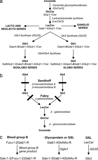
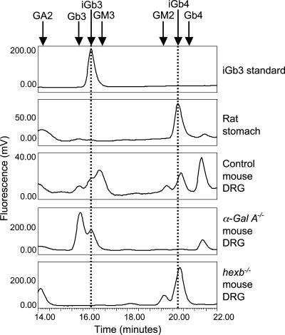
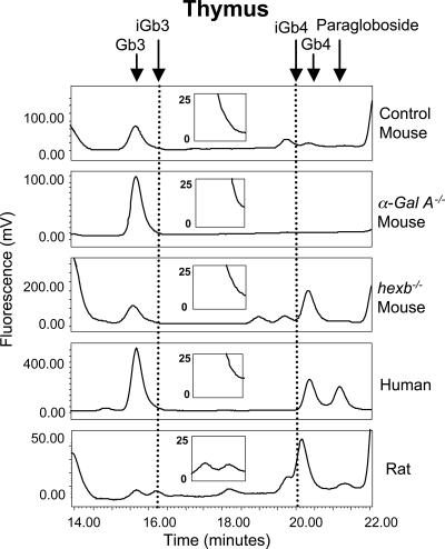
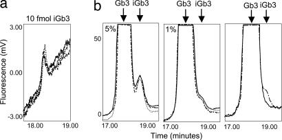
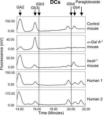
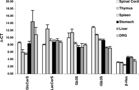
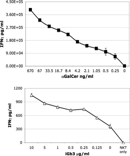
Development of invariant natural killer T (iNKT) cells requires the presentation of lipid ligand(s) by CD1d molecules in the thymus. The glycosphingolipid (GSL) isoglobotrihexosylceramide (iGb3) has been proposed as the natural iNKT cell-selecting ligand in the thymus and to be involved in peripheral activation of iNKT cells by dendritic cells (DCs). However, there is no direct biochemical evidence for the presence of iGb3 in mouse or human thymus or DCs. Using a highly sensitive HPLC assay, the only tissue where iGb3 could be detected in mouse was the dorsal root ganglion (DRG). iGb3 was not detected in other mouse or any human tissues analyzed, including thymus and DCs. Even in mutant mice that store isoglobo-series GSLs in the DRG, we were still unable to detect these GSLs in the thymus. iGb3 is therefore unlikely to be a physiologically relevant iNKT cell-selecting ligand in mouse and humans. A detailed study is now warranted to better understand the nature of iNKT cell-selecting ligand(s) in vivo.
Conflict of interest statement
The authors declare no conflict of interest.
Figures
Comment in
-
Natural killer T cells: know thyself.Proc Natl Acad Sci U S A. 2007 Apr 3;104(14):5713-4. doi: 10.1073/pnas.0701493104. Epub 2007 Mar 27. Proc Natl Acad Sci U S A. 2007. PMID: 17389403 Free PMC article. No abstract available.
Similar articles
-
Normal development and function of invariant natural killer T cells in mice with isoglobotrihexosylceramide (iGb3) deficiency.Proc Natl Acad Sci U S A. 2007 Apr 3;104(14):5977-82. doi: 10.1073/pnas.0611139104. Epub 2007 Mar 19. Proc Natl Acad Sci U S A. 2007. PMID: 17372206 Free PMC article.
-
Globosides but not isoglobosides can impact the development of invariant NKT cells and their interaction with dendritic cells.J Immunol. 2012 Sep 15;189(6):3007-17. doi: 10.4049/jimmunol.1201483. Epub 2012 Aug 8. J Immunol. 2012. PMID: 22875802 Free PMC article.
-
CD1d protein structure determines species-selective antigenicity of isoglobotrihexosylceramide (iGb3) to invariant NKT cells.Eur J Immunol. 2013 Mar;43(3):815-25. doi: 10.1002/eji.201242952. Epub 2013 Jan 31. Eur J Immunol. 2013. PMID: 23280365 Free PMC article.
-
Chewing the fat on natural killer T cell development.J Exp Med. 2006 Oct 2;203(10):2229-32. doi: 10.1084/jem.20061787. Epub 2006 Sep 25. J Exp Med. 2006. PMID: 17000869 Free PMC article. Review.
-
Turned on by danger: activation of CD1d-restricted invariant natural killer T cells.Immunology. 2012 Sep;137(1):20-7. doi: 10.1111/j.1365-2567.2012.03612.x. Immunology. 2012. PMID: 22734667 Free PMC article. Review.
Cited by
-
Invariant NKT cells regulate the CD8 T cell response during Theiler's virus infection.PLoS One. 2014 Jan 30;9(1):e87717. doi: 10.1371/journal.pone.0087717. eCollection 2014. PLoS One. 2014. PMID: 24498175 Free PMC article.
-
CD1 mediated T cell recognition of glycolipids.Curr Opin Struct Biol. 2007 Oct;17(5):521-9. doi: 10.1016/j.sbi.2007.09.010. Epub 2007 Oct 24. Curr Opin Struct Biol. 2007. PMID: 17951048 Free PMC article. Review.
-
NKT cell ligand recognition logic: molecular basis for a synaptic duet and transmission of inflammatory effectors.J Immunol. 2011 Aug 1;187(3):1081-9. doi: 10.4049/jimmunol.1001910. J Immunol. 2011. PMID: 21772035 Free PMC article. Review.
-
Recognition of β-linked self glycolipids mediated by natural killer T cell antigen receptors.Nat Immunol. 2011 Jul 31;12(9):827-33. doi: 10.1038/ni.2076. Nat Immunol. 2011. PMID: 21804559 Free PMC article.
-
Lack of iGb3 and Isoglobo-Series Glycosphingolipids in Pig Organs Used for Xenotransplantation: Implications for Natural Killer T-Cell Biology.J Carbohydr Chem. 2013 Jan 1;32(1):44-67. doi: 10.1080/07328303.2012.741637. Epub 2013 Jan 11. J Carbohydr Chem. 2013. PMID: 23378701 Free PMC article.
References
-
- Brigl M, Brenner MB. Annu Rev Immunol. 2004;22:817–890. - PubMed
-
- Kinjo Y, Wu D, Kim G, Xing GW, Poles MA, Ho DD, Tsuji M, Kawahara K, Wong CH, Kronenberg M. Nature. 2005;434:520–525. - PubMed
-
- Mattner J, Debord KL, Ismail N, Goff RD, Cantu C, III, Zhou D, Saint-Mezard P, Wang V, Gao Y, Yin N, et al. Nature. 2005;434:525–529. - PubMed
-
- Godfrey DI, MacDonald HR, Kronenberg M, Smyth MJ, Van Kaer L. Nat Rev Immunol. 2004;4:231–237. - PubMed
Publication types
MeSH terms
Substances
Grants and funding
LinkOut - more resources
Full Text Sources
Molecular Biology Databases
