. 2007 Mar 27;104(13):5413-8.
doi: 10.1073/pnas.0700809104. Epub 2007 Mar 19.

A critical role for the E3-ligase activity of c-Cbl in VEGFR-2-mediated PLCgamma1 activation and angiogenesis

Affiliations

A critical role for the E3-ligase activity of c-Cbl in VEGFR-2-mediated PLCgamma1 activation and angiogenesis

Amrik J Singh et al. Proc Natl Acad Sci U S A. .

Abstract

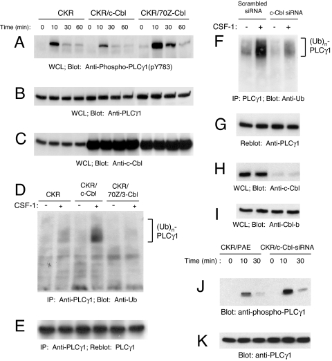
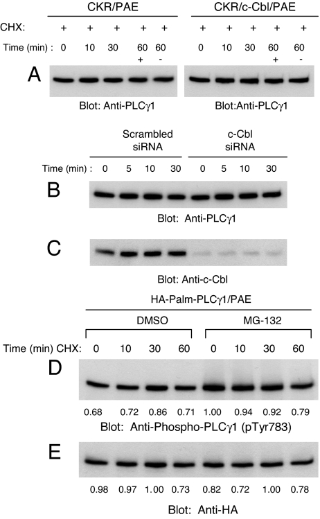
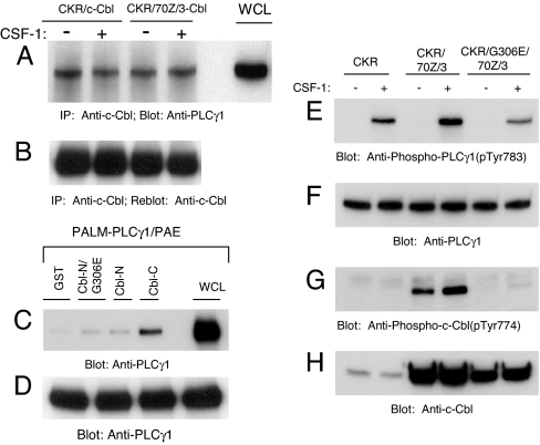
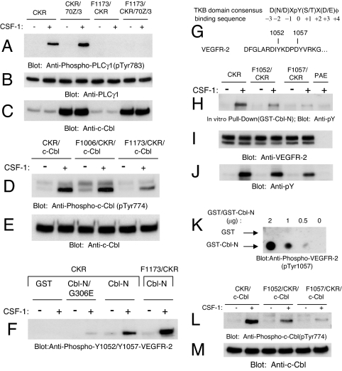
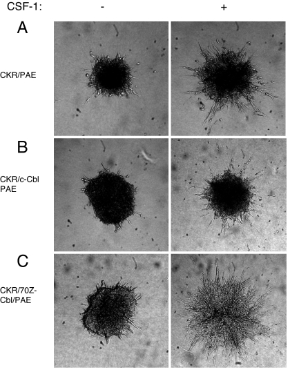
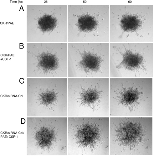
Activation of phospholipase Cgamma1 (PLCgamma1) by vascular endothelial growth factor receptor-2 (VEGFR-2) in endothelial cells in part is responsible for angiogenesis in vivo. The cellular mechanisms exerting negative control over PLCgamma1 activation, however, remain unaddressed. Here by using in vitro and in vivo binding assays, we show that the Casitas B-lineage lymphoma (c-Cbl) E3 ubiquitin ligase constitutively associates with PLCgamma1 via its C-terminal domain and conditionally interacts with VEGFR-2 via the N-terminal/TKB domain. Site-directed mutagenesis of VEGFR-2 showed that full activation of c-Cbl requires its direct association with phospho-tyrosines 1052 and 1057 of VEGFR-2 via its TKB domain and indirect association with phospho-tyrosine 1173 of VEGFR-2 via PLCgamma1. The tertiary complex formation between VEGFR-2, PLCgamma1 and c-Cbl selectively promotes ubiquitylation and suppression of tyrosine phosphorylation of PLCgamma1 by a proteolysis-independent mechanism. Further analysis showed that association of c-Cbl with VEGFR-2 does not impact ubiquitylation, down-regulation, or tyrosine phosphorylation of VEGFR-2. Silencing of c-Cbl by siRNA revealed that endogenous c-Cbl plays an inhibitory role in angiogenesis. Our data demonstrate that corecruitment of c-Cbl and PLCgamma1 to VEGFR-2 serves as a mechanism to fine-tune the angiogenic signal relay of VEGFR-2.


Conflict of interest statement

The authors declare no conflict of interest.

Figures

Fig. 1.
Fig. 1.
c-Cbl inhibits PLCγ1 tyrosine phosphorylation and promotes its ubiquitylation in endothelial cells. (A) PAE cells expressing either CKR (chimeric VEGFR-2) alone, coexpressing the wild-type Cbl or 70Z/3-Cbl were either unstimulated or stimulated with CSF-1 for various times as indicated. PLCγ1 activation in normalized whole cell lysates (WCL) was analyzed by immunoblotting with a phospho-specific (pY783) PLCγ1 antibody. (B) PLCγ1 expression in whole cell lysates was determined in a parallel immunoblot using an anti-PLCγ1 antibody. (C) overexpression of c-Cbl and 70Z/3-Cbl proteins was determined by immunoblotting with an anti-c-Cbl antibody. (D) PLCγ1 was immunoprecipitated (IP) from RIPA whole cell lysates and was assessed by immunoblotting with an anti-ubiquitin (Ub) antibody. (E) The blot shown in D was stripped and reprobed with an anti-PLCγ1 antibody to demonstrate equal amounts of immunoprecipitated PLCγ1. (F) CKR/PAE cells were retrovirally transduced with either constructs expressing a control siRNA (Scrambled siRNA) or a siRNA targeting c-Cbl (c-Cbl siRNA). The aforementioned cell lines were left either unstimulated or stimulated for 10 min, immunoprecipitated with anti-PLCγ1 antibody, and immunoblotted with anti-ubiquitin antibody. (G) The same membrane was immunoblotted with anti-PLCγ1 antibody. (H and I) Whole cell lysates were immunoblotted with anti-c-Cbl (H) or anti-Cbl-b (I) antibodies. (J and K) CKR/PAE cells and CKR/PAE cells expressing c-Cbl-siRNA were stimulated with CSF-1 for various times as indicated, and whole cell lysates was analyzed by immunoblotting with a phospho-specific (pY783) PLCγ1 antibody (J) or with an anti-PLCγ1 antibody (K).
Fig. 2.
Fig. 2.
c-Cbl E3 ligase activity is not associated with proteolysis of PLCγ1. (A) PAE cells either expressing wild-type CKR alone or coexpressing c-Cbl were treated with CHX (20 μg/ml) and CSF-1 for indicated times and whole cell lysates were immunoblotted with an anti-PLCγ1 antibody. (B) PAE and CKR/PAE cells expressing either a control siRNA (CKR/Scrambled siRNA) or a siRNA targeting c-Cbl (CKR/c-Cbl siRNA) were unstimulated (−) or stimulated (+) with CSF-1 for indicated periods of time, and whole cell lysates were immunoblotted with an anti-PLCγ1 antibody. (C) A parallel immunoblot of whole cell lysate aliquots was probed with an anti-c-Cbl antibody. (D) PAE cells expressing constitutively active PLCγ1 (HA-Palm-PLCγ1) were preincubated for 2 h with either DMSO or MG-132 (50 μM) followed by a 30-min preincubation with CHX (20 μg/ml). Cells were then either lysed (0 min) or incubated in the continued presence of CHX for the indicated periods with or without MG-132. At each time point, whole cell lysates were immunoblotted with an anti-phospho-PLCγ1 antibody (pTyr783). (E) A parallel immunoblot of whole cell lysates was probed with an anti-HA antibody.
Fig. 3.
Fig. 3.
c-Cbl constitutively associates with PLCγ1 via its carboxyl domain. (A) CKR/c-Cbl/PAE and CKR/70Z/3-Cbl/PAE cells were either unstimulated (−) or stimulated (+) for 10 min with CSF-1. Normalized whole cell lysates were immunoprecipitated (IP) with an anti-c-Cbl antibody and immunoprecipitates were immunoblotted with an anti-PLCγ1 antibody. Positive controls consist of whole cell lysate (WCL) aliquots and are indicated as such. (B) The same membrane was stripped and reprobed with an anti-c-Cbl antibody. (C) PAE cells ectopically expressing constitutively active PLCγ1 (Palm-PLCγ1) were serum-starved overnight. Normalized whole cell lysates from each of the four dishes were incubated separately with equal amounts of GST, GST-Cbl-N/G306E, GST-Cbl-N, and GST-Cbl-C fusion proteins as indicated in an in vitro GST pull-down assay. Precipitated PLCγ1 was analyzed by immunoblotting with an anti-PLCγ1 antibody. Positive controls consist of whole cell lysate aliquots and are indicated as such. (D) A parallel blot of whole cell lysates was probed with an anti-PLCγ1 antibody as a loading control. (E) PAE cells either expressing wild-type CKR alone or with the indicated Cbl constructs were either unstimulated (−) or stimulated (+) for 10 min with CSF-1 and normalized whole cell lysates were immunoblotted with an anti-phospho-PLCγ1 antibody (pTyr783). (F) Parallel blot of whole cell lysates were probed with an anti-PLCγ1 antibody. (G and H) Phosphorylation of 70Z/3-Cbl and G306E-70Z/3-Cbl and their expression.
Fig. 4.
Fig. 4.
Role of tyrosines 1173, 1052, and 1057 of VEGFR-2 in the recruitment and tyrosine phosphorylation of c-Cbl. (A) PAE cells expressing CKR, F1173/CKR alone or coexpressing F1173/CKR with 70Z/3-Cbl were either unstimulated (−) or stimulated (+) for 10 min with CSF-1, and whole cell lysates were immunoblotted with an anti-phospho-PLCγ1 antibody (pTyr783). (B and C) Parallel blots of whole cell lysates were probed with an anti-PLCγ1 (B) and an anti-c-Cbl (C) antibodies to show their expression. (D) PAE cells coexpressing CKR with c-Cbl, F1006/CKR with c-Cbl, and F1173/CKR with c-Cbl were either unstimulated (−) or stimulated (+) for 10 min with CSF-1, and whole cell lysates were immunoblotted with an anti-phospho-c-Cbl antibody (pTyr774). (E) Parallel blot of whole cell lysates was probed with an anti-c-Cbl antibody. (F) CKR/PAE and F1173/CKR/PAE cells were treated as in A. Whole cell lysates were incubated with equal amounts of GST, GST-Cbl-N/G306E, and GST-Cbl-N fusion proteins as indicated in an in vitro GST pull-down assay. Precipitated CKR was analyzed by immunoblotting with an anti-phospho-VEGFR-2 antibody that detects VEGFR-2 autophosphorylated at Y1052 and Y1057 (pTyr1052/pTyr1057). (G) The c-Cbl TKB domain consensus binding sequence and a partial alignment of the activation loop primary amino acid sequence of VEGFR-2. (H) PAE cells expressing either wild-type CKR or the indicated activation loop mutants were unstimulated (−) or stimulated (+) for 10 min with CSF-1. Whole cell lysates were incubated with equal amounts of a GST-Cbl-N fusion protein in an in vitro GST pull-down assay. Cell lysates from CSF-1-stimulated PAE cells were used as a negative control. CKR·GST-Cbl-N complexes were analyzed by immunoblotting with an anti-phosphotyrosine antibody (pY). (I) A parallel blot of whole cell lysates was probed with an anti-VEGFR-2 antibody (1412) as a control for receptor levels. (J) A parallel blot of whole cell lysates was probed with an anti-phosphotyrosine antibody (pY). (K) To detect a direct interaction between the Cbl TKB domain and VEGFR-2 activation loop tyrosines, the indicated quantities of purified recombinant GST control (Upper) and GST-Cbl-N fusion proteins (Lower) were dot blotted as described in Materials and Methods. (L and M) PAE cells coexpressing wild-type c-Cbl and either wild-type CKR or the indicated mutant receptors were treated as described in H. Whole cell lysates were immunoblotted in parallel with anti-phospho-c-Cbl and anti-c-Cbl antibodies.
Fig. 5.
Fig. 5.
c-Cbl regulates VEGFR-2-driven angiogenesis in cell culture system. (A–C) PAE cells expressing either CKR alone, coexpressing wild-type c-Cbl, or coexpressing 70Z/3-Cbl were prepared as spheroids and subjected to an in vitro angiogenesis/tubulogenesis assay as described in Materials and Methods.
Fig. 6.
Fig. 6.
Silencing the expression of c-Cbl in endothelial cells enhances VEGFR-2-driven angiogenesis. PAE cells expressing either CKR alone or coexpressing c-Cbl-siRNA were prepared as spheroids and subjected to an in vitro angiogenesis/tubulogenesis as described in Fig. 5. Cells were either unstimulated, or stimulated with 40 ηg/ml CSF-1 (+CSF-1).

Similar articles

Cited by

References

    1. Jain R. Nat Med. 2003;9:685–693. - PubMed
    1. Rahimi N. Front Biosci. 2006;11:818–829. - PMC - PubMed
    1. Takahashi T, Yamaguchi S, Chida K, Shibuya M. EMBO J. 2001;20:2768–2778. - PMC - PubMed
    1. Meyer RD, Latz C, Rahimi N. J Biol Chem. 2003;278:16347–16355. - PMC - PubMed
    1. Sakurai Y, Ohgimoto K, Kataoka Y, Yoshida N, Shibuya M. Proc Natl Acad Sci USA. 2005;102:1076–1081. - PMC - PubMed

Publication types