

Recent advances in ophthalmic anterior segment imaging: a new era for ophthalmic diagnosis?
- PMID: 17372341
- PMCID: PMC1994765
- DOI: 10.1136/bjo.2006.103408
Recent advances in ophthalmic anterior segment imaging: a new era for ophthalmic diagnosis?
Abstract
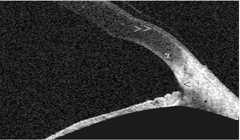
Anterior segment imaging is a rapidly advancing field of ophthalmology. New imaging modalities, such as rotating Scheimpflug imaging (Pentacam-Scheimpflug) and anterior segment optical coherence tomography (Visante OCT and Slit-Lamp OCT), have recently become commercially available. These new modalities supplement the more established imaging devices of Orbscan scanning slit topography and ultrasound biomicroscopy (UBM). All devices promise quantitative information and qualitative imaging of the cornea and anterior chamber. They provide a quantitative angle estimation by calculating the angle between the iris surface and the posterior corneal surface. Direct angle visualisation is possible with the OCT devices and UBM; they provide images of the scleral spur, ciliary body, ciliary sulcus and even canal of Schlemm in some eyes. Pentacam-Scheimpflug can measure net corneal power, a feature particularly useful for cataract patients having undergone previous corneal surgery. Anterior segment OCT can measure corneal flap depth following LASIK and anterior chamber width prior to phakic intraocular lens implantation. The arrival of the new imaging devices may herald the dawn of a new era for ophthalmic diagnosis, particularly in view of the ease and non-contact nature of examination.
Similar articles
-
Comparison of anterior segment optical coherence tomography and ultrasound biomicroscopy for assessment of the anterior segment.J Cataract Refract Surg. 2007 May;33(5):837-40. doi: 10.1016/j.jcrs.2007.01.021. J Cataract Refract Surg. 2007. PMID: 17466858
-
[New diagnostic methods for imaging the anterior segment of the eye to enable treatment modalities selection].Nippon Ganka Gakkai Zasshi. 2011 Mar;115(3):297-322; discussion 323. Nippon Ganka Gakkai Zasshi. 2011. PMID: 21476312 Review. Japanese.
-
Technologies for anatomical and geometric characterization of the corneal structure and anterior segment: a review.Semin Ophthalmol. 2015 May;30(3):161-70. doi: 10.3109/08820538.2013.835844. Epub 2013 Oct 31. Semin Ophthalmol. 2015. PMID: 24175646 Review.
-
Anterior segment imaging for iris melanocytic tumors.Eur J Ophthalmol. 2011 Sep-Oct;21(5):608-14. doi: 10.5301/EJO.2011.6214. Eur J Ophthalmol. 2011. PMID: 21218392
-
Comparison of anterior chamber depth measurements by 3-dimensional optical coherence tomography, partial coherence interferometry biometry, Scheimpflug rotating camera imaging, and ultrasound biomicroscopy.J Cataract Refract Surg. 2012 Jul;38(7):1207-13. doi: 10.1016/j.jcrs.2012.02.036. Epub 2012 May 19. J Cataract Refract Surg. 2012. PMID: 22613688
Cited by
-
Imaging analysis of the biological parameters of the lens in patients with cortical age-related cataracts using ultrasound biomicroscopy.BMC Ophthalmol. 2023 Nov 22;23(1):480. doi: 10.1186/s12886-023-03227-2. BMC Ophthalmol. 2023. PMID: 37993828 Free PMC article.
-
Intraocular endoscopy for the evaluation and treatment of hypotony due to a traumatic cyclodialysis: a case report.BMC Ophthalmol. 2020 Mar 23;20(1):117. doi: 10.1186/s12886-020-01375-3. BMC Ophthalmol. 2020. PMID: 32293350 Free PMC article.
-
Corneal biomechanical assessment using corneal visualization scheimpflug technology in keratoconic and normal eyes.J Ophthalmol. 2014;2014:147516. doi: 10.1155/2014/147516. Epub 2014 Mar 30. J Ophthalmol. 2014. PMID: 24800059 Free PMC article.
-
Corneal Changes After Collagen Crosslinking for Keratoconus Using Dual Scheimpflug Imaging.J Ophthalmic Vis Res. 2015 Oct-Dec;10(4):358-63. doi: 10.4103/2008-322X.176894. J Ophthalmic Vis Res. 2015. PMID: 27051478 Free PMC article.
-
Effect of Proparacaine 0.375%-Sodium Fluorescein 0.25% Eye Drop Mixture and Fluorescein Strip on Anterior Segment Parameters.J Ophthalmol. 2018 Sep 5;2018:5926508. doi: 10.1155/2018/5926508. eCollection 2018. J Ophthalmol. 2018. PMID: 30254756 Free PMC article.
References
-
- Masters B R. Three‐dimensional microscopic tomographic imaging of the cataract in a human lens in vivo. Optics Express 19983332–338. - PubMed
-
- Brezinski M E, Fujimoto J G. Optical coherence tomography: High‐resolution imaging in non‐transparent tissue. IEEE J Select Topics in Quantum Electron 199951185–1192.
Publication types
MeSH terms
LinkOut - more resources
Full Text Sources
Other Literature Sources
Medical