

Decreased fragile histidine triad expression in colorectal cancer and its association with apoptosis inhibition
- PMID: 17373735
- PMCID: PMC4146863
- DOI: 10.3748/wjg.v13.i7.1018
Decreased fragile histidine triad expression in colorectal cancer and its association with apoptosis inhibition
Abstract
Aim: To detect the expression of fragile histidine triad (FHIT) in normal colorectal tissue, colorectal adenoma and colorectal cancer (CRC) tissue, and to analyze its relationship with the clinicopathological features of CRC, and apoptosis-associated proteins (Bcl-2, Bax, survivin) and apoptosis in colorectal cancer.
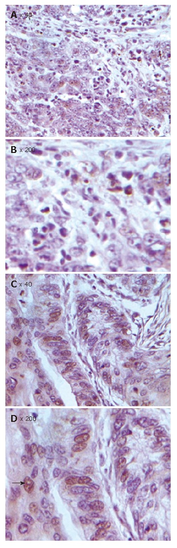
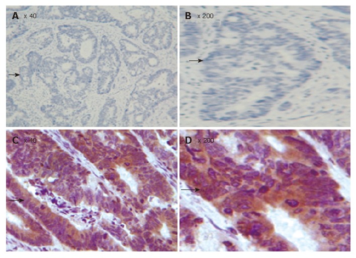
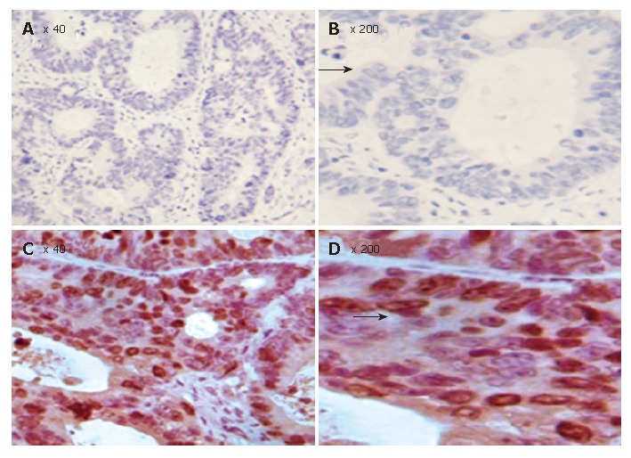
Methods: FHIT mRNA analysis was performed by nested reverse transcription-polymerase chain reaction (RT-PCR) assay. Tissue microarray (TMA) was established to detect the expression of FHIT, Bcl-2, Bax and survivin genes in 80 CRC tissue specimens, 16 colorectal adenoma tissue specimens and 16 hemorrhoid (PPH) tissue specimens during the same period of time as the control. Citrate-microwave-SP was used as immunohistochemical method. The relationship between clinicopathological factors, such as differentiation grades and 5-year survival rate was observed. TUNEL assay was used to detect the apoptosis index in 80 CRC tissue specimens.
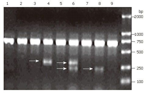
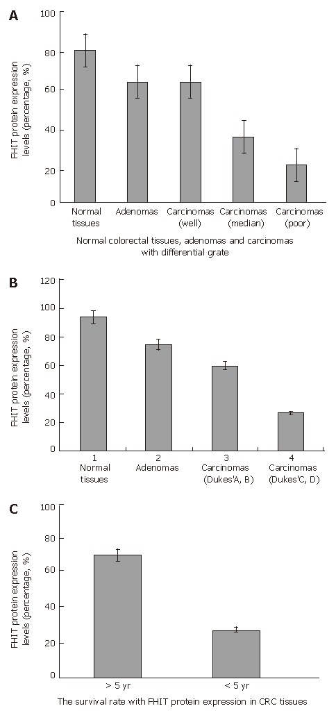
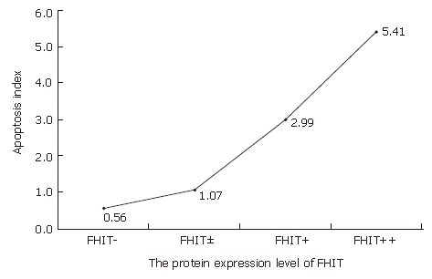
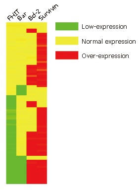
Results: Ten out of 26 (38.5%) CRC tissue specimens expressed aberrant FHIT transcripts, none of the aberrant FHIT transcripts was observed in the matched normal tissue and colorectal adenoma tissue by nested RT-PCR assay. The positive rate of FHIT gene expression in normal colorectal tissue, colorectal adenoma and carcinoma tissue was 93.75%, 68.75% and 46.25%, respectively. Clinicopathological analysis of patients showed that the decreased FHIT gene expression was not associated with age, sex, serum CEA levels, tumor site and size, histological classification. However, the expression of FHIT was correlated with differentiation grades, pathological stages, lymph node metastases and 5-year survival rate after operation. The positive rate of apoptosis-associated proteins (Bax, Bcl-2 and survivin) in CRC tissue was 72.50%, 51.25% and 77.50%, respectively. The expression of these apoptosis-associated proteins in CRC tissue was correlated with the expression of FHIT. The mean apoptosis index in FHIT negative tumors was significantly lower than that in FHIT positive tumors (5.41 +/- 0.23 vs 0.56 +/- 0.10, P < 0.01).
Conclusion: The FHIT gene plays an important role in the regulation of apoptosis and decreased FHIT expression plays a key role in the initiation and progression of colorectal carcinoma.
Figures


Similar articles
-
Down-regulation of FHIT inhibits apoptosis of colorectal cancer: mechanism and clinical implication.Surg Oncol. 2006 Dec;15(4):223-33. doi: 10.1016/j.suronc.2007.01.006. Epub 2007 Mar 26. Surg Oncol. 2006. PMID: 17382535
-
[Fragile histidine triad protein expression and correlation with apoptosis in rectal carcinoma].Zhonghua Wei Chang Wai Ke Za Zhi. 2007 Mar;10(2):177-81. Zhonghua Wei Chang Wai Ke Za Zhi. 2007. PMID: 17380463 Chinese.
-
Fragile histidine triad (FHIT) gene is overexpressed in colorectal cancer.J Physiol Pharmacol. 2009 Oct;60 Suppl 4:63-70. J Physiol Pharmacol. 2009. PMID: 20083853
-
BCL-2 family deregulation in colorectal cancer: potential for BH3 mimetics in therapy.Apoptosis. 2020 Jun;25(5-6):305-320. doi: 10.1007/s10495-020-01601-9. Apoptosis. 2020. PMID: 32335811 Free PMC article. Review.
-
FHIT: doubts are clear now.ScientificWorldJournal. 2010 Jun 16;10:1142-51. doi: 10.1100/tsw.2010.110. ScientificWorldJournal. 2010. PMID: 20563537 Free PMC article. Review.
Cited by
-
Livin serves as a prognostic marker for mid-distal rectal cancer and a target of mid-distal rectal cancer treatment.Oncol Lett. 2017 Dec;14(6):7759-7766. doi: 10.3892/ol.2017.7230. Epub 2017 Oct 20. Oncol Lett. 2017. PMID: 29344221 Free PMC article.
-
Coordinate loss of fragile gene expression in pancreatobiliary cancers: correlations among markers and clinical features.Ann Surg Oncol. 2009 Aug;16(8):2331-8. doi: 10.1245/s10434-009-0507-4. Epub 2009 May 12. Ann Surg Oncol. 2009. PMID: 19434452 Free PMC article.
-
Reduced FHIT expression is associated with mismatch repair deficient and high CpG island methylator phenotype colorectal cancer.J Histochem Cytochem. 2013 Sep;61(9):627-38. doi: 10.1369/0022155413497367. Epub 2013 Jun 24. J Histochem Cytochem. 2013. PMID: 23797051 Free PMC article.
-
Aberrant crypt focus and fragile histidine triad protein in sporadic colorectal carcinoma.World J Gastrointest Oncol. 2012 Dec 15;4(12):250-8. doi: 10.4251/wjgo.v4.i12.250. World J Gastrointest Oncol. 2012. PMID: 23443232 Free PMC article.
-
Fhit down-regulation is an early event in pancreatic carcinogenesis.Virchows Arch. 2017 Jun;470(6):647-653. doi: 10.1007/s00428-017-2105-3. Epub 2017 Mar 13. Virchows Arch. 2017. PMID: 28289900 Free PMC article.
References
-
- Hawk ET, Levin B. Colorectal cancer prevention. J Clin Oncol. 2005;23:378–391. - PubMed
-
- Ohta M, Inoue H, Cotticelli MG, Kastury K, Baffa R, Palazzo J, Siprashvili Z, Mori M, McCue P, Druck T, et al. The FHIT gene, spanning the chromosome 3p14.2 fragile site and renal carcinoma-associated t(3; 8) breakpoint, is abnormal in digestive tract cancers. Cell. 1996;84:587–597. - PubMed
-
- Boldog F, Gemmill RM, West J, Robinson M, Robinson L, Li E, Roche J, Todd S, Waggoner B, Lundstrom R, et al. Chromosome 3p14 homozygous deletions and sequence analysis of FRA3B. Hum Mol Genet. 1997;6:193–203. - PubMed
-
- Hayashi S, Tanimoto K, Hajiro-Nakanishi K, Tsuchiya E, Kurosumi M, Higashi Y, Imai K, Suga K, Nakachi K. Abnormal FHIT transcripts in human breast carcinomas: a clinicopathological and epidemiological analysis of 61 Japanese cases. Cancer Res. 1997;57:1981–1985. - PubMed
Publication types
MeSH terms
Substances
LinkOut - more resources
Full Text Sources
Medical
Research Materials
