

Pancreatic metastasis of leiomyosarcoma in the right thigh: a case report
- PMID: 17373753
- PMCID: PMC4146881
- DOI: 10.3748/wjg.v13.i7.1135
Pancreatic metastasis of leiomyosarcoma in the right thigh: a case report
Abstract
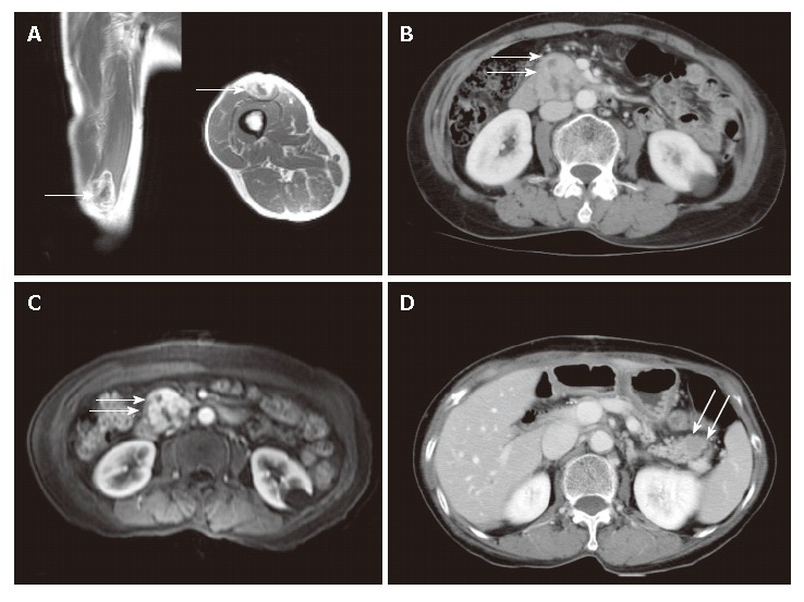
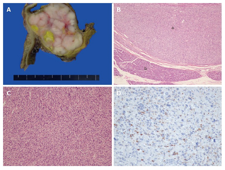
Pancreatic tumors are primary in most of the cases. Pancreatic metastases associated with other primary malignancies, especially pancreatic metastasis of leiomyosarcoma, are uncommon. A 66-year-old woman underwent surgical resection of malignant mesenchymoma (70% osteosarcoma and 30% leiomyosarcoma) in the right thigh. In the postoperative period, a pancreatic mass was identified radiologically by abdominal computed tomography. Pylorus-preserving pancreaticoduodenectomy was performed. The surgical specimen revealed leiomyosarcoma metastasized to the pancreas. A metastatic nodule on the remnant pancreatic tail was discovered 9 mo after the first pancreatic resection, and distal pancreatectomy was performed. Cases of pancreatic metastasis from leiomyosarcoma are extremely rare, especially when the tumor was resectable. We report here a unique case of pancreatic metastasis from a leiomyosarcoma in the right thigh that had been treated surgically.
Figures
References
-
- Crippa S, Angelini C, Mussi C, Bonardi C, Romano F, Sartori P, Uggeri F, Bovo G. Surgical treatment of metastatic tumors to the pancreas: a single center experience and review of the literature. World J Surg. 2006;30:1536–1542. - PubMed
-
- Adsay NV, Andea A, Basturk O, Kilinc N, Nassar H, Cheng JD. Secondary tumors of the pancreas: an analysis of a surgical and autopsy database and review of the literature. Virchows Arch. 2004;444:527–535. - PubMed
-
- Sperti C, Pasquali C, Liessi G, Pinciroli L, Decet G, Pedrazzoli S. Pancreatic resection for metastatic tumors to the pancreas. J Surg Oncol. 2003;83:161–166; discussion 166. - PubMed
-
- Roland CF, van Heerden JA. Nonpancreatic primary tumors with metastasis to the pancreas. Surg Gynecol Obstet. 1989;168:345–347. - PubMed
-
- Hiotis SP, Klimstra DS, Conlon KC, Brennan MF. Results after pancreatic resection for metastatic lesions. Ann Surg Oncol. 2002;9:675–679. - PubMed
Publication types
MeSH terms
LinkOut - more resources
Full Text Sources
Medical
