. 2007 Apr;5(4):e81.
doi: 10.1371/journal.pbio.0050081.

H2A.Z-mediated localization of genes at the nuclear periphery confers epigenetic memory of previous transcriptional state

Affiliations

H2A.Z-mediated localization of genes at the nuclear periphery confers epigenetic memory of previous transcriptional state

Donna Garvey Brickner et al. PLoS Biol. 2007 Apr.

Abstract

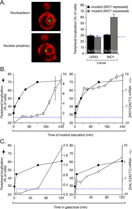
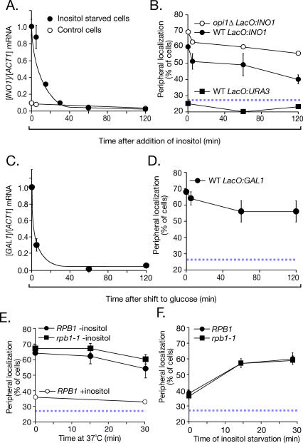
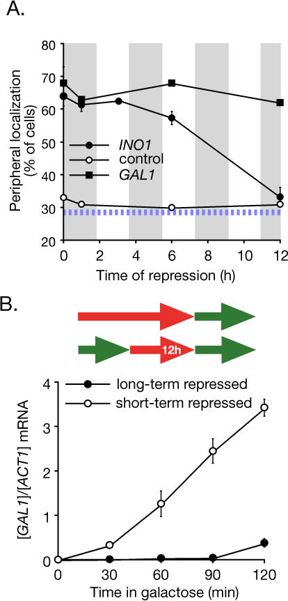
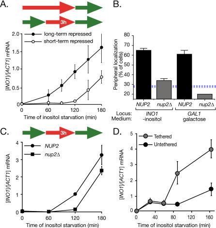
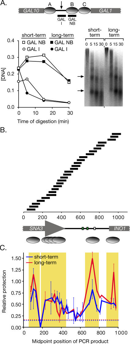
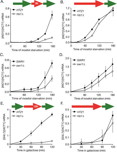
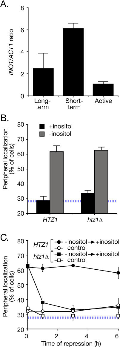
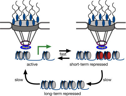
Many genes are recruited to the nuclear periphery upon transcriptional activation. The mechanism and functional significance of this recruitment is unclear. We find that recruitment of the yeast INO1 and GAL1 genes to the nuclear periphery is rapid and independent of transcription. Surprisingly, these genes remain at the periphery for generations after they are repressed. Localization at the nuclear periphery serves as a form of memory of recent transcriptional activation, promoting reactivation. Previously expressed GAL1 at the nuclear periphery is activated much more rapidly than long-term repressed GAL1 in the nucleoplasm, even after six generations of repression. Localization of INO1 at the nuclear periphery is necessary and sufficient to promote more rapid activation. This form of transcriptional memory is chromatin based; the histone variant H2A.Z is incorporated into nucleosomes within the recently repressed INO1 promoter and is specifically required for rapid reactivation of both INO1 and GAL1. Furthermore, H2A.Z is required to retain INO1 at the nuclear periphery after repression. Therefore, H2A.Z-mediated localization of recently repressed genes at the nuclear periphery represents an epigenetic state that confers memory of transcriptional activation and promotes reactivation.


Conflict of interest statement

Competing interests. The authors have declared that no competing interests exist.

Figures

Figure 1
Figure 1. Recruitment of INO1 and GAL1 to the Nuclear Periphery Is Rapid
(A) Left: merged confocal micrographs of cells stained for Lac I-GFP (green) and Sec63-myc (red), and scored as peripheral or nucleoplasmic. Right: cells having the lac repressor array integrated either at URA3 (strain JBY409) or INO1 (JBY397) were grown in the presence or absence of inositol, and scored for peripheral localization as described [6]. Data are averages of multiple replicates (indicated as N) from independent cultures. Each replicate represents 30–50 cells. The hatched blue line represents the baseline level of peripheral localization for the URA3 gene. (B) At the indicated times after removal of inositol, cells were scored for peripheral localization of INO1 (filled circles, extrapolated to 300 min [Figure S1]; two replicates of 30–50 cells). Also, INO1 mRNA abundance was quantified using RT Q-PCR and expressed relative to ACT1 mRNA (open circles; [58]). Left panel: both datasets plotted on a linear scale. Right panel: the mRNA abundance was plotted on a logarithmic scale, and the localization was plotted on a linear scale. (C) The localization of the GAL1 gene (two replicates of 30–50 cells) and the GAL1 mRNA abundance were quantified in strain DBY32 and plotted as in (B).
Figure 2
Figure 2. Gene Recruitment Is Maintained after Repression
(A and B) The abundance of the INO1 mRNA ([A], filled circles) and its peripheral localization ([B], filled circles; five replicates of 30–50 cells) in strain JBY397 were quantified at the indicated times after adding 100 μM myo-inositol. In (A), a control strain that was grown continuously in the presence of inositol (open circles) was included for comparison. In (B), the localization of INO1 in the opi1Δ mutant (strain JBY404, open circles; two replicates of 30–50 cells), which lacks the repressor of INO1, and the localization of URA3 (strain JBY409, filled squares; one replicate of 50 cells) were included for comparison. WT, wild type. (C and D) Strain DBY32 was shifted from galactose medium to glucose medium, and the abundance of the GAL1 mRNA (C) and its peripheral localization (two replicates of 30–50 cells (D) were quantified. In (A) and (C), the mRNA ratios were normalized to the initial, fully induced levels. (E and F) Wild-type (strain JBY397) or rpb1–1 (strain JBY461-r2) cells having the lac repressor array integrated at INO1 were grown at 25 °C in the absence (E) or presence (F) of inositol. In (E), cells were shifted to 37 °C for the indicated times and scored for peripheral localization (two replicates of 30–50 cells). In (F), cells were first incubated at 37 °C for 15 min before shifting into medium lacking inositol at 37 °C (two replicates of 30–50 cells). Peripheral localization of the INO1 gene was quantified as in Figure 1. The hatched blue line represents the baseline level of peripheral localization for the URA3 gene.
Figure 3
Figure 3. Memory of Recent Transcription.
(A) Localization of INO1 and GAL1 at the nuclear periphery persists for generations after repressing transcription. Strains JBY397 (lac operator array at INO1) and DBY32 (lac operator array at GAL1) were shifted from the activating condition to the repressing condition, and the localization at the nuclear periphery was quantified at the indicated times (two replicates of 30–50 cells). Cells were maintained in log phase by continual dilution, and the doubling time was approximately 110 min, indicated as grey vertical bars along the x-axis. The hatched blue line represents the baseline level of peripheral localization for the URA3 gene. (B) Top: schematic of the growth conditions: green arrows indicate growth under activating conditions, red arrows indicate growth under repressing conditions, and inset time indicates the duration of repression. Bottom: GAL1 activation versus reactivation. Strain BY4741 cultures grown under long-term or short-term (12 h) repressing conditions were shifted into galactose medium. GAL1 mRNA abundance was quantified at the indicated times using RT Q-PCR and expressed relative to ACT1 mRNA. Note that strain BY4741 activates GAL1 more slowly than the CRY1-derived strain used in Figure 1 (Figure S5).
Figure 4
Figure 4. Localization at the Nuclear Periphery Is Necessary and Sufficient to Promote Reactivation of INO1
(A) INO1 activation versus reactivation. Strain BY4741 cultures grown under long-term or short-term (3 h) repressing conditions were shifted into medium lacking inositol. INO1 mRNA abundance was quantified at the indicated times of inositol starvation using RT Q-PCR and expressed relative to ACT1 mRNA. (B) Wild-type (JBY397 for INO1, DBY32 for GAL1; four replicates of 30–50 cells) and nup2Δ (JBY462 for INO1, JBY467 for GAL1; two replicates of 30–50 cells) strains having the lac repressor–binding site array integrated at INO1 or GAL1 were scored for peripheral localization under activating conditions. The hatched blue line represents the baseline level of peripheral localization for the URA3 gene. (C) Schematic of the growth conditions: green arrows indicate growth under activating conditions; red arrows indicate growth under repressing conditions. After 3 h of repression with 100 μM inositol, wild-type (CRY1) or nup2Δ (JBY451-r1) mutant cells were shifted to medium lacking inositol, and INO1 mRNA levels were quantified at the indicated times. (D) Tethering of INO1 to the nuclear periphery enhances the rate of activation. Strains having the lac operator array integrated upstream of the INO1 gene were transformed with either wild-type Lac I-GFP (JBY397) or Lac I-FFAT-GFP (JBY399) to target the gene to the nuclear membrane [6]. These strains were shifted into medium lacking inositol for the indicated times, and INO1 mRNA levels were quantified.
Figure 5
Figure 5. Nucleosome Positioning and Relative Occupancy in the Long-Term Repressed and Short-Term Repressed INO1 Promoters
(A) Top panel: a map of three known, well-positioned nucleosomes, called “A,” “B,” and “C” (grey ovals) within the GAL1–10 promoter [33]. PCR products to monitor the concentration of sequences protected by nucleosome “B” (GAL NB) or sequences from the inter-nucleosomal region (arrow; GAL I) are indicated below. Bottom panel: DNA from either long-term repressed (open symbols) or short-term repressed (1 h; filled symbols) strain CRY1 was digested with micrococcal nuclease. The concentration of the templates for GAL NB (squares) and GAL I (circles) was determined relative to intact yeast genomic DNA by Q-PCR and plotted against different time points of digestion. Right: inverted image of ethidium bromide–stained gel of the digestion reactions. (B) A map of the INO1 promoter and flanking sequences, along with the positions of PCR products quantified to analyze nuclease protection and nucleosome occupancy. Green boxes represent UASINO elements, and the white box represents the TATA box. Below the map, well-positioned nucleosomes are indicated as single ovals, and poorly positioned nucleosomes are indicated as an overlapping series. (C) DNA from short-term and long-term repressed cells digested with micrococcal nuclease for 30 min (A) was analyzed by Q-PCR. Relative protection of the templates for each PCR product in (B) was calculated as a ratio of the concentration of the GAL NB template and mapped using the midpoint of the PCR product. Error bars represent standard error. The hatched lines represent the relative protection of the non-nucleosomal GAL I for each sample. Yellow boxes highlight protected sequences.
Figure 6
Figure 6. Htz1 Is Required for Transcriptional Memory
(A and B) Strains BY4741 and BY4741 htz1Δ from either short-term (3 h) repressing conditions (A) or long-term repressing conditions (B) were shifted into medium without inositol and collected at the indicated time points. The INO1 and ACT1 mRNA levels were quantified by RT Q-PCR. (C and D) Strains BY4741 and BY4741 swr1Δ from either short-term (3 h) repressing conditions (A) or long-term repressing conditions (B) were shifted into medium without inositol and collected at the indicated time points. The INO1 and ACT1 mRNA levels were quantified by RT Q-PCR. (E and F) Strains BY4741 and BY4741 htz1Δ from either short-term (12 h) repressing conditions (E) or long-term repressing conditions (F) were shifted into galactose medium and collected at the indicated time points. The GAL1 and ACT1 mRNA levels were quantified by RT Q-PCR.
Figure 7
Figure 7. H2A.Z Associates Specifically with the Recently Repressed INO1 Promoter and Is Specifically Required to Maintain INO1 at the Nuclear Periphery after Repression
(A) Chromatin immunoprecipitation analysis of H2A.Z association with the INO1 promoter. Strain CRY1 was grown under activating, long-term repressing or short-term repressing (1 h) conditions, fixed with formaldehyde, and processed for immunoprecipitation using anti-Htz1 antibodies (Abcam). Recovered INO1 promoter was quantified by Q-PCR [6] and expressed relative to recovered ACT1 coding sequence. (B) H2A.Z is not required for gene recruitment. Wild-type (JBY397) and htz1Δ (DBY50) strains were grown either in the presence or absence of inositol and scored for localization at the nuclear periphery. Data are averages of five (HTZ1) or three (htz1Δ) replicates of 30–50 cells. (C) H2A.Z is required for transcriptional memory. Wild-type and htz1Δ strains were grown in the absence of inositol to activate INO1. Inositol was added to 100 μM and cells were collected for immunofluorescence at the indicated times after repressing transcription. Each time point represents an average of two replicates of 30–50 cells. The hatched line in (B) and (C) represents the baseline level of peripheral localization for the URA3 gene.
Figure 8
Figure 8. A Model for Transcriptional Memory
The long-term repressed state of INO1 and GAL1 localizes randomly within the nucleoplasm and is activated slowly. Upon activation, these genes are recruited to the nuclear periphery through interaction with NPC-associated Nup2, with full transcriptional activation following recruitment. Upon repression, INO1 and GAL1 remain at the nuclear periphery. Specific incorporation of the histone variant H2A.Z into the recently repressed promoter mediates retention at the nuclear periphery and rapid reactivation.

References

    1. Cockell M, Gasser SM. Nuclear compartments and gene regulation. Curr Opin Genet Dev. 1999;9:199–205. - PubMed
    1. Isogai Y, Tjian R. Targeting genes and transcription factors to segregated nuclear compartments. Curr Opin Cell Biol. 2003;15:296–303. - PubMed
    1. Brown KE, Guest SS, Smale ST, Hahm K, Merkenschlager M, et al. Association of transcriptionally silent genes with Ikaros complexes at centromeric heterochromatin. Cell. 1997;91:845–854. - PubMed
    1. Csink AK, Henikoff S. Genetic modification of heterochromatic association and nuclear organization in Drosophila . Nature. 1996;381:529–531. - PubMed
    1. Dernburg AF, Broman KW, Fung JC, Marshall WF, Philips J, et al. Perturbation of nuclear architecture by long-distance chromosome interactions. Cell. 1996;85:745–759. - PubMed

Publication types

MeSH terms