. 2007 Apr;5(4):e92.
doi: 10.1371/journal.pbio.0050092.

Protein oxidation implicated as the primary determinant of bacterial radioresistance

Affiliations

Protein oxidation implicated as the primary determinant of bacterial radioresistance

Michael J Daly et al. PLoS Biol. 2007 Apr.

Abstract

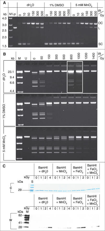
In the hierarchy of cellular targets damaged by ionizing radiation (IR), classical models of radiation toxicity place DNA at the top. Yet, many prokaryotes are killed by doses of IR that cause little DNA damage. Here we have probed the nature of Mn-facilitated IR resistance in Deinococcus radiodurans, which together with other extremely IR-resistant bacteria have high intracellular Mn/Fe concentration ratios compared to IR-sensitive bacteria. For in vitro and in vivo irradiation, we demonstrate a mechanistic link between Mn(II) ions and protection of proteins from oxidative modifications that introduce carbonyl groups. Conditions that inhibited Mn accumulation or Mn redox cycling rendered D. radiodurans radiation sensitive and highly susceptible to protein oxidation. X-ray fluorescence microprobe analysis showed that Mn is globally distributed in D. radiodurans, but Fe is sequestered in a region between dividing cells. For a group of phylogenetically diverse IR-resistant and IR-sensitive wild-type bacteria, our findings support the idea that the degree of resistance is determined by the level of oxidative protein damage caused during irradiation. We present the case that protein, rather than DNA, is the principal target of the biological action of IR in sensitive bacteria, and extreme resistance in Mn-accumulating bacteria is based on protein protection.


Conflict of interest statement

Competing interests. The authors have declared that no competing interests exist.

Figures

Figure 1
Figure 1. Model of IR-Driven Mn and Fe Redox Cycling
(H2O)n + γ-IR, radiolysis of water exposed to ionizing radiation [15]: H2O→ HO + H+ + e (IR-induced solvated electron); Primary radiolytic reaction [15]: 2 HO→ H2O2; IR-induced superoxide [15]: O2 + e → O2 •−; Fenton reaction [15]: Fe(II) + H2O2 → Fe(III) + HO + OH (hydroxide ion); Haber-Weiss reaction [15]: Fe(III) + HO2 → Fe(II) + O2 + H+ and 2 Fe(III) + H2O2 → 2 Fe(II) + O2 + 2 H+; Mn oxidation [13]: Mn(II) + O2 •− + 2 H+→ Mn(III) + H2O2; Mn reduction [13]: 2 Mn(III) + H2O2 → 2 Mn(II) + O2 + 2 H+. Under IR, Fe(II,III) redox cycling is predicted to generate HO and O2 •−, whereas Mn(II,III) redox cycling is predicted to favor O2 •− scavenging without HO production.
Figure 2
Figure 2. Mn(II) Protects Proteins, but Not DNA, during In Vitro Irradiation
(A) DMSO-mediated DNA protection. pUC19 plasmid DNA was irradiated aerobically to the indicated doses in dH2O, 1% DMSO (HO scavenger), or 5 mM MnCl2, followed by agarose gel electrophoresis (AGE). MnCl2 and DMSO were prepared in dH2O. IR, 60Co, aerobic at 0 °C. L, linear (2,686 base pairs); Lp, pUC19 + BamHI; M, size markers; OC, open circular; SC, supercoiled. (B) Mn(II)-mediated protein protection. BamHI enzyme was irradiated aerobically to the indicated doses in dH2O, 1% DMSO, or 5 mM MnCl2, and then incubated with λ-phage DNA for 1 h at 37 °C, followed by AGE. Inset (white border), top gel (dH2O): BamHI irradiated anaerobically. M, size markers; U, uncut λ-DNA. (C) Western blot immunoassay of protein-bound carbonyl groups in BamHI irradiated aerobically to the indicated doses in the presence or absence of 5 mM MnCl2 and/or 200 μM FeCl2. Approximately 220-ng BamHI were loaded per lane in the Western blot (W) and in the Coomassie-stained polyacrylamide denaturing gel (C); M, mixture of artificial IgG-binding protein standards; S, wide-range protein standards.
Figure 3
Figure 3. In Vivo IR-Induced Oxidative Protein Damage
Samples were treated (+) or not treated (−) with DNPH. For the Western blot (W) and for the Coomassie-stained polyacrylamide denaturing gel (C), 20-μg protein samples were loaded per lane. DR/Fe/0.4 kGy indicates that the D. radiodurans (non-irradiated) cell extract was adjusted to 500 μM FeCl2 and exposed to 0.4 kGy in vitro. Values for intracellular Mn/Fe concentration ratios and D10 at the bottom of the figure, as reported previously [1]. M, mixture of artificial IgG-binding protein standards; O, oxidized protein standards; S, wide-range protein standards; SO, S. oneidensis.
Figure 4
Figure 4. Additional Oxidative Protein Damage Assays
(A) In vitro IR-induced oxidative protein damage. Western blot (W) immunoassay of protein-bound carbonyl groups in D. radiodurans (non-irradiated) cell extract adjusted to 500 μM FeCl2 and irradiated to the indicated doses. A total of 20 μg of protein extract loaded per lane. (B) Mn-depleted, radiosensitive D. radiodurans cells [1] are highly susceptible to oxidative protein damage during irradiation. D. radiodurans was grown in defined rich medium without Mn supplementation (no-Mn DRM) [1] to OD600 0.8 and exposed aerobically to 10 kGy. A total of 20 μg of protein extract loaded per lane. (C) Decreased survival of D. radiodurans irradiated at pH 10.5 correlates with oxidative protein damage. D. radiodurans was grown to OD600 approximately 0.9 in TGY (pH 7), adjusted to pH 10.5, and exposed aerobically to the indicated doses. A total of 20 μg of protein extract loaded per lane. (D) P. putida proteins are similarly susceptible to oxidative protein damage when cells are irradiated anaerobically (+Ar) or aerobically (+O2). P. putida was grown to OD600 approximately 0.9 in TGY, purged with ultra-high purity Ar, and irradiated in sealed tubes to 4 kGy. Values for intracellular Mn/Fe concentration ratios and D10 at the bottom of (B), (C), and (D), as reported previously [1]. A total of 20 μg of protein extract loaded per lane. C, Coomassie-stained polyacrylamide denaturing gel; M, mixture of artificial IgG-binding protein standards; O, oxidized protein standards; S, wide-range protein standards; +, DNPH treated;−, DNPH untreated.
Figure 5
Figure 5. Mn-Dependent IR-Driven O2 and H2O2 Generation
(A) Column I. No IR, aerobic; II. No IR, anaerobic (pre-conditioned by purging with Ar [ultra-high purity]); III. 10 kGy, anaerobic; IV. 10 kGy, anaerobic, followed by incubation at 32 °C for 60 min; V. As for column III, but re-purged with Ar after irradiation; and VI. As for column III, but treated with catalase (15,000 units) after irradiation and then re-purged with Ar. O2/H2O2 concentration determined by the Rhodazine D assay. See Figure S2 for additional assays. Irradiations (60Co) were at 0 °C. MnCl2 solutions and bacteria (1.6 × 10⁹ cells/ml) were prepared in dH2O (pH ∼6). (B) Standards.
Figure 6
Figure 6. IR Resistance and Mn Profiles of D. radiodurans
(A) XANES absorption spectra. Top, Mn standards: MnCl2 (Mn(II)), γ-MnOOH (Mn(III)), and MnO2 (Mn(IV)). Bottom, D. radiodurans. Control: No IR, analyzed frozen (−14 °C). DR-1: +10 kGy (−78 °C), analyzed frozen (−14 °C). DR-2: +10 kGy (−78 °C), analyzed thawed (5 °C). (B) pH-dependent IR survival of D. radiodurans. Top, cells were grown in TGY (pH 7), irradiated (60Co) in TGY (0 °C) at the indicated pH, neutralized, and then recovered on TGY (pH 7). Bottom, survival of non-irradiated D. radiodurans grown in TGY (pH 7), then held in TGY (0 °C) at the indicated pH for 16 h, neutralized, and then plated on TGY (pH 7). IR survival assays as described previously [1]. (C) XRF elemental distribution maps of Mn and Fe in D. radiodurans. The D. radiodurans diplococcus (designation: no. 109) was isolated from the mid-logarithmic growth phase (OD600 0.3). For additional XRF microprobe analyses (P, Cl, Mn, Fe, Co, Ni, and Cr), see Figure S3. (D) Transparent image overlay of TEM, LM, and XRF measurements, and ppm contour lines displayed in (C).

Comment in

References

    1. Daly MJ, Gaidamakova EK, Matrosova VY, Vasilenko A, Zhai M, et al. Accumulation of Mn(II) in Deinococcus radiodurans facilitates gamma-radiation resistance. Science. 2004;306:1025–1028. - PubMed
    1. Gerard E, Jolivet E, Prieur D, Forterre P. DNA protection mechanisms are not involved in the radioresistance of the hyperthermophilic archaea Pyrococcus abyssi and P. furiosus . Mol Genet Genomics. 2001;266:72–78. - PubMed
    1. Cox MM, Battista JR. Deinococcus radiodurans—The consummate survivor. Nat Rev Microbiol. 2005;3:882–892. - PubMed
    1. Ghosal D, Omelchenko MV, Gaidamakova EK, Matrosova VY, Vasilenko A, et al. How radiation kills cells: Survival of Deinococcus radiodurans and Shewanella oneidensis under oxidative stress. FEMS Microbiol Rev. 2005;29:361–375. - PubMed
    1. Qiu X, Daly MJ, Vasilenko A, Omelchenko MV, Gaidamakova EK, et al. Transcriptome analysis applied to survival of Shewanella oneidensis MR-1 exposed to ionizing radiation. J Bacteriol. 2006;188:1199–1204. - PMC - PubMed

Publication types