. 2007 Jul 15;405(2):331-40.
doi: 10.1042/BJ20070299.

Mechanism of membrane redistribution of protein kinase C by its ATP-competitive inhibitors

Affiliations

Mechanism of membrane redistribution of protein kinase C by its ATP-competitive inhibitors

Hideyuki Takahashi et al. Biochem J. .

Abstract

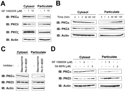
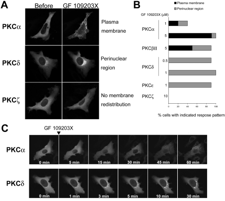
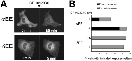
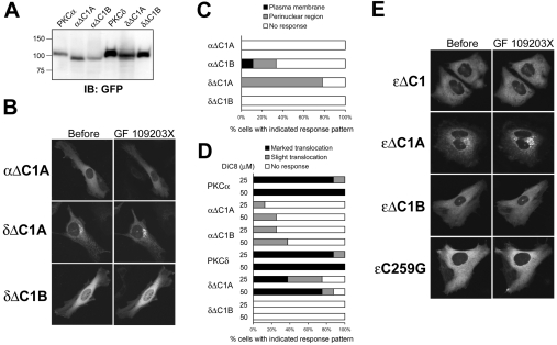
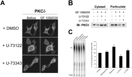
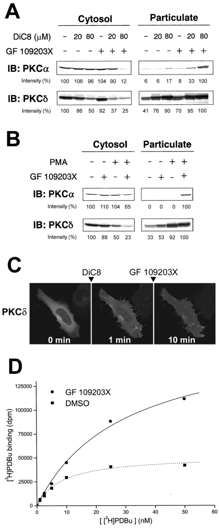
ATP-competitive inhibitors of PKC (protein kinase C) such as the bisindolylmaleimide GF 109203X, which interact with the ATP-binding site in the PKC molecule, have also been shown to affect several redistribution events of PKC. However, the reason why these inhibitors affect the redistribution is still controversial. In the present study, using immunoblot analysis and GFP (green fluorescent protein)-tagged PKC, we showed that, at commonly used concentrations, these ATP-competitive inhibitors alone induced redistribution of DAG (diacylglycerol)-sensitive PKCalpha, PKCbetaII, PKCdelta and PKCepsilon, but not atypical PKCzeta, to the endomembrane or the plasma membrane. Studies with deletion and point mutants showed that the DAG-sensitive C1 domain of PKC was required for membrane redistribution by these inhibitors. Furthermore, membrane redistribution was prevented by the aminosteroid PLC (phospholipase C) inhibitor U-73122, although an ATP-competitive inhibitor had no significant effect on acute DAG generation. Immunoblot analysis showed that an ATP-competitive inhibitor enhanced cell-permeable DAG analogue- or phorbol-ester-induced translocation of endogenous PKC. Furthermore, these inhibitors also enhanced [3H]phorbol 12,13-dibutyrate binding to the cytosolic fractions from PKCalpha-GFP-overexpressing cells. These results clearly demonstrate that ATP-competitive inhibitors cause redistribution of DAG-sensitive PKCs to membranes containing endogenous DAG by altering the DAG sensitivity of PKC and support the idea that the inhibitors destabilize the closed conformation of PKC and make the C1 domain accessible to DAG. Most importantly, our findings provide novel insights for the interpretation of studies using ATP-competitive inhibitors, and, especially, suggest caution about the interpretation of the relationship between the redistribution and kinase activity of PKC.


Figures

Figure 1
Figure 1. Translocation of endogenous PKCs to the particulate fraction by ATP-competitive inhibitors
(A) HeLa cells were treated with 1 or 10 μM GF 109203X for 20 min. (B) HeLa cells were treated with 5 μM GF 109203X for the indicated times. (C) HeLa cells were treated with 0.1 μM staurosporine or 5 μM Ro-31-8220 for 30 min. (D) HeLa cells were treated with the indicated concentrations of GF 109203X or Gö 6976 for 15 min. Subcellular fractions were obtained as described in the Experimental section and subjected to SDS/PAGE, followed by immunoblotting with anti-PKCα, anti-PKCδ or anti-PKCζ and with anti-actin antibody. Actin was used as a loading control. Results are representative of at least three separate experiments.
Figure 2
Figure 2. ATP-competitive inhibitors selectively induce membrane redistribution of DAG-sensitive PKCs
(A) HeLa cells transiently expressing PKCα–, PKCδ– or PKCζ–GFP were treated with GF 109203X at 5 μM for 60 min, 1 μM for 30 min or 10 μM for 60 min respectively. (B) Percentage of cells displaying GF 109203X-induced membrane redistribution of PKC. HeLa cells transiently expressing GFP-tagged cPKCs (PKCα and βII), nPKCs (PKCδ and ϵ), aPKC (PKCζ) were treated with GF 109203X at the indicated concentrations for 60, 15 or 60 min respectively. A total of 10–14 cells were observed. (C) HeLa cells transiently expressing PKCα- or PKCδ–GFP were treated with GF 109203X at 5 or 1 μM respectively for the indicated times.
Figure 3
Figure 3. ATP-competitive inhibitors induce membrane redistribution of GFP-tagged αEE and δEE mutants
(A) HeLa cells transiently expressing GFP-tagged αEE or δEE were treated with 10 or 1 μM GF 109203X respectively for the indicated times. (B) Percentage of cells displaying GF 109203X-induced membrane redistribution of PKC. HeLa cells transiently expressing GFP-tagged αEE or δEE were treated with GF 109203X at the indicated concentrations for 60 or 15 min respectively. A total of 10–12 cells were observed.
Figure 4
Figure 4. PKC membrane redistribution by an ATP-competitive inhibitor requires its DAG-sensitive C1 domain
(A) Whole-cell lysates were obtained from COS-7 cells transfected with the indicated plasmids using ice-cold homogenization buffer containing 1% Triton X-100 and subjected to SDS/PAGE, followed by immunoblotting with anti-GFP antibody. (B) HeLa cells transiently expressing GFP-tagged αΔC1A, δΔC1A and δΔC1B were treated with GF 109203X at 5 μM for 60 min (αΔC1A) or 1 μM for 20 min (δΔC1A and δΔC1B). A total of seven to nine cells were observed. (C) HeLa cells transiently expressing GFP-tagged αΔC1A, αΔC1B, δΔC1A and δΔC1B were treated with GF 109203X at 5 μM for 60 min (αΔC1A and αAC1B) or 1 μM for 20 min (δΔC1A and δΔC1B). A total of eight cells were observed. (D) HeLa cells transiently expressing GFP-tagged PKCα, αΔC1A, αΔC1B, PKCδ, δΔC1A and δΔC1B were treated with 25 or 50 μM DiC8 for 10 min. The translocation of δΔC1A indicates the decrease from the perinuclear region. (E) HeLa cells transiently expressing GFP-tagged ϵΔC1, ϵΔC1A, ϵΔC1B and ϵC259G were treated with 1 μM GF 109203X for 20 min. Results are representative of at least seven separate experiments.
Figure 5
Figure 5. PKC membrane redistribution by ATP-competitive inhibitors requires endogenous DAG generated by the spontaneous action of PLC
(A) HeLa cells transiently expressing PKCδ–GFP were pretreated with 2 μM U-73122 or U-73343 for 10 min and treated with 1 μM GF 109203X for 20 min. (B) HeLa cells were pretreated with 4 μM U-73122 or U-73343 for 30 min and treated with 1 μM GF 109203X for 30 min. Subcellular fractions were obtained as described in the Experimental section and subjected to SDS/PAGE, followed by immunoblotting (IB) with anti-PKCδ antibody. Results are representative of at least three separate experiments. (C) The DAG level in staurosporine (1 μM, 15 min)-, histamine (100 μM, 5 min)-, U-73122 and U-73343 (4 μM, 60 min)-treated cells was analysed by using the DAG kinase assay. The DAG content was determined by quantifying the amount of 32P-labelled phosphatidic acid (PA) using the BAS-2500 Phosphor Imager. Results are means±S.E.M. for four separate experiments. *P<0.01 versus control cells (unpaired Student's t test). We used U-73122 at a lower concentration (2 μM) in the confocal analysis because higher concentrations of U-73122 seemed to affect cell adhesion to some extent, perhaps as a result of the lower cell density.
Figure 6
Figure 6. GF 109203X enhances DiC8- and PMA-induced translocation of endogenous PKC and [3H]PDBu binding to the cytosolic fraction prepared from PKCα–GFP-overexpressing cells
(A) HL-60 cells were pretreated with 0.5 μM GF 109203X for 20 min and then stimulated with 20 or 80 μM DiC8 for 5 min. (B) HL-60 cells were pretreated with 1 μM GF109203X for 20 min and then stimulated with 10 nM PMA for 15 min. Subcellular fractions were obtained as described in the Experimental section and subjected to SDS/PAGE, followed by immunoblotting (IB) with anti-PKCα and anti-PKCδ antibodies. Densitometric analysis was performed using Scion Image software. In the cytosolic fraction the intensity of the protein bands is shown as a percentage of that of the control cells. In the particulate fraction the intensity of the protein bands is shown as a percentage of that of the 80 μM DiC8-plus-GF 109203X (A)- or PMA-plus-GF 109203X (B)-treated cells. Results are representative of at least three separate experiments. (C) HeLa cells transiently expressing PKCδ–GFP were pretreated with 10 μM DiC8 for 1 min, and then treated with 1 μM GF 109203X for 9 min. Results are representative of at least three separate experiments. (D) The PDBu binding was evaluated by the method described in the Experimental section in the presence of DMSO or 10 μM GF 109203X. The expression of PKCα–GFP was confirmed by immunoblotting with anti-GFP and anti-PKCα antibodies. Results are representative of at least three separate experiments, with duplicate determinations in each experiment.

Similar articles

Cited by

References

    1. Newton A. C. Regulation of protein kinase C. Curr. Opin. Cell Biol. 1997;9:161–167. - PubMed
    1. Nishizuka Y. Intracellular signaling by hydrolysis of phospholipids and activation of protein kinase C. Science. 1992;258:607–614. - PubMed
    1. Farrar W. L., Anderson W. B. Interleukin-2 stimulates association of protein kinase C with plasma membrane. Nature. 1985;315:233–235. - PubMed
    1. Oancea E., Meyer T. Protein kinase C as a molecular machine for decoding calcium and diacylglycerol signals. Cell. 1998;95:307–318. - PubMed
    1. Sakai N., Sasaki K., Ikegaki N., Shirai Y., Ono Y., Saito N. Direct visualization of the translocation of the γ-subspecies of protein kinase C in living cells using fusion proteins with green fluorescent protein. J. Cell Biol. 1997;139:1465–1476. - PMC - PubMed

MeSH terms