

The CXCR4/CXCL12 (SDF-1) signalling pathway protects non-obese diabetic mouse from autoimmune diabetes
- PMID: 17374136
- PMCID: PMC1941939
- DOI: 10.1111/j.1365-2249.2007.03370.x
The CXCR4/CXCL12 (SDF-1) signalling pathway protects non-obese diabetic mouse from autoimmune diabetes
Abstract
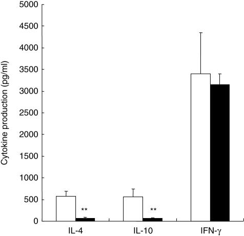
Chemokines and their receptors are part of polarized T helper 1 (Th1)- and Th2-mediated immune responses which control trafficking of immunogenic cells to sites of inflammation. The chemokine stromal cell-derived factor-1 CXCL-12 (SDF-1) and its ligand the CXCR4 chemokine receptor are important regulatory elements. CXCR4 is expressed on the surface of CD4(+) T cells, dendritic cells and B lymphocytes. Levels of CXCR4 mRNA were increased in pancreatic lymph nodes (PLNs) of 4-week-old non-obese diabetic (NOD) mice in comparison to Balb/C mice. However, a significant reduction of CXCR4 was noticed at 12 weeks both at the mRNA and protein levels while expression increased in the inflamed islets. The percentage of SDF-1 attracted splenocytes in a transwell chemotaxis assay was significantly increased in NOD versus Balb/c mice. SDF-1 attracted T cells completely abolished the capacity of diabetogenic T cells to transfer diabetes in the recipients of an adoptive cell co-transfer. When T splenocytes from NOD females treated with AMD3100, a specific CXCR4 antagonist, were mixed with diabetogenic T cells during adoptive cell co-transfer experiments, prevalence of diabetes in the recipients rose from 33% to 75% (P < 0.001). This effect was associated with an increase of interferon (IFN)-gamma mRNA and a reduction of interleukin (IL)-4 mRNA levels both in PLNs and isolated islets. AMD3100 also reduced IL-4 and IL-10 production of plate-bound anti-CD3 and anti-CD28-stimulated splenocytes. Immunofluorescence studies indicated that AMD3100 reduced the number of CXCR4(+) and SDF-1 positive cells in the inflamed islets. We can conclude that the CXCL-12/CXCR4 pathway has protective effects against autoimmune diabetes.
Figures


References
-
- Yang Y, Santamaria P. Lessons on autoimmune diabetes from animal models. Clin Sci (Lond) 2006;110:627–39. - PubMed
-
- Atkinson MA, Leiter EH. The NOD mouse model of type 1 diabetes: as good as it gets? Nat Med. 1999;5:601–4. - PubMed
-
- O'Reilly LA, Hutchings PR, Crocker PR, et al. Characterization of pancreatic islet cell infiltrates in NOD mice: effect of cell transfer and transgene expression. Eur J Immunol. 1991;21:1171–80. - PubMed
-
- Fabien N, Bergerot I, Maguer-Satta V, et al. Pancreatic lymph nodes are early targets of T cells during adoptive transfer of diabetes in NOD mice. J Autoimmun. 1995;8:323–34. - PubMed
MeSH terms
Substances
LinkOut - more resources
Full Text Sources
Other Literature Sources
Medical
Molecular Biology Databases
Research Materials
