. 2007 Mar 20:4:21.
doi: 10.1186/1742-4690-4-21.

In-Silico docking of HIV-1 integrase inhibitors reveals a novel drug type acting on an enzyme/DNA reaction intermediate

Affiliations

In-Silico docking of HIV-1 integrase inhibitors reveals a novel drug type acting on an enzyme/DNA reaction intermediate

Andrea Savarino. Retrovirology. .

Abstract

Background: HIV-1 integrase (IN) is an emerging drug target, as IN strand transfer inhibitors (INSTIs) are proving potent antiretroviral agents in clinical trials. One credible theory sees INSTIs as docking at the cellular (acceptor) DNA-binding site after IN forms a transitional complex with viral (donor) DNA. However, mapping of the DNA and INSTI binding sites within the IN catalytic core domain (CCD) has been uncertain.

Methods: Structural superimpositions were conducted using the SWISS PDB and Cn3D free software. Docking simulations of INSTIs were run by a widely validated genetic algorithm (GOLD).

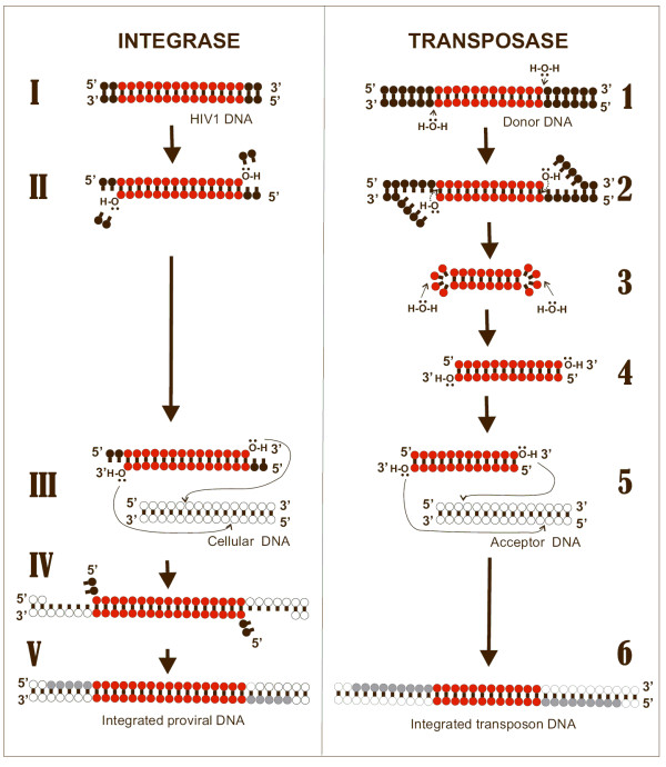
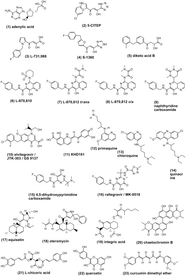
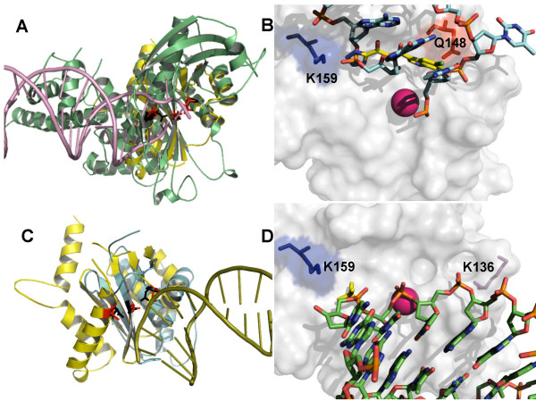
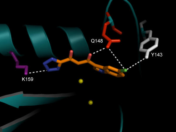
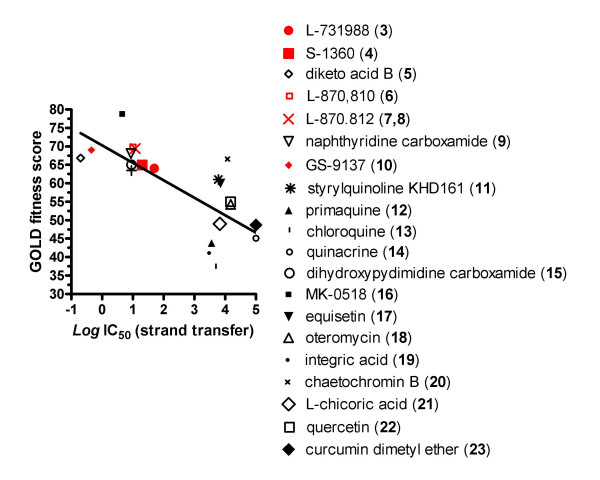
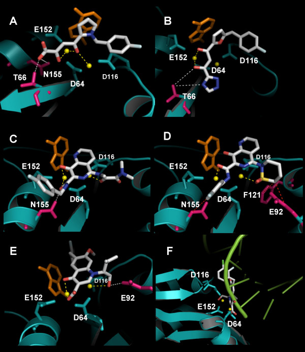
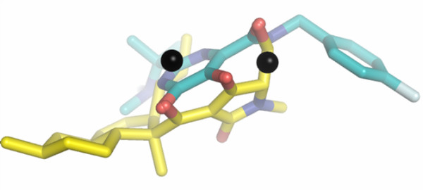
Results: Structural superimpositions suggested that a two-metal model for HIV-1 IN CCD in complex with small molecule, 1-(5-chloroindol-3-yl)-3-(tetrazoyl)-1,3-propandione-ene (5CITEP) could be used as a surrogate for an IN/viral DNA complex, because it allowed replication of contacts documented biochemically in viral DNA/IN complexes or displayed by a crystal structure of the IN-related enzyme Tn5 transposase in complex with transposable DNA. Docking simulations showed that the fitness of different compounds for the catalytic cavity of the IN/5CITEP complex significantly (P < 0.01) correlated with their 50% inhibitory concentrations (IC50s) in strand transfer assays in vitro. The amino acids involved in inhibitor binding matched those involved in drug resistance. Both metal binding and occupation of the putative viral DNA binding site by 5CITEP appeared to be important for optimal drug/ligand interactions. The docking site of INSTIs appeared to overlap with a putative acceptor DNA binding region adjacent to but distinct from the putative donor DNA binding site, and homologous to the nucleic acid binding site of RNAse H. Of note, some INSTIs such as 4,5-dihydroxypyrimidine carboxamides/N-Alkyl-5-hydroxypyrimidinone carboxamides, a highly promising drug class including raltegravir/MK-0518 (now in clinical trials), displayed interactions with IN reminiscent of those displayed by fungal molecules from Fusarium sp., shown in the 1990s to inhibit HIV-1 integration.

Conclusion: The 3D model presented here supports the idea that INSTIs dock at the putative acceptor DNA-binding site in a IN/viral DNA complex. This mechanism of enzyme inhibition, likely to be exploited by some natural products, might disclose future strategies for inhibition of nucleic acid-manipulating enzymes.


Figures

Figure 1
Figure 1
Sequence of events in HIV-1 integration (left) and Tn5 transposition (right). HIV-1: I) donor DNA; II) integrase-catalyzed 3' processing; III) integrase-catalyzed strand transfer; IV) product of strand transfer; V) DNA repair by cellular enzymes. Tn5 transposon: 1) donor DNA; 2) 3'processing; 3–4) 5' processing, consisting of loop formation (3) and generation of blunt-ended DNA (4); 5) strand transfer; 6) repaired strand transfer product. Portions of the donor DNA that become integrated are shown in red. Acceptor DNA is shown in white. Portions of acceptor DNA repaired following the strand transfer reaction are shown in grey.
Figure 2
Figure 2
Compounds mentioned in the present study. Note that the structure of 8-hydroxy-1,6-naphthyridine carboxamide, L-870,810 is presented both in trans and cis forms (the latter also referred to L-870,8125) as described in Refs [37] and [31], respectively. The structure of raltegravir/MK-0518 was retrieved from Ref. [54]. All other structures are available in the NCBI website [45].
Figure 3
Figure 3
Mapping of the nucleic acid-binding sites within the HIV-1 integrase (IN) catalytic site. Panel A: Structural superimposition between the crystal structures of HIV-1 IN catalytic core domain (PDB accession code: 1QS4; in yellow) and Tn5 transposase in complex with donor DNA (PDB: 1MM8; protein in green; DNA: in violet). The catalytic triads of IN and Tn5 transposase are shown in red and black, respectively. Panel B: transposition of Tn5 donor DNA (carbon backbone in cyan) to a crystal structure (PDB:1QS4) of HIV-1 IN in complex with 1-(5-chloroindol-3-yl)-3-(tetrazoyl)-1,3-propandione-ene (5CITEP; carbon backbone in yellow). HIV-1 DNA-interacting residues Q148 and K159 are shown as sticks. Hydrogens have been removed for better clarity. The metal ion crystallized with IN is shown in magenta. Panel C: Structural superimposition between the crystal structures of HIV-1 IN catalytic core domain (in yellow), and Bacillus halodurans RNAse H in complex with an RNA/DNA hybrid (PDB: 1ZBL; protein: in cyan; nucleic acid: in smudge green). The catalytic triads of IN and RNAse H are shown in red and black, respectively. Panel D: Transposition of B. halodurans RNA/DNA hybrid (carbon backbone in green) to HIV-1 IN. Putative DNA-interacting residues, K136 and K159 (from right to left) are shown as sticks. A phosphate (in yellow) co crystallized with IN (PDB: 2B4J) had been added by superimposing the 2B4J structure to the IN structure (PDB: 1QS4) used as reference structure in this part of the present study.
Figure 4
Figure 4
Interaction of integrase (IN) 3' processing inhibitor, 5CITEP with HIV-1 DNA-interacting residues. 5CITEP is shown in CPK with an orange carbon backbone. Donor DNA-interacting residues are shown in color as sticks. The putative metal ions within the IN active site are shown as yellow spheres. Possible hydrogen bonds are shown as dashed lines. Hydrogens have been removed for better clarity.
Figure 5
Figure 5
Correlation between the inhibitory potency of different compounds on HIV-1 integrase strand transfer and in-silico fitness for a two-metal HIV-1 integrase core domain in complex with 5CITEP. x axis: the in-vitro inhibitory potency of the compounds is presented as a Log transform of the IC50 value retrieved from the NCBI database (Ref. [45]). y axis: the in-silico fitness is presented as a score automatically attributed by the GOLD program. The regression line best fitting the data points is shown as a solid line. Compounds are numbered as in Fig. 2.
Figure 6
Figure 6
In-silicodocking of integrase strand transfer inhibitors (INSTIs) at the integrase (IN) active site. The structure of INSTIs L-731,988 (Panel A), S-1360 (B), L-870,812 (C), L-870,810 (D), GS-9137 (E, F) is shown in CPK. The catalytic triad (D64, D116 and E152) is shown in the same color as the protein backbone. Metal ions are presented as yellow balls. Amino acids responsible for drug resistance are colored in magenta. Significant enzyme/ligand interactions are shown as dashed lines (hydrogen bonds in white, metal coordination in yellow, Van-der-Waals forces in red). An adenine (in orange), marking the terminal portion of 3' processed viral DNA has been inserted by superimposition with the indole ring of 5CITEP. The adenine is shown for purely representative reasons, as the docking experiments were conducted in the presence of 5CITEP. A full 3D view of complexes in panels A-E can be obtained using the 3D coordinates provided as additional material [see Additional files 1, 2, 3, 4, 56]. In panel F, superimposition between the IN/inhibitor complex and a crystal structure of RNAse H in complex with an RNA/DNA hybrid results in an overlap between the INSTI (GS-9137 is shown as an example) and the nucleic acid (evidenced in pale green).
Figure 7
Figure 7
Superimposition of the best docking solutions for natural product integrase inhibitor equisetin from Fusarium sp. and a 4,5-dihydroxypyrimidine carboxamide strand transfer inhibitor. Compounds are shown in CPK. The carbon backbone of equisetin is displayed in yellow, that of the dihydroxypyrimidine carboxamide is in cyan. Metal ions are shown in black.

References

    1. Pommier Y, Johnson AA, Marchand C. Integrase inhibitors to treat HIV/AIDS. Nat Rev Drug Discov. 2005;4:236–248. doi: 10.1038/nrd1660. - DOI - PubMed
    1. Savarino A. A historical sketch of the discovery and development of HIV-1 integrase inhibitors. Expert Opin Investig Drug. 2006;12:1507–522. doi: 10.1517/13543784.15.12.1507. - DOI - PubMed
    1. DeJesus E, Berger D, Markowitz M, Cohen C, Hawkins T, Ruane P, Elion R, Farthing C, Zhong L, Cheng AK, McColl D, Kearney BP, for the 183-0101 Study Team Antiviral activity, pharmacokinetics, and dose response of the HIV-1 integrase inhibitor GS-9137 (JTK-303) in treatment-naive and treatment-experienced patients. J Acquir Immune Defic Syndr. 2006;1:1–5. - PubMed
    1. Markowitz M, Morales-Ramirez JO, Nguyen BY, Kovacs CM, Steigbigel RT, Cooper DA, Liporace R, Schwartz R, Isaacs R, Gilde LR, Wenning L, Zhao J, Teppler H, the Protocol 004 Study Team Antiretroviral Activity, Pharmacokinetics, and Tolerability of MK-0518 a Novel Inhibitor of HIV-1 Integrase, Dosed As Monotherapy for 10 Days in Treatment-Naive HIV-1-Infected Individuals. J Acquir Immune Defic Syndr. 2006;5:509–515. 2006 Dec 15. - PubMed
    1. Sharkey M, Triques K, Kuritzkes DR, Stevenson M. In vivo evidence for instability of episomal human immunodeficiency virus type 1 cDNA. J Virol. 2005;8:5203–5210. doi: 10.1128/JVI.79.8.5203-5210.2005. - DOI - PMC - PubMed

MeSH terms