. 2007 Apr 23;96(8):1293-301.
doi: 10.1038/sj.bjc.6603696. Epub 2007 Mar 20.

Identification of SOX2 as a novel glioma-associated antigen and potential target for T cell-based immunotherapy

Affiliations

Identification of SOX2 as a novel glioma-associated antigen and potential target for T cell-based immunotherapy

M Schmitz et al. Br J Cancer. .

Erratum in

  • Br J Cancer. 2007 Jun 18;96(12):1928

Abstract

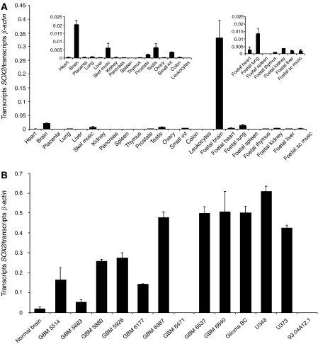
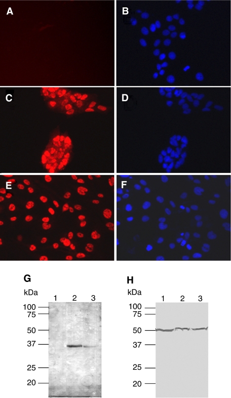
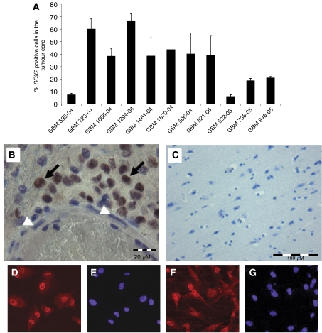
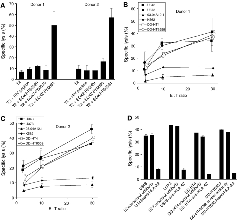
Prognosis for patients suffering from malignant glioma has not substantially improved. Specific immunotherapy as a novel treatment concept critically depends on target antigens, which are highly overexpressed in the majority of gliomas, but the number of such antigens is still very limited. SOX2 was identified by screening an expression database for transcripts that are overexpressed in malignant glioma, but display minimal expression in normal tissues. Expression of SOX2 mRNA was further investigated in tumour and normal tissues by real-time PCR. Compared to cDNA from pooled normal brain, SOX2 was overexpressed in almost all (9 out of 10) malignant glioma samples, whereas expression in other, non-malignant tissues was almost negligible. SOX2 protein expression in glioma cell lines and tumour tissues was verified by Western blot and immunofluorescence. Immunohistochemistry demonstrated SOX2 protein expression in all malignant glioma tissues investigated ranging from 6 to 66% stained tumour cells. Human leucocyte antigen-A(*)0201-restricted SOX2-derived peptides were tested for the activation of glioma-reactive CD8+ cytotoxic T lymphocytes (CTLs). Specific CTLs were raised against the peptide TLMKKDKYTL and were capable of lysing glioma cells. The abundant and glioma-restricted overexpression of SOX2 and the generation of SOX2-specific and tumour-reactive CTLs may recommend this antigen as target for T-cell-based immunotherapy of glioma.


Figures

Figure 1
Figure 1
Dot blot analysis of SOX2 mRNA expression pattern in normal tissues. A 112 bp cDNA fragment of the SOX2 transcript was radioactively labelled and hybridised to the human MTE Array 2 that provides poly(A)+ RNA from 58 human adult tissues, eight tumour cell lines and seven human fetal tissues normalised to eight housekeeping genes. Strong hybridisation signals were revealed only in fetal brain. A1, whole brain; B1, cerebral cortex; C1, frontal lobe; D1, parietal lobe; E1, occipital lobe; F1, temporal lobe; F1, paracentral gyrus of the cerebral cortex; H1, pons; A2, cerebellum left; B2, cerebellum right; C2, corpus callosum; D2, amygdala; E2, nucleus caudatus; F2, hippocampus; G2, medulla oblongata, H2, putamen; B3, nucleus accumbens, C3, thalamus; E3, spinal cord; A4, heart; B4, aorta; C4, atrium left; D4, atrium right; E4, ventricle left; F4, ventricle, right; G4, interventricular septum; H4, apex of the heart; A5, oesophagus; B5, stomach; C5, duodenum; D5, jejunum; E5, ileum; F5, ilocaecum; G5, appendix; H5, colon ascendens; A6, colon transversum; B6, colon descendens; C6, rectum; A7, kidney; B7, skeletal muscle; C7, spleen; D7, thymus; E7, peripheral blood leucocyte; F7, lymph node; G7, bone marrow; H7, trachea; A8, lung; B8, placenta; C8; bladder; D8, uterus; E8, prostate; F8, testis; G8, ovary; A9, liver; B9, pancreas; C9, adrenal gland; D9, thyroid gland; E9, salivary gland; A10, leukaemia, HL-60; B10, HeLa S3; C10, leukaemia, K-562; D10, leukaemia, MOLT-4; E10, Burkitt's lymphoma, Raji; F10, Burkitt's lymphoma, Daudi; G10, colorectal adenocarcinoma, SW480; H10, lung carcinoma, A549; A11, fetal brain; B11; fetal heart; C11, fetal kidney; D11; fetal liver; E11, fetal spleen; F11, fetal thymus; G11, fetal lung.
Figure 2
Figure 2
Real-time PCR analysis of SOX2 expression in normal adult and fetal tissues and in brain tumours. (A) The tissue specificity of SOX2 mRNA was determined by an SYBR Green I-based quantitative PCR assay in cDNA samples derived from 16 different pooled human adult tissues and eight different pooled human fetal tissues. The amount of SOX2 transcripts was normalised to the quantity of transcripts of the housekeeping gene β-actin. High transcript levels of SOX2 were found only in pooled fetal brain, whereas transcripts levels detected in adult brain, testis and skeletal muscle, the adult tissues with highest SOX2 expression, were 16.5-, 52.9- and 54.1-fold lower, respectively (see insets). (B) The same assay was applied for the quantification of SOX2 transcripts in brain tumours. SOX2 was upregulated in almost all (nine out of 10) GBM samples, with eight of the samples displaying more than five-fold overexpression compared to pooled normal adult brain cDNA. The pooled adult brain sample from (A) is inserted for comparison. The sample marked ‘BC’ is commercially available from Biocat (see Materials and Methods). The results represent the means of two independent LC runs, bars indicate s.e. GBM, glioblastoma multiforme.
Figure 3
Figure 3
SOX2 protein expression in the glioma cell lines U343 and U373. Indirect immunofluorescence analyses of SOX2 expression. (A) 93.04A12.1 melanoma cells; (C) U343 and (D) U373 glioma cells. Predominant nuclear SOX2 expression was detected only in the glioma cells. (B, D and F) Appropriate counterstaining of cell nuclei with Hoechst33342. (G and H) Western blot analysis of total protein lysates from 93.04A12.1-melanoma cells (lane 1); U343 (lane 2) and U373 glioma cells (lane 3). (G) SOX2 was detected only in lysates of glioma cell lines. (H), equal loading of the gels was confirmed by using a monoclonal antibody against α-tubulin.
Figure 4
Figure 4
Immunohistochemistry of GBM tissue and primary GBM cells. Tumour cell nuclei positively stained for SOX2 protein by immunohistochemical analysis of glioblastoma tissue were quantified. Percentage of positive cells refers to the area of the compact tumour core and is the mean value (±s.e.m.) from three randomly selected high-power fields. (A) Overexpression of SOX2 protein in GBM can be detected by immunohistochemical analysis of tumour specimens. (B) In the neoplastic tissue, nuclei of tumour cells are intensely stained for SOX2 (arrows), whereas vascular structures of pathologic blood vessels remained unstained (arrowheads). (C) SOX2 staining is absent in normal cortex tissue from control cases without pathological findings. Bars=20 μm in (B) and 100 μm in (C). (DG) The primary GBM cell lines DD-HT4 and DD-HT6559 express SOX2 protein. (D and F) SOX2 staining. (E and G) Counterstaining of nuclei using Hoechst33342.
Figure 5
Figure 5
(A) In vitro generation of cytotoxic effector T cells specifically recognising the SOX2-derived peptide 60031. Purified CD8+ T lymphocytes of healthy donors were weekly stimulated by SOX2 peptide-pulsed autologous DCs. After four stimulations, T-cell cultures were tested for the activation of peptide-specific CTLs. The stimulated T cells were added to 3 × 10³ peptide-pulsed, 51Cr-labelled T2 target cells per well at an effector cell to target cell ratio of 20 : 1. Unloaded T2 cells and T2 cells pulsed with an irrelevant peptide from HIV reverse transcriptase served as controls. The results represent the mean values of triplicate determinations, bars indicate s.e.m. (B and C) SOX2-specific lysis of glioma cells by in vitro-generated cytotoxic effector cells. After four rounds of stimulation activated CD8+ T cells from the two donors were co-cultured with 51Cr-labelled U343, U373, DD-HT4, DD-HT6559, 93.04A12.1 or K562 tumour cells per well at various effector cell (E) to target cell (T) ratios (3 : 1, 10 : 1, 30 : 1). After 4 h of incubation, chromium release was determined. (D) HLA-A2-restricted recognition of glioma cells by SOX2 peptide-stimulated cytotoxic effector cells. Inhibition of T-cell-mediated cytotoxicity against U343, U373, DD-HT4 and DD-HT6559 cells was tested in the presence of a monoclonal anti-HLA-A2 antibody or an isotype-matched control antibody at an E : T ratio of 30 : 1. Columns represent mean±s.e.m. of results obtained from two different donors.

Similar articles

Cited by

References

    1. Biernat W, Huang H, Yokoo H, Kleihues P, Ohgaki H (2004) Predominant expression of mutant EGFR (EGFRvIII) is rare in primary glioblastomas. Brain Pathol 14: 131–136 - PMC - PubMed
    1. Butowski NA, Sneed PK, Chang SM (2006) Diagnosis and treatment of recurrent high-grade astrocytoma. J Clin Oncol 24: 1273–1280 - PubMed
    1. Chi DD, Merchant RE, Rand R, Conrad AJ, Garrison D, Turner R, Morton DL, Hoon DS (1997) Molecular detection of tumour-associated antigens shared by human cutaneous melanomas and gliomas. Am J Pathol 150: 2143–2152 - PMC - PubMed
    1. Dong C, Wilhelm D, Koopman P (2004) Sox genes and cancer. Cytogenet Genome Res 105: 442–447 - PubMed
    1. Ehtesham M, Black KL, Yu JS (2004) Recent progress in immunotherapy for malignant glioma: treatment strategies and results from clinical trials. Cancer Control 11: 192–207 - PubMed

Publication types