. 2007 May;151(2):195-205.
doi: 10.1038/sj.bjp.0707225. Epub 2007 Mar 20.

Melatonin inhibits nitric oxide production by microvascular endothelial cells in vivo and in vitro

Affiliations

Melatonin inhibits nitric oxide production by microvascular endothelial cells in vivo and in vitro

C L M Silva et al. Br J Pharmacol. 2007 May.

Abstract

Background and purpose: We have previously shown that melatonin inhibits bradykinin-induced NO production by endothelial cells in vitro. The purpose of this investigation was to extend this observation to an in vivo condition and to explore the mechanism of action of melatonin.

Experimental approach: RT-PCR assays were performed with rat cultured endothelial cells. The putative effect of melatonin upon arteriolar tone was investigated by intravital microscopy while NO production by endothelial cells in vitro was assayed by fluorimetry, and intracellular Ca(2+) measurements were assayed by confocal microscopy.

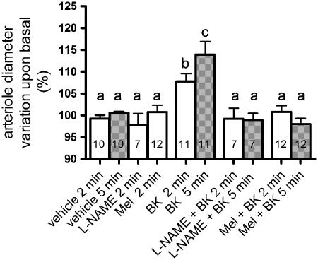
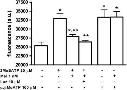
Key results: No expression of the mRNA for the melatonin synthesizing enzymes, arylalkylamine N-acetyltransferase and hydroxyindole-O-methyltransferase, or for the melatonin MT(2) receptor was detected in microvascular endothelial cells. Melatonin fully inhibited L-NAME-sensitive bradykinin-induced vasodilation and also inhibited NO production induced by histamine, carbachol and 2-methylthio ATP, but did not inhibit NO production induced by ATP or alpha, beta-methylene ATP. None of its inhibitory effects was prevented by the melatonin receptor antagonist, luzindole. In nominally Ca(2+)-free solution, melatonin reduced intracellular Ca(2+) mobilization induced by bradykinin (40%) and 2-methylthio ATP (62%) but not Ca(2+) mobilization induced by ATP.

Conclusions and implications: We have confirmed that melatonin inhibited NO production both in vivo and in vitro. In addition, the melatonin effect was selective for some G protein-coupled receptors and most probably reflects an inhibition of Ca(2+) mobilization from intracellular stores.


Figures

Figure 1
Figure 1
RT-PCR analysis of MT2 melatonin receptor mRNA expression in pineal gland (P; positive control) and two separate cultures of endothelial cells (E1, E2). N=negative control (no template added); MW=molecular weight standards. RT-PCR transcripts were obtained in two independent PCR steps and separated by electrophoresis in agarose gel (see Methods).
Figure 2
Figure 2
Effects of melatonin on L-NAME-sensitive, bradykinin-induced, rat arteriolar vasodilation. Arteriolar diameter (mean basal diameter: 19.2 μm) was measured 2 (clear bars) and 5 min (grey bars) after vehicle, bradykinin (BK, 1 μM), L-NAME (500 μM) or melatonin (Mel, 1 nM). The effect of BK was determined in the absence or presence of L-NAME or Mel which were added 2 min before and maintained throughout the exposure to BK. Data are expressed as mean±s.e.m. of the individual measurements indicated in the columns. Different letters indicate significant differences between the experimental conditions (P<0.05; ANOVA, followed by Newman–Keuls test). The number of rats per group was vehicle (7), BK (8); L-NAME (2) and Mel/BK (6).
Figure 3
Figure 3
Effects of melatonin on agonist-induced nitric oxide production by endothelial cells. Data are expressed as mean and s.e.m. (a) histamine 10 μM (His, n=18), melatonin 1 nM (Mel, n=18), luzindole (Luz, n=15). *P<0.01 vs basal (n=18); **P<0.01 vs histamine (one-way ANOVA), from four different cultures. (b) carbachol 100 μM (CCh, n=10), melatonin 1 nM (Mel, n=13). *P<0.01 vs basal (n=11); **P<0.01 vs carbachol (one-way ANOVA), from three different cultures. (c) ATP 100 μM (n=14), melatonin 1 nM (Mel, n=13), suramin 300 μM (Sur, n=6). *P<0.01 vs basal (n=12) and **P<0.05 vs ATP alone (one-way ANOVA), from three different cultures (except for Sur; two cultures).
Figure 4
Figure 4
Investigation of the effects of melatonin on endothelial cell nitric oxide production induced by agonists of P2 receptors. Data are expressed as mean and s.e.m. 2-Methylthio ATP 30 μM (2MeSATP, n=10), 2-methylthio ATP/melatonin 1 nM (Mel, n=11), 2-methylthio ATP/melatonin/luzindole 10 μM (Luz, n=8), α,β-methylene ATP 100 μM (α,β MeATP, n=9), α,β-methylene ATP/melatonin (n=9). *P<0.01 vs basal; **P<0.01 vs 2-methylthio ATP (one-way ANOVA).
Figure 5
Figure 5
Melatonin effect upon 0.1 μM bradykinin- (a), 100 μM ATP- (b) and (c) 30 μM 2-methylthio ATP-induced increase of Ca2+ in cultured endothelial cells in nominally Ca2+-free solution. The agonists were added at 30 s. In (a) and (c), the upper and lower traces represent data obtained in the absence and presence of melatonin, respectively. In (b), the upper trace represents data obtained in the absence of melatonin, the middle trace represents data obtained in the presence of melatonin and the lower trace represents the stimulation of endothelial cells with 100 μM α,β-methylene ATP (n=20 cells). Data are expressed as mean (c; n=16–17 cells) or mean and s.e.m. (a; n=7–8 cultures, b; n=8 cultures). See Methods for details.
Figure 6
Figure 6
Effect of sequential stimulation of endothelial cells with Ca2+ mobilizing agonists in nominally Ca2+-free solution. (a) 0.1 μM bradykinin (upper trace) was added at 30 s and, after 6 min, 100 μM ATP was added and the increase in fluorescence recorded (lower trace). (b) 100 μM ATP (upper trace) was added at 30 s. Then 6 min later, 0.1 μM bradykinin was added and the increase in fluorescence recorded (lower trace). Data are expressed as means from 35 and 33 cells for (a) and (b), respectively, from three experiments performed with two different cultures. The s.e.m. value was omitted for clarity from upper traces.

References

    1. Balcerczyk A, Soszynski M, Bartosz G. On the specificity of 4-amino-5-methylamino-2′,7′-difluorofluorescein as a probe for nitric oxide. Free Radic Biol Med. 2005;39:327–335. - PubMed
    1. Bauer PM, Fulton D, Boo YC, Sorescu GP, Kemp BE, Jo H, et al. Compensatory phosphorylation and protein–protein interactions revealed by loss of function and gain of function mutants of multiple serine phosphorylation sites in endothelial nitric-oxide synthase. J Biol Chem. 2003;278:14841–14849. - PubMed
    1. Benitez-King G, Huerto-Delgadillo L, Anton-Tay F. Binding of 3H-melatonin to calmodulin. Life Sci. 1993;53:201–207. - PubMed
    1. Beutelspacher SC, Tan PH, McClure MO, Larkin DF, Lechler RI, George AJ. Expression of indoleamine 2,3-dioxygenase (IDO) by endothelial cells: implications for the control of alloresponses. Am J Transplant. 2006;6:1320–1330. - PubMed
    1. Boutin JA, Audinot V, Ferry G, Delagrange P. Molecular tools to study melatonin pathways and actions. Trends Pharmacol Sci. 2005;26:412–419. - PubMed

Publication types

MeSH terms