

Molecular classification of breast carcinoma in situ
- PMID: 17375183
- PMCID: PMC1828915
- DOI: 10.2174/138920206779315719
Molecular classification of breast carcinoma in situ
Abstract
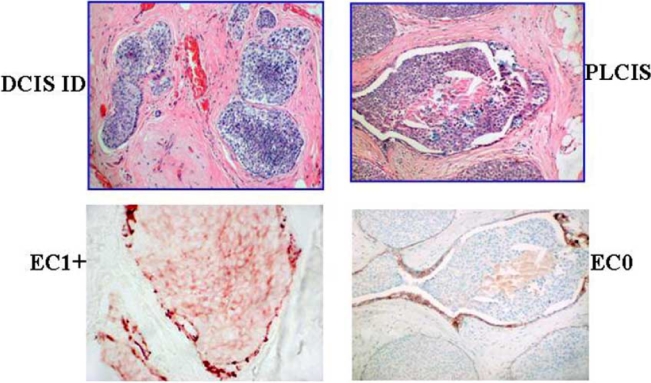
Pleomorphic variant of invasive lobular carcinoma (PILC) is an aggressive variant of invasive lobular carcinoma (ILC). Its in situ counterpart, pleomorphic lobular carcinoma in situ (PLCIS) is a recently described entity. Morphologically it has the typical architectural pattern of LCIS, but the neoplastic cells resemble intermediate grade DCIS. Molecular signatures that distinguish PLCIS from DCIS and LCIS would provide additional tools to aid in the histopathologic classification of PLCIS as a lesion distinct from LCIS and DCIS. CIS lesions, obtained from a study cohort of 38 breast cancer patients, were divided into 18 DCIS, 14 PLCIS and 6 LCIS. DNA from microdissected archival tissue was interrogated for loss or gain of 112 breast-cancer-specific genes using the Multiplex Ligation-dependent Probe Amplification Assay (MLPA). Classification Regression Tree (CART) analysis was employed to develop a gene-based molecular classification to distinguish or separate out PLCIS from DCIS and LCIS. Molecular classification via CART, based on gene copy number, agreed with histopathology in 34/38 CIS cases. Loss of CASP1 was predictive of LCIS (n=4) with one misclassified PLCIS. Gain of RELA predicted only the LCIS classification (n=2 cases). STK15 and TNFRSF1B were predictive only for DCIS with no misclassifications. Gain of EHF and TNFRSF1B and loss of NCOA3 were predictive of PLCIS, but not without misclassification. Molecular reclassification by CART was accomplished in 4 CIS cases: 1 PLCIS was reclassified as LCIS, 1 LCIS reclassified as PLCIS, and 2 DCIS cases as PLCIS. This study provides additional rationale for molecular modeling strategies in the evaluation of CIS lesions. This diagnostic aid may serve to minimize misclassification between PLCIS and DCIS, and PLCIS and LCIS, aiding to increase accuracy in the differential diagnosis of CIS lesions.
Figures


References
-
- Fisher ER, Costantino J, Fisher B, Palekar AS, Paik SM, Suarez CM, Wolmark N. Pathologic findings from the National Surgical Adjuvant Breast Project (NSABP) Protocol B-17. Five-year observations concerning lobular carcinoma in situ. Cancer. 1996;78:1403–16. - PubMed
-
- Schnitt SJ, Morrow M. Lobular carcinoma in situ: current concepts and controversies. Semin Diagn Pathol. 1999;16:209–23. - PubMed
-
- Bentz JS, Yassa N, Clayton F. Pleomorphic lobular carcinoma of the breast: clinicopathologic features of 12 cases. Mod Pathol. 1998;11:814–22. - PubMed
-
- Frost AR, Tsangaris TN, Silverberg SG. Pleomorphic lobular carcinoma in situ. Pathol Case Rev. 1961;1:27.
-
- Reis-Filho JS, Simpson PT, Jones C, Steele D, Mackay A, Iravani M, Fenwick K, Valgeirsson H, Lambros M, Ashworth A, Palacios J, Schmitt F, Lakhani SR. Pleomorphic lobular carcinoma of the breast: role of comprehensive molecular pathology in characterization of an entity. J Pathol. 2005;207:1–13. - PubMed
Grants and funding
LinkOut - more resources
Full Text Sources
Other Literature Sources
Miscellaneous