. 2007 Feb 27;40(1):9-17.
doi: 10.1267/ahc.06011. Epub 2007 Feb 5.

Role of heat shock protein 70 in induction of stress fiber formation in rat arterial endothelial cells in response to stretch stress

Affiliations

Role of heat shock protein 70 in induction of stress fiber formation in rat arterial endothelial cells in response to stretch stress

Shan-Shun Luo et al. Acta Histochem Cytochem. .

Abstract

We investigated the mechanism by which endothelial cells (ECs) resist various forms of physical stress using an experimental system consisting of rat arterial EC sheets. Formation of actin stress fibers (SFs) and expression of endothelial heat-shock stress proteins (HSPs) in response to mechanical stretch stress were assessed by immunofluorescence microscopy. Stretch stimulation increased expression of HSPs 25 and 70, but not that of HSP 90. Treatment with SB203580, a p38 MAP kinase inhibitor that acts upstream of the HSP 25 activation cascade, or with geldanamycin, an inhibitor of HSP 90, had no effect on the SF formation response to mechanical stretch stress. In contrast, treatment with quercetin, an HSP 70 inhibitor, inhibited both upregulation of endothelial HSP 70 and formation of SFs in response to tensile stress. In addition, treatment of stretched ECs with cytochalasin D, which disrupts SF formation, did not adversely affect stretch-induced upregulation of endothelial HSP 70. Our data suggest that endothelial HSP 70 plays an important role in inducing SF formation in response to tensile stress.


Figures

Fig. 1
Fig. 1
Rhodamine-phalloidin staining in non-stretch-stressed (A) and stretch-stressed (B) ECs from the common iliac artery of rat. The ECs formed many long SFs (arrows) in response to application of mechanical stretch stress at 30% amplitude for 1 hr. Images A and B are shown at the same magnification; bar=20 µm.
Fig. 2
Fig. 2
Quantitative analysis of SF-positive ECs before (gray bar) and after (black bar) mechanical stretch stress applied at 30% amplitude for 1 hr. SF formation was induced in the rat arterial ECs from the middle parts of the abdominal aorta (Ab) and the common iliac artery (Cil), but not in the proximal part of the thoracic aorta (Th-p) or the common carotid artery (Cc). Values shown are means±SD. *Differences between before and after stretch in Ab segment, and in Cil segment are significant (p<0.01).
Fig. 3
Fig. 3
Quantitative analysis of the expression of HSPs 25, 70, and 90 in rat arterial ECs in vivo. No significant differences in HSP immunofluorescence intensity were evident among the straight portions from the four anatomical sources examined. The abbreviations are the same as those used in Fig. 2. Values shown are means±SD.
Fig. 4
Fig. 4
HSP 70 immunohistochemistry in rat arterial ECs from Th-p (A, B) and Cil (C, D) under non-stretch-stressed (A, C) and stretch-stressed (B, D) conditions. HSP 70 expression was upregulated by application of stretch stress (30% amplitude, 1 hr) in Cil ECs (D) but not in Th-p ECs (B). The abbreviations used here are the same as those used in Fig. 2. Images A–D are shown at the same magnification; bar=20 µm.
Fig. 5
Fig. 5
Quantitative analysis of HSP upregulation in response to stretch stress. Rat arterial ECs were subjected to application of mechanical stretch stress at 30% amplitude for 1 hr. The relative activities of HSPs 25 (gray bar), 70 (black bar), and 90 (white bar) are shown as the ratios of fluorescence intensities before and after cells were subjected to stretch stimulation. In Ab and Cil ECs, expression of HSPs 25 and 70 increased after stretch stimulation, and the level of HSP 70 induction was greater than that of HSP 25. In Th-p and Cc ECs, expression levels of HSPs 25 and 70 after stretch stimulation was about the same as or slightly less than those in the non-stretched controls. Endothelial HSP 90 was not upregulated by tensile stress in any of the samples. The abbreviations used here are the same as those used in Fig. 2. Values shown are means±SD. *Differences in HSP 70 upregulation between Ab and Th-p, Ab and Cc, Cil and Th-p, and Cil and Cc segments are significant (p<0.01). #Differences in HSP 25 upregulation between Ab and Th-p, Ab and Cc, Cil and Th-p, and Cil and Cc segments are significant (p<0.01).
Fig. 6
Fig. 6
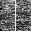
Effects of HSP inhibitors on stretch-stress-induced SF formation in ECs from the common iliac artery as revealed by rhodamine-phalloidin staining. The arterial segments were treated with inhibitors while in the stretched state (30% amplitude, 1 hr). (A) Vehicle control (DMSO); (B) 5 µg/ml geldanamycin; (C) 50 µM SB203580; (D) 1 µM quercetin; (E) 50 µM quercetin; (F) 100 µM quercetin. Stretch-induced SF formation was not affected by the presence of either geldanamycin (B) or SB203580 (C). In contrast, quercetin inhibited SF formation in a concentration-dependent manner (D, E, F). Images A–F are shown at the same magnification; bar=20 µm.
Fig. 7
Fig. 7
Quantitative analysis of the effect of quercetin on stretch-stress-induced SF formation. Quercetin was administered to the arterial segments while they were in the stretched state (30% amplitude, 1 hr). Gray and black bars indicate the relative frequencies of SF-positive ECs observed in DMSO- and 100 µM quercetin-treated samples, respectively. Quercetin treatment significantly reduced the frequency of SF-positive ECs in Ab and Cil ECs. The abbreviations used here are the same as those used in Fig. 2. Values shown are means±SD. *Differences between with quercetin and with DMSO in Ab segment, and in Cil segment are significant (p<0.01).
Fig. 8
Fig. 8
Effects of quercetin and cytochalasin D on upregulation of HSP 70 in stretch-stressed ECs from the common iliac artery of rat. Results shown are from HSP 70 immunohistochemistry of ECs that were (A) DMSO-treated and non-stretched, (B) DMSO-treated and stretch-stressed (30% amplitude 1 hr), (C) 100 µM quercetin-treated and stretch-stressed, or (D) 2 µg/ml cytochalasin D-treated and stretch-stressed. Upregulation of HSP 70 was blocked by quercetin but not by cytochalasin D. Images A–D are shown at the same magnification; bar=20 µm.
Fig. 9
Fig. 9
Quantitative analysis of HSP 70 expression in rat arterial ECs after treatment with 100 µM quercetin or 2 µg/ml cytochalasin D under stretch-stress conditions (30% amplitude, 1 hr). The relative HSP 70 activities of ECs treated with the vehicle control DMSO (gray bars), cytochalasin D (black bars), or quercetin (white bars) are shown as the ratios of the fluorescence intensities before and after stretch stimulation. In Ab and Cil ECs, upregulation of HSP 70 was largely blocked by quercetin but not by cytochalasin D. The abbreviations used here are the same as those used in Fig. 2. Values shown are means±SD. *Differences between with quercetin and with DMSO, and with quercetin and with cytochalasin D in each segment tested are significant (p<0.01).

Similar articles

Cited by

References

    1. Azuma N., Akasaka N., Kito H., Ikeda M., Gahtan V., Sasajima T., Sumpio B. E. Role of p38 MAP kinase in endothelial cell alignment induced by fluid shear stress. Am. J. Physiol. 2001;280:H189–197. - PubMed
    1. Burridge K., Fath K., Kelly T., Nuckollos G. C., Turner C. Focal adhesions: Transmembrane junctions between the extracellular matrix and the cytoskeleton. Ann. Rev. Cell Biol. 1988;4:487–525. - PubMed
    1. Chan Y. C., Shukla N., Abdus-Samee M., Berwanger C. S., Stanford J., Singh M., Mansfield A. O., Stansby G. Anti-heat-shock protein 70 kDa antibodies in vascular patients. Eur. J. Vasc. Endovasc. Surg. 1999;18:381–385. - PubMed
    1. Chien S. S., Li S., Shyy Y. J. Effects of mechanical forces on signal transduction and gene expression in endothelial cells. Hypertension. 1998;31:162–169. - PubMed
    1. Craig E. A., Weissman J. S., Horwich A. L. Heat shock proteins and molecular chaperones: mediators of protein conformation and turnover in the cell. Cell. 1994;78:365–372. - PubMed