. 2007 Feb 27;40(1):27-34.
doi: 10.1267/ahc.06015. Epub 2007 Feb 5.

Localization of estrogen receptors alpha and beta in the articular surface of the rat femur

Affiliations

Localization of estrogen receptors alpha and beta in the articular surface of the rat femur

Yasushi Oshima et al. Acta Histochem Cytochem. .

Abstract

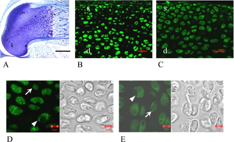
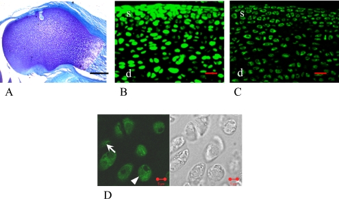
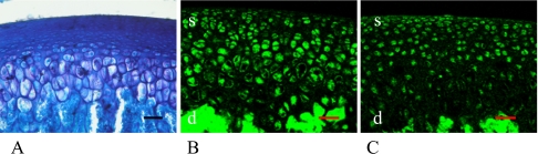
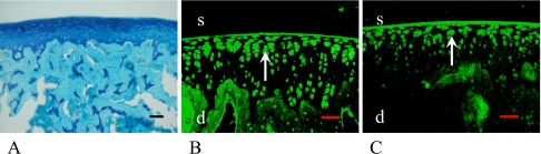
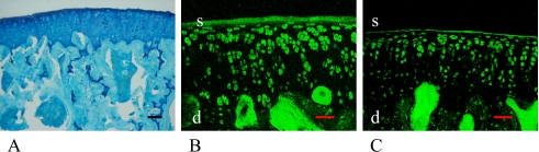
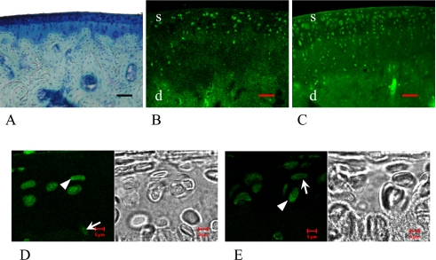
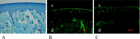
It has been suggested that the degradation of the articular cartilage and osteoarthritis (OA) are associated with gender and the estrogen hormone. Although many investigators have reported the presence of the estrogen receptors (ERs) alpha and beta in the articular cartilage, the localization of these receptors and the difference in their in vivo expression have not yet been clearly demonstrated. We performed immunofluorescence staining of ERalpha and ERbeta to elucidate the localization of the ERs and to note the effects of gender and the aging process on these receptors. The results revealed that ERalpha and ERbeta were expressed in the articular cartilage and subchondral bone layers of adult rats of both sexes. We also observed the high expression of these receptors in immature rats. In contrast, their expression levels decreased in an ovariectomised model, as a simulation of postmenopause, and in aged female rats. Therefore, this study suggests the direct effects of estrogen and ER expression on articular surface metabolism.


Figures

Fig. 1
Fig. 1
1-day-old female rat. Femoral epiphyseal areas were stained by toluidine blue (A), indicating the existence of cartilage matrix. In the low magnification images, almost all of the cells in both the superficial (s) and the deep (d) layers of epiphyseal areas were stained with ERα (B) and ERβ (C). In the high magnification images, both nucleus and cytoplasm were stained diffusely with ERα (D) and ERβ (E). Arrowheads show the cells that were stained with ERs mainly in the cytoplasm, and arrows show the cells that were stained with ERs mainly in the nucleus. Bars=5 µm (A, D, E), 20 µm (B, C).
Fig. 2
Fig. 2
1-week-old female rat. In the low magnification images, femoral epiphysial areas still stained by toluidine blue (A), in which almost all of the cells in both the superficial (s) and the deep (d) layers were stained with ERα (B) and ERβ (C). In the high magnification image, arrowhead shows the cell that was stained with ERs mainly in the cytoplasm, and arrow shows the cell that was stained with ERs mainly in the nucleus (D). Bars=5 µm (A, D), 20 µm (B, C).
Fig. 3
Fig. 3
1-month-old female rat. Articular surface was constructed with articular cartilage and subchondral bone layers (A). Almost all of the cells in both the articular cartilage and subchondral bone layers were stained with ERα (B) and ERβ (C). Bar=50 µm.
Fig. 4
Fig. 4
3-month-old female rat. Cartilage and bone formations were mature (A). In female, almost all of the cells in the superficial (s) layer (arrows) of the articular cartilage were stained with ERα (B) and ERβ (C). Bar=50 µm.
Fig. 5
Fig. 5
3-month-old male rat. Similar to female, almost all the cells, especially in the superficial (s) layer of articular cartilage (A) expressed both ERα (B) and ERβ (C). Bar=50 µm.
Fig. 6
Fig. 6
24-month-old female rat. There was no remarkable degenerative change on the articular surface; however, the cartilage thickness was thinner than that of the younger models (A). In the low magnification images, some cells in the superficial (s) and a few cells in the deeper (d) layer of the articular cartilage were stained with ERα (B) and ERβ (C). In the high magnification images, both nucleus and cytoplasm were stained diffusely with ERα (D) and ERβ (E). Arrowheads show the cells that were stained with ERs mainly in the cytoplasm, and arrows show the cells that were stained with ERs mainly in the nucleus. Bars=50 µm (A–C), 5 µm (D, E).
Fig. 7
Fig. 7
4-month-old female rat 1 month after OVX. There was no remarkable degradational or osteoarthritic change (A). Similar to the results of aged rat models, some cells were stained with ERα (B) and ERβ (C). Bar=50 µm.

Similar articles

Cited by

References

    1. Barrett-Connor E., Stuenkel C. A. Hormone replacement therapy (HRT)—risks and benefits. Int. J. Epidemiol. 2001;30:423–426. - PubMed
    1. Brandenberger A. W., Tee M. K., Lee J. Y., Chao V., Jaffe R. B. Tissue distribution of estrogen receptors alpha (ER-alpha) and beta (ER-beta) mRNA in the midgestational human fetus. J. Clin. Endocrinol. Metab. 1997;82:3509–3512. - PubMed
    1. Creutz L. M., Kritzer M. F. Mesostriatal and mesolimbic projections of midbrain neurons immunoreactive for estrogen receptor beta or androgen receptors in rats. J. Comp. Neurol. 2004;476:348–362. - PubMed
    1. Felson D. T., Nevitt M. C. Estrogen and osteoarthritis: how do we explain conflicting study results? Prev. Med. 1999;28:445–448. 449–450. discussion . - PubMed
    1. Hoegh-Andersen P., Tanko L. B., Andersen T. L., Lundberg C. V., Mo J. A., Heegaard A. M., Delaisse J. M., Christgau S. Ovariectomized rats as a model of postmenopausal osteoarthritis: validation and application. Arthritis Res. Ther. 2004;6:R169–180. - PMC - PubMed