

Preparation and observation of fresh-frozen sections of the green fluorescent protein transgenic mouse head
- PMID: 17375207
- PMCID: PMC1828081
- DOI: 10.1267/ahc.05051
Preparation and observation of fresh-frozen sections of the green fluorescent protein transgenic mouse head
Abstract
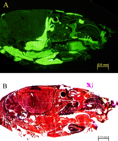
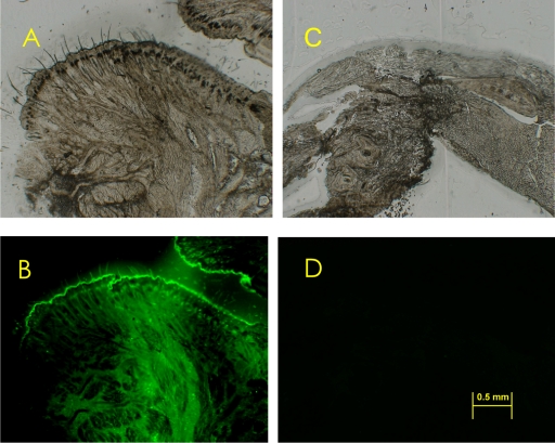
Hard tissue decalcification can cause variation in the constituent protein characteristics. This paper describes a method of preparating of frozen mouse head sections so as to clearly observe the nature of the constituent proteins. Frozen sections of various green fluorescent protein (GFP) transgenic mouse heads were prepared using the film method developed by Kawamoto and Shimizu. This method made specimen dissection without decalcification possible, wherein GFP was clearly observed in an undamaged state. Conversely, using the same method with decalcification made GFP observation in the transgenic mouse head difficult. This new method is suitable for observing GFP marked cells, enabling us to follow the transplanted GFP marked cells within frozen head sections.
Figures
Similar articles
-
Rapid preparation of fresh frozen tissue-engineered bone sections for histological, histomorphological and histochemical analyses.Biomed Mater Eng. 2006;16(6):405-13. Biomed Mater Eng. 2006. PMID: 17119279
-
Fluorescence imaging preparation methods for tissue scaffolds implanted into a green fluorescent protein porcine model.Transgenic Res. 2015 Oct;24(5):911-9. doi: 10.1007/s11248-015-9891-7. Epub 2015 Jun 25. Transgenic Res. 2015. PMID: 26109094
-
Transgenic nude mouse with ubiquitous green fluorescent protein expression as a host for human tumors.Cancer Res. 2004 Dec 1;64(23):8651-6. doi: 10.1158/0008-5472.CAN-04-3118. Cancer Res. 2004. PMID: 15574773
-
Use of a new adhesive film for the preparation of multi-purpose fresh-frozen sections from hard tissues, whole-animals, insects and plants.Arch Histol Cytol. 2003 May;66(2):123-43. doi: 10.1679/aohc.66.123. Arch Histol Cytol. 2003. PMID: 12846553 Review.
-
Color-coded fluorescent protein imaging of angiogenesis: the AngioMouse models.Curr Pharm Des. 2008;14(36):3810-9. doi: 10.2174/138161208786898644. Curr Pharm Des. 2008. PMID: 19128234 Review.
References
-
- Aaron J. E., Carter D. H. Rapid preparation of fresh-frozen undecalcified bone for histological and histochemical analysis. J. Histochem. Cytochem. 1987;35:361–369. - PubMed
-
- Chalfie M., Tu Y., Euskirchen G., Ward W. W., Prasher D. C. Green fluorescent protein as a marker for gene expression. Science. 1994;263:802–805. - PubMed
-
- Goodwin P. C. GFP biofluorescence: imaging gene expression and protein dynamics in living cells. Design considerations for a fluorescence imaging laboratory. Methods Cell Biol. 1999;58:343–367. - PubMed
-
- Kawamoto T., Shimizu M. A method for preparing 2- to 50-micron-thick fresh-frozen sections of large samples and undecalcified hard tissues. Histochem. Cell Biol. 2000;113:331–339. - PubMed
-
- Kawamoto T. Use of a new adhesive film for the preparation of multi-purpose fresh-frozen sections from hard tissues, whole-animals, insects and plants. Arch. Histol. Cytol. 2003;66:123–143. - PubMed
LinkOut - more resources
Full Text Sources
Molecular Biology Databases
Research Materials