. 2007 Apr;64(1):111-24.
doi: 10.1111/j.1365-2958.2007.05630.x.

Corynebacterium diphtheriae employs specific minor pilins to target human pharyngeal epithelial cells

Affiliations

Corynebacterium diphtheriae employs specific minor pilins to target human pharyngeal epithelial cells

Anjali Mandlik et al. Mol Microbiol. 2007 Apr.

Abstract

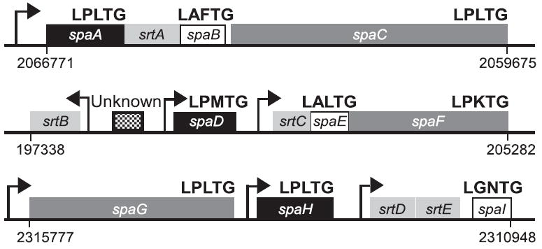
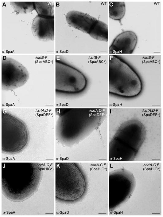
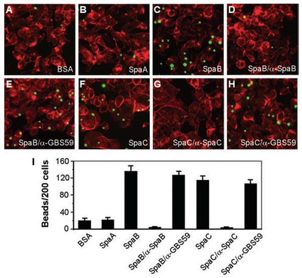
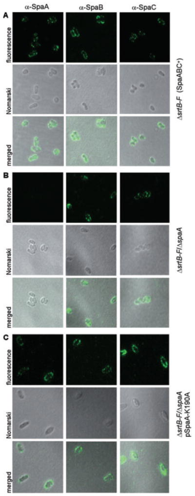
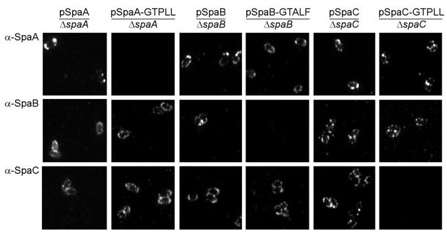
Adherence to host tissues mediated by pili is pivotal in the establishment of infection by many bacterial pathogens. Corynebacterium diphtheriae assembles on its surface three distinct pilus structures. The function and the mechanism of how various pili mediate adherence, however, have remained poorly understood. Here we show that the SpaA-type pilus is sufficient for the specific adherence of corynebacteria to human pharyngeal epithelial cells. The deletion of the spaA gene, which encodes the major pilin forming the pilus shaft, abolishes pilus assembly but not adherence to pharyngeal cells. In contrast, adherence is greatly diminished when either minor pilin SpaB or SpaC is absent. Antibodies directed against either SpaB or SpaC block bacterial adherence. Consistent with a direct role of the minor pilins, latex beads coated with SpaB or SpaC protein bind specifically to pharyngeal cells. Therefore, tissue tropism of corynebacteria for pharyngeal cells is governed by specific minor pilins. Importantly, immunoelectron microscopy and immunofluorescence studies reveal clusters of minor pilins that are anchored to cell surface in the absence of a pilus shaft. Thus, the minor pilins may also be cell wall anchored in addition to their incorporation into pilus structures that could facilitate tight binding to host cells during bacterial infection.


Figures

Fig. 1
Fig. 1
Three gene clusters in the chromosome of C. diphtheriae NCTC13129 that encode sortase genes (srtA–E) and the sortase-mediated pilus assembly genes (spaA–I). A sixth sortase (srtF) is located between positions 2363388 and 2364209 in the chromosome. Arrows indicate the position of predicted promoters as well as direction of transcription. Similarity and homology between spa gene products are indicated as shared patterns. Numbers show the location of pilus gene clusters in the chromosome.
Fig. 2
Fig. 2
A pilin-specific sortase required for pilus assembly. Wild-type C. diphtheriae (A–C) and its isogenic derivatives ΔsrtB–F (designated as SpaABC+) (D–F), ΔsrtA,D–F (designated as SpaDEF+) (G–I) or ΔsrtA-C,F (designated as SpaHIG+) (J–L) were immobilized on carbon grids, stained with a specific antiserum against SpaA, SpaD or SpaH and goat anti-rabbit IgG conjugated to 12 nm gold particles. Samples were viewed by transmission electron microscopy. Scale bars indicate the length of 0.2 μm.
Fig. 3
Fig. 3
Pilus-mediated adherence of C. diphtheriae to human epithelial cells. A. Adherence of corynebacterial variants to human lung (A549), laryngeal (HEp2) and pharyngeal (D562) epithelial cells. Confluent cell monolayers were infected with the wild-type C. diphtheriae and its isogenic deletion mutants, and adherent bacteria were then enumerated. Data are presented as percentage of adhesion relative to that of wild type. Mean adhesion percentage of wild-type to A549, HEp2 and D562 cells was ~20%, ~25% and ~9% respectively. The results are presented as averages (with standard deviations ± SD) from at least three independent experiments performed in triplicates. B. Differential binding of corynebacterial pili to epithelial cells. Similar experiments were carried out as described in (A) with strains ΔsrtB–F, ΔsrtA,D–F and ΔsrtA–C,F designated as SpaABC+, SpaDEF+ and SpaHIG+ respectively.
Fig. 4
Fig. 4
Pilus polymerization of corynebacteria. The wild-type strain and its isogenic derivatives carrying deletions of srt or spa genes were treated with muramidase prior to extraction with hot SDS sample buffer. Proteins were separated on 4–12% Tris-Glycine gradient gels and detected by immunoblotting with the specific antisera α-SpaA (A), α-SpaB (B) and α-SpaC (C). Strain ΔspaA, ΔspaB or ΔspaC strain was generated from the parental strain ΔsrtB–F (designated as SpaABC+). A recombinant plasmid harbouring the wild-type SpaA, SpaB or SpaC was used to complement a respective deletion mutant. The positions of molecular mass markers (M) are indicated. Note the slower mobility of the SpaC band in ΔspaA strain (C, lane 3).
Fig. 5
Fig. 5
Minor pilins SpaB and SpaC required for bacterial adherence. A and B. The parental strain SpaABC+srtB–F) and its isogenic deletion mutants were subjected to adhesion assays using pharyngeal epithelial cells (A) or lung epithelial cells (B). Data are presented as percentage of adhesion relative to that of the parental strain. The results are presented as averages (with standard deviations ± SD) from at least three independent experiments performed in triplicates. Statistical analysis was performed using Student’s t-test. C. In a similar experiment as described in (A), monolayer of pharyngeal epithelial cells were infected with the parental strain SpaABC+ pre-incubated with a specific antiserum at a dilution of 1:100 for 1 h at 37°C. Pre-immune sera and an unrelated antibody against a pilus protein of Streptococcus agalactiae, α-GBS59, were used as controls.
Fig. 6
Fig. 6
SpaB- or SpaC-mediated adherence of latex beads to pharyngeal cells. Semi-confluent cells grown on coverslips were infected with protein-coated beads for 1 h. The washed cells were fixed and stained with Texas-Red-X phalloidin. A–H. Shown here are the fluorescence images of cells incubated with fluorescent beads bound to BSA (A), SpaA (B), SpaB (C), SpaB blocked with α-SpaB (D), SpaC (F) and SpaC blocked with α-SpaC (G). Beads with bound protein incubated with an unrelated antibody against a pilin of Streptococcus agalactiae, α-GBS59, were used as controls (E and H). I. The results are shown as an average of the numbers of beads bound per 200 cells of three independent experiments (with standard deviation ± SD).
Fig. 7
Fig. 7
Assembly of minor pilins SpaB and SpaC on bacterial surface. Corynebacteria were immobilized on carbon grids, stained with specific antiserum against SpaB (α-SpaB) (A, F and G) or SpaC (α-SpaC) (B, E and H) and goat anti-rabbit IgG conjugated to 12 nm gold particles. For double labelling (C), cells were first reacted with α-SpaC, followed by IgG-conjugated 18 nm gold particles (open arrow in D), and then reacted with α-SpaB, followed by IgG-conjugated 12 nm gold particles (filled arrow in D). Samples were viewed by transmission electron microscopy. An enlarged area of (C) is shown in (D). Scale bars indicate the length of 0.2 μm.
Fig. 8
Fig. 8
Immunofluorescent detection of SpaB and SpaC pilins on the bacterial surface. Corynebacteria were stained with a specific antibody against SpaA (α-SpaA), SpaB (α-SpaB) or SpaC (α-SpaC) and AlexaFluor 488 chicken anti-rabbit IgG. Shown are the fluorescent, the Nomarski DIC and the merged images of strain ΔsrtB–F (A), its isogenic derivative ΔspaA (B) or its isogenic derivative ΔspaA transformed with a plasmid expressing the SpaA protein mutated at the K190 in the pilin motif (C). The samples were observed on a Zeiss LSM 510 confocal microscope.
Fig. 9
Fig. 9
Anchoring of minor pilins SpaB and SpaC required the conserved LPXTG motif. Strain ΔsrtB–F with a deletion of spaA, spaB or spaC gene was transformed with a plasmid expressing the wild-type or mutated SpaA, SpaB or SpaC respectively. Corynebacteria were stained with a specific antibody against SpaA (α-SpaA), SpaB (α-SpaB) or SpaC (α-SpaC) and AlexaFluor 488 chicken anti-rabbit IgG. The samples were observed on a Zeiss LSM 510 confocal microscope. Only fluorescent images are shown.

Similar articles

Cited by

References

    1. Ankri S, Reyes O, Leblon G. Electrotransformation of highly DNA-restrictive corynebacteria with synthetic DNA. Plasmid. 1996;35:62–66. - PubMed
    1. Barocchi MA, Ries J, Zogaj X, Hemsley C, Albiger B, Kanth A, et al. A pneumococcal pilus influences virulence and host inflammatory responses. Proc Natl Acad Sci USA. 2006;103:2857–2862. - PMC - PubMed
    1. Braun L, Ohayon H, Cossart P. The InIB protein of Listeria monocytogenes is sufficient to promote entry into mammalian cells. Mol Microbiol. 1998;27:1077–1087. - PubMed
    1. Cerdeno-Tarraga AM, Efstratiou A, Dover LG, Holden MT, Pallen M, Bentley SD, et al. The complete genome sequence and analysis of Corynebacterium diphtheriae NCTC13129. Nucleic Acids Res. 2003;31:6516–6523. - PMC - PubMed
    1. Cisar JO, Vatter AE, Clark WB, Curl SH, Hurst-Calderone S, Sandberg AL. Mutants of Actinomyces viscosus T14V lacking type 1, type 2, or both types of fimbriae. Infect Immun. 1988;56:2984–2989. - PMC - PubMed

Publication types

MeSH terms