. 2007 May;50(3):466-74.
doi: 10.1111/j.1365-313X.2007.03061.x. Epub 2007 Mar 21.

Function of the anion transporter AtCLC-d in the trans-Golgi network

Affiliations

Function of the anion transporter AtCLC-d in the trans-Golgi network

Jennifer von der Fecht-Bartenbach et al. Plant J. 2007 May.

Abstract

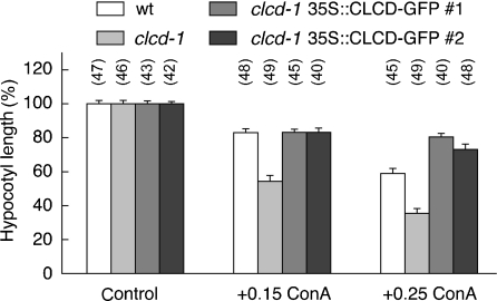
Anion transporting proteins of the CLC type are involved in anion homeostasis in a variety of organisms. CLCs from Arabidopsis have been shown to participate in nitrate accumulation and storage. In this study, the physiological role of the functional chloride transporter AtCLC-d from Arabidopsis was investigated. AtCLC-d is weakly expressed in various tissues, including the root. When transiently expressed as a GFP fusion in protoplasts, it co-localized with the VHA-a1 subunit of the proton-transporting V-type ATPase in the trans-Golgi network (TGN). Stable expression in plants showed that it co-localized with the endocytic tracer dye FM4-64 in a brefeldin A-sensitive compartment. Immunogold electron microscopy confirmed the localization of AtCLC-d to the TGN. Disruption of the AtCLC-d gene by a T-DNA insertion did not affect the nitrate and chloride contents. The overall morphology of these clcd-1 plants was similar to that of the wild-type, but root growth on synthetic medium was impaired. Moreover, the sensitivity of hypocotyl elongation to treatment with concanamycin A, a blocker of the V-ATPase, was stronger in the clcd-1 mutant. These phenotypes could be complemented by overexpression of AtCLC-d in the mutant background. The results suggest that the luminal pH in the trans-Golgi network is adjusted by AtCLC-d-mediated transport of a counter anion such as Cl(-) or NO(3)(-).


Figures

Figure 1
Figure 1
Expression pattern of AtCLC-d in Arabidopsis. Staining pattern of homozygous T2 plants expressing a 2 kb promoter fragment fused to GUS in (a, b) 2-day-old, (e) 5-day-old and (f) 10-day-old seedlings, plus (c) hydathodes and (d) flowers. (g) Overall expression pattern in 28-day-old plants. The numbers indicate days after germination.
Figure 2
Figure 2
Isolation of a T-DNA insertion line for AtCLC-d. (a) Schematic position of the insertion in the 4th intron and location of primers. Exons are indicated as black boxes. The indicated primers were used for genotypic screening. (b) RT-PCR showing absence of the full transcript but expression of the 3′ fragment of the transcript in the clcd-1/clcd-1 line. (c) Northern blot hybridization with total RNA from wt and homozygous clcd-1 plants confirming loss of the 5′ part of the transcript in the mutant line.
Figure 3
Figure 3
Anion profile of 19-day-old Col-0 and homozygous clcd-1 plants grown on various synthetic agar media. Nitrate (a) and chloride (b) concentrations per fresh weight for plants grown in the absence of NaCl on 1.5 mm NH4NO3 or 6 mm NO3 as the sole nitrogen source or on 1.5 mm NH4NO3 supplemented with 60 mm NaCl. The results are means and SE for triplicate determinations. Similar data were obtained in an independent experiment.
Figure 4
Figure 4
Co-localization of AtCLC-d with TGN markers in protoplasts. (a, e, i, m) Confocal laser scanning microscopy of AtCLC-d–GFP showing punctate fluorescence. (b, f, j, n) Staining patterns of co-transformed (b) ARA7–mRFP, (f) ST–mRFP, (j) Syp41–mRFP and (n) the mRFP-labeled VHA-a1 subunit of V-ATPase. Overlays are shown in (c, g, k, o) and bright-field images in (d, h, l, p). Scale bars = 10 µm. (q) Maximum projection of serial sections of part of a protoplast co-expressing AtCLC-d–GFP and VHA-a1–mRFP.
Figure 5
Figure 5
Immunogold labeling of ultra-thin root cryosections of plants stably expressing AtCLC-d–GFP. (a, b) Anti-GFP antibodies were detected with silver-enhanced Nanogold. Gold particles accumulated in the TGN region close to the trans-Golgi cisternae of cortex and epidermal cells of root tips. The trans-Golgi side is marked with (T) and is always shown on the lower side of the Golgi stack (G). (c) No labeling above background was seen in controls not expressing AtCLC-d–GFP. (d) Labeling in plants stably expressing VHA-a1–GFP. Scale bar = 200 nm.
Figure 6
Figure 6
Brefeldin A sensitivity and co-localization with the endocytic marker FM4-64 in root cells. Fluorescence (a, c) and bright-field (b, d) images of stably AtCLC-d–GFP-expressing plant roots. (a, b) Untreated control, (c, d) treatment with BFA (50 µm). Scale bar = 50 µm. Green AtCLC-d–GFP fluorescence (e, h), red FM4-64 labeling (f, i) and overlay (g, j) taken after 6 min (e–g) and in a different experiment after about 10 min (h–j). Scale bars = 10 µm.
Figure 7
Figure 7
Root growth at acidic pH in the clcd-1/clcd-1 line and complementation by ectopic overexpression of AtCLC-d–GFP. Representative root length of seedlings after 8 days (upper panels). Lower panel: statistical analysis of primary root length. The root length was calculated from three independent experiments (means ± SE). More than 50 seedlings were analyzed in each experiment.
Figure 8
Figure 8
Impaired hypocotyl elongation by loss of AtCLC-d and by treatment with concanamycin A. Normalized hypocotyl length of wild-type 5-day-old seedlings (white bar), clcd-1 mutant (light gray bar) and two ectopic AtCLC-d–GFP overexpressers in the clcd-1 background (dark gray bars) without concanamycin A (left), and with 0.15 (middle) or 0.25 µm concanamycin A (right). The number of hypocotyls analyzed in this particular experiment is shown in parentheses on the top of each bar. Means and SE are given and were statistically different between clcd-1 and the other lines upon treatment with concanamycin A (P < 0.001, Student’s t-test). Similar differences between wild-type and clcd-1 were observed in four independent experiments; the results for one of these are shown. Although absolute values of hypocotyl length were almost identical for wild-type and clcd-1 in the absence on concanamycin A, normalized values are given here for better display. Absolute hypocotyl lengths (in mm) were: wild-type = 11.5; clcd-1 = 10.4; complemented clcd-1 = 11.5 and 10.8.

References

    1. Accardi A. Secondary active transport mediated by a prokaryotic homologue of ClC Cl− channels. Nature. 2004;427:803–807. - PubMed
    1. Accardi A. Separate ion pathways in a Cl−/H+ exchanger. J. Gen. Physiol. 2005;126:563–570. - PMC - PubMed
    1. Bassham D C. AtVPS45 complex formation at the trans-Golgi network. Mol. Biol. Cell. 2000;11:2251–2265. - PMC - PubMed
    1. Bolte S. FM-dyes as experimental probes for dissecting vesicle trafficking in living plant cells. J. Microsci. 2004;214:159–173. - PubMed
    1. Campbell R E. A monomeric red fluorescent protein. Proc. Natl Acad. Sci. USA. 2002;99:7877–7882. - PMC - PubMed

Publication types

MeSH terms