. 2007 May 15;305(2):397-410.
doi: 10.1016/j.ydbio.2007.02.024. Epub 2007 Feb 24.

Progression from mitotic catastrophe to germ cell death in Caenorhabditis elegans lis-1 mutants requires the spindle checkpoint

Affiliations

Progression from mitotic catastrophe to germ cell death in Caenorhabditis elegans lis-1 mutants requires the spindle checkpoint

Edgar A Buttner et al. Dev Biol. .

Abstract

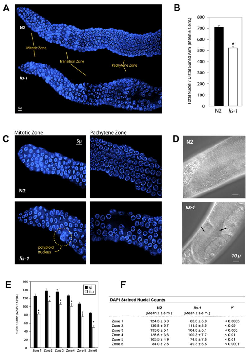
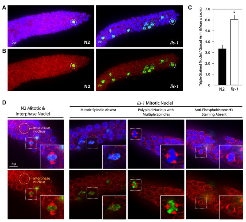
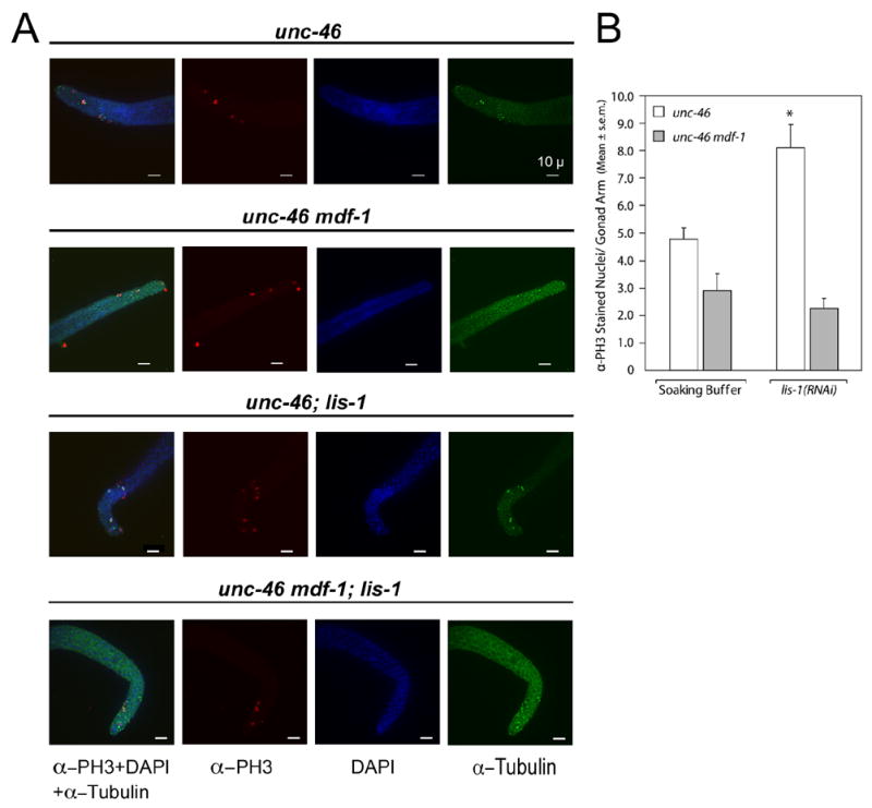
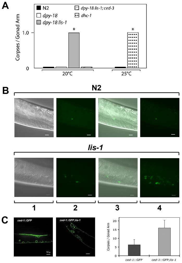
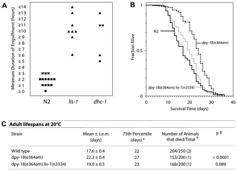
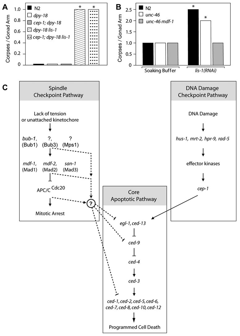
Deletion of the lissencephaly disease gene LIS-1 in humans causes an extreme disorganization of the brain associated with significant reduction in cortical neurons. Here we show that deletion or RNA interference (RNAi) of Caenorhabditis elegans lis-1 results in a reduction in germline nuclei and causes a variety of cellular, developmental, and neurological defects throughout development. Our analysis of the germline defects suggests that the reduction in nuclei number stems from dysfunctional mitotic spindles resulting in cell cycle arrest and eventually programmed cell death (apoptosis). Deletion of the spindle checkpoint gene mdf-1 blocks lis-1(lf)-induced cell cycle arrest and germline apoptosis, placing the spindle checkpoint pathway upstream of the programmed cell death pathway. These results suggest that apoptosis may contribute to the cell-sparse pathology of lissencephaly.


Figures

Fig. 1
Fig. 1
LIS-1 protein alignments and lis-1 gene structure. (A) The C. elegans LIS-1 protein is 58% identical to the human Lis1 protein. Both proteins contain an amino-terminal LISH domain followed by seven WD40 domains. The WD40 repeat domains were located using the WD40 repeat portion of Li and Roberts’ regular expression for the structure of a typical WD-repeat protein propeller blade (Li and Roberts, 2001). (B) Two deletions were obtained, both of which are completely encompassed within the lis-1 genomic sequence. n3334 is an ~2 kb out-of-frame deletion removing exons 4–6, and n3346 is an ~1.5 kb in-frame deletion removing exons 4–5. Homozygous progeny of heterozygous parents are Egl and mildly Unc. Homozygous progeny of homozygous parents display a Mel phenotype.
Fig. 2
Fig. 2
lis-1(lf) disrupts development throughout the germline. (A) Hoechst staining reveals defects in the mitotic, transition, and pachytene zones of lis-1(lf) distal gonads. (B) lis-1(n3334) mutants contain fewer germ cells per distal gonad arm than the N2 strain. lis-1 homozygotes were identified by selecting GFP-negative progeny of strain MT12272. The data are averages ± s.e.m. for N2 (n=8) and lis-1 (n=4) animals. N2 distal gonad arms contain an average of 711.1 ± 12.4 nuclei, whereas lis-1 distal gonad arms contain an average of 521.3 ± 18.1 nuclei (P<0.0001, unpaired t-test). (C) The mitotic zone of lis-1(n3334) mutants contains polyploid nuclei. The pachytene zone of lis-1(n3334) mutants contains large cell-sparse gaps. (D) Arrows point to cell-sparse gaps in the lis-1 germline visible by Nomarski DIC. (E, F) We counted germ cells in 40 μm zones from the distal tip (Zone 1) to the bend of the gonad (Zone 6). Cell counts were significantly lower in each zone of the lis-1(n3334) distal gonad than in the corresponding N2 zones.
Fig. 3
Fig. 3
lis-1(lf) disrupts the mitotic spindle and produces cell cycle arrest. (A) lis-1(n3334) and N2 gonads stained with Hoechst, α-tubulin antibody, and α-phosphohistone H3 antibody (α-PH3). lis-1 gonads contain an increased number of α-PH3-positive nuclei compared to N2 gonads. Representative mitotic nuclei are circled for both strains. Photographs represent a z-stack through the width of the gonad, flattened using deconvolution microscopy. (B) Photographs of the same animals presented in (a), showing only α-tubulin antibody and α-PH3 staining. (C) The mean number of triple-stained nuclei per distal gonad arm is significantly greater in lis-1(n3334) than in the N2 control (P<0.0001, unpaired t-test). The data are averages ± s.e.m. of N2 (n=47) and lis-1 (n=61). (D) N2 mitotic nuclei were easily distinguished by their symmetrical bipolar spindles and distinct centralized Hoechst and α-PH3 staining. An N2 interphase nuclei is circled above for comparison. lis-1 nuclei, on the other hand, display a range of defects including spindle abnormalities, polyploidy, and lack of α-PH3 staining. Representative nuclei are indicated by dashed-squares and are magnified in the inset images.
Fig. 4
Fig. 4
mdf-1(gk2) blocks lis-1(RNAi)-induced cell cycle arrest. (A) Triple-staining of dissected gonads with α-tubulin antibody, α-PH3 antibody, and DAPI. Gonads are from four groups: unc-46 in soaking buffer alone, unc-46 mdf-1 in soaking buffer alone, unc-46; lis-1(RNAi), unc-46 mdf-1;lis-1(RNAi). unc-46; lis-1(RNAi) gonads display an increased number of mitotic nuclei compared to unc-46 gonads in soaking buffer. Staining of unc-46 mdf-1;lis-1(RNAi) gonads displays no such increase in the number of mitotic nuclei when compared to unc-46 mdf-1 gonads in soaking buffer. Photographs represent z-stacks through the width of the gonad, flattened using deconvolution microscopy. (B) The average number of α-PH3-stained nuclei per gonad arm in unc-46 gonads in soaking buffer (4.8 ± 0.4, n=21) is significantly less than in unc-46;lis-1(RNAi) gonads (8.2. ±0.9, n=25) (P<0.005, Tukey test). There is no difference between the average number of α-PH3 stained nuclei per gonad arm in unc-46 mdf-1 gonads in soaking buffer (2.9 ± 0.6, n=17) and unc-46 mdf-1;lis-1(RNAi) gonads (2.3 ± 0.4, n=30).
Fig. 5
Fig. 5
lis-1(lf) triggers apoptosis in the C. elegans germline. (A) dpy-18 lis-1 animals contain significantly more corpses per gonad arm than dpy-18 control animals (P<0.001, Mann-Whitney test). ced-3 blocks the lis-1(lf)-induced increase in apoptosis, as evidenced by the reduced number of corpses per gonad arm in dpy-18 lis-1; ced-3 animals. At the restrictive temperature dhc-1ts mutants exhibit a similar increase compared to N2 (P<0.05, Mann-Whitney test). dpy-18 data and dpy-18 lis-1 data were pooled from 2 datasets, one with and one without a lin-11::GFP marker used for strain construction. The data presented are medians. (B) Corpse number is increased in the lis-1 germline compared to N2. 1) Nomarski DIC alone. 2) SYTO-12-positive corpses in a single focal plane. 3) Nomarski DIC and SYTO-12 images together in a single focal plane. 4) SYTO-12 positive corpses visible in a flattened z-stack. (C) A CED-1::GFP reporter reveals a marked increase in corpse number in lis-1(n3334) gonads.
Fig. 6
Fig. 6
lis-1 and dhc-1 mutants are defective in engulfment but not in aging. (A) lis-1(lf) and dhc-1 exhibit significantly increased engulfment kinetics compared to N2 (P<0.0005 and P<0.005 respectively, Mann-Whitney test). The average minimum engulfment times for lis-1(n3334) (10.9 hours ± 0.9, n=8) and dhc-1 (9.4 hours ± 1.7, n=5) were greater than the N2 average (2.3 hr ± 0.2, n=14). Statistical comparisons using the Mann-Whitney test were conducted between pooled N2 data (unstained mobile, stained mobile and stained immobile worms), and stained mobile lis-1(lf) and dhc-1 worms (see Supplementary data, Fig. S1). (B) Kaplan-Meier lifespan curve analysis confirms that the increase in apoptosis seen in lis-1(n3334) is not a result of accelerated aging. (c) There was no significant difference between the lifespans of wild-type and dpy-18 lis-1 animals; they exhibited similar average lifespans, 75th percentiles, and number of deaths relative to the number of animals observed. For unknown reasons, dpy-18 animals exhibited a longer than wild-type average lifespan (P<0.0001, log-rank test). * The 75th percentile is the age when the fraction of animals alive reaches 0.25. † The total number of observations equals the number of animals that died plus the number censored. The number of independent times the lifespan was determined is in parentheses. Animals that crawled off the plate, exploded or bagged were censored at the time of the event.‡ Compared with wild-type control in all experiments.
Fig. 7
Fig. 7
The spindle checkpoint lies upstream of the programmed cell death pathway. (A) dpy-18 lis-1 and cep-1; dpy-18 lis-1 mutants exhibit no significant difference in the average number of corpses per gonad arm when stained with SYTO 12. On the other hand, they both contain a significantly greater average number of corpses per gonad arm than N2, dpy-18 and cep-1; dpy-18 (P<0.001, for all comparisons, Mann-Whitney test). The data presented are medians. (B) Treatment of N2 and unc-46 with lis-1 dsRNA produces a significant increase in the average number of corpses per gonad arm, compared to soaking buffer alone (P<0.05 and P<0.001 respectively, Mann-Whitney test). The unc-46 mdf-1 double mutant contains a similar average number of corpses per gonad arm as N2 and unc-46 in soaking buffer alone but does not show an increase in corpse number upon treatment with lis-1 dsRNA. The data are medians, while the averages ± the s.e.m. and are presented in Table 1. Because mdf-1 homozygotes display variable phenotypes (Kitagawa and Rose, 1999), we excluded animals exhibiting tumerous gonads (tum phenotype) or abnormal gonad development (gon phenotype) from our analysis. (C) The spindle checkpoint gene mdf-1 is required for lis-1(lf)-induced apoptosis, suggesting that the spindle checkpoint lies upstream of the programmed cell death pathway and in parallel to the DNA damage checkpoint. Genes linking the spindle checkpoint to the programmed cell death pathway remain unknown.

Similar articles

Cited by

References

    1. Basu J, Bousbaa H, Logarinho E, Li Z, Williams BC, Lopes C, Sunkel CE, Goldberg ML. Mutations in the essential spindle checkpoint gene bub1 cause chromosome missegregation and fail to block apoptosis in Drosophila. J Cell Biol. 1999;146:13–28. - PMC - PubMed
    1. Bhalla KN. Microtubule-targeted anticancer agents and apoptosis. Oncogene. 2003;22:9075–9086. - PubMed
    1. Blocker A, Severin FF, Burkhardt JK, Bingham JB, Yu H, Olivo JC, Schroer TA, Hyman AA, Griffiths G. Molecular requirements for bi-directional movement of phagosomes along microtubules. J Cell Biol. 1997;137:113–129. - PMC - PubMed
    1. Cockell MM, Baumer K, Gonczy P. lis-1 is required for dynein-dependent cell division processes in C. elegans embryos. J Cell Sci. 2004;117:4571–4582. - PubMed
    1. Davidson FF, Steller H. Blocking apoptosis prevents blindness in Drosophila retinal degeneration mutants. Nature. 1998;391:587–591. - PubMed

Publication types

MeSH terms

Substances

LinkOut - more resources