. 2007 May 1;313(8):1639-51.
doi: 10.1016/j.yexcr.2007.02.018. Epub 2007 Feb 28.

Organization of cytokeratin cytoskeleton and germ plasm in the vegetal cortex of Xenopus laevis oocytes depends on coding and non-coding RNAs: three-dimensional and ultrastructural analysis

Affiliations

Organization of cytokeratin cytoskeleton and germ plasm in the vegetal cortex of Xenopus laevis oocytes depends on coding and non-coding RNAs: three-dimensional and ultrastructural analysis

Malgorzata Kloc et al. Exp Cell Res. .

Abstract

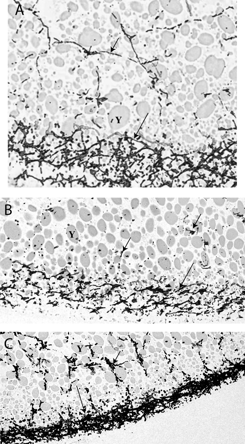
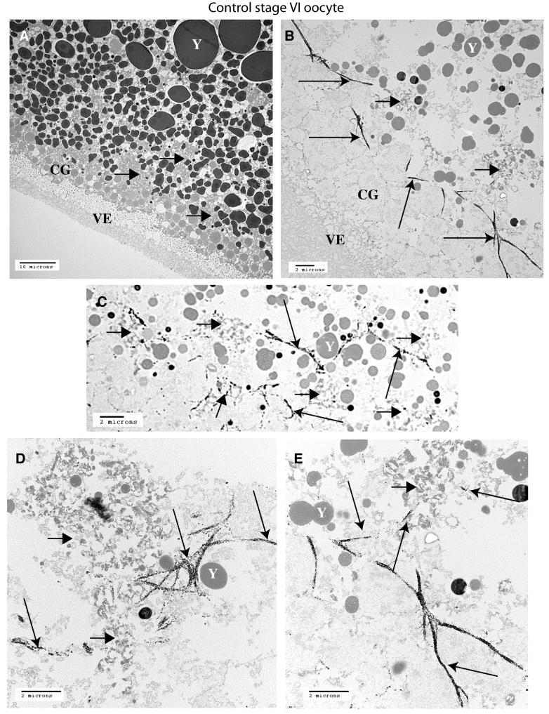
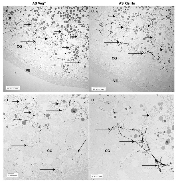
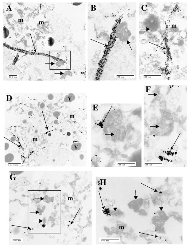
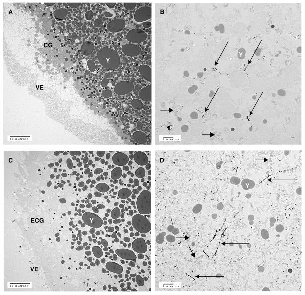
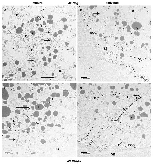
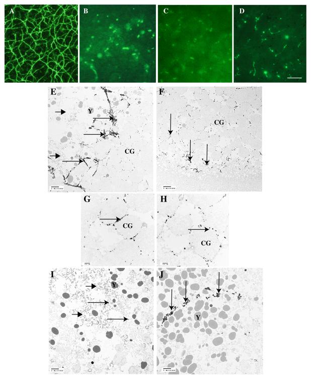
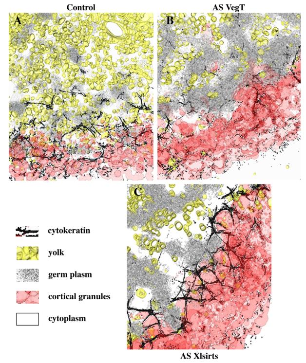
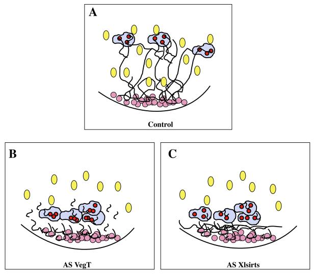
Recent studies discovered a novel structural role of RNA in maintaining the integrity of the mitotic spindle and cellular cytoskeleton. In Xenopus laevis, non-coding Xlsirts and coding VegT RNAs play a structural role in anchoring localized RNAs, maintaining the organization of the cytokeratin cytoskeleton and germinal granules in the oocyte vegetal cortex and in subsequent development of the germline in the embryo. We studied the ultrastructural effects of antisense oligonucleotide driven ablation of Xlsirts and VegT RNAs on the organization of the cytokeratin, germ plasm and other components of the vegetal cortex. We developed a novel method to immunolabel and visualize cytokeratin at the electron microscopy level, which allowed us to reconstruct the ultrastructural organization of the cytokeratin network relative to the components of the vegetal cortex in Xenopus oocytes. The removal of Xlsirts and VegT RNAs not only disrupts the cytokeratin cytoskeleton but also has a profound transcript-specific effect on the anchoring and distribution of germ plasm islands and their germinal granules and the arrangement of yolk platelets within the vegetal cortex. We suggest that the cytokeratin cytoskeleton plays a role in anchoring of germ plasm islands within the vegetal cortex and germinal granules within the germ plasm islands.


Figures

Figure 1
Figure 1
Changes in the organization of the cytokeratin network in the vegetal cortex of stage VI oocytes, after Xlsirts and VegT RNAs ablation. (A-C) Three-dimensional reconstruction of 3 sets of 10 semithin (0.7μm) sections of the fragments of the vegetal cortex of oocytes stained with anti-cytokeratin antibody, nanogold-conjugated secondary antibody, silver enhanced and stained lightly with 1% toluidine blue diluted in 1% borax. (A) In control oocytes, long filaments of the cytokeratin network are visible at the cortex (long arrow) and also away from the cortex between the yolk platelets (short arrow). (B) Oocytes injected with AS VegT ODN show fragmentation of filaments at the cortex (long arrow) and lack of long filaments between the yolk and only granular foci of cytokeratin (short arrows) visible between the yolk. (C) Oocytes injected with AS Xlsirts ODNs show a prominent narrow layer of cytokeratin at the cortex (long arrow) and also the presence of long but granular in appearance filaments between the yolk (short arrow). Yolk platelets, Y.
Figure 2
Figure 2
Ultrastructural analysis of cytokeratin distribution in the vegetal cortex of stage VI oocytes. Oocytes were immunolabeled with anti-cytokeratin antibody and nanogold conjugated secondary antibody and silver enhanced. (A) Low magnification view of the fragment of the oocyte showing the arrangement of the components of the vegetal cortex. The vitelline envelope is followed by the layer of cortical granules and yolk. The islands of germ plasm (arrows) are distributed at the boundary between the germinal granules and yolk. Cytokeratin is not visible at this magnification. (B) Fragment of the vegetal cortex showing the germ plasm islands (short arrows) and the long filaments of cytokeratin network (long arrows) situated between the layer of cortical granules and yolk and in contact with the periphery of germ plasm islands. (C) Fragment of the vegetal cortex showing several (visibly separated from each other) germ plasm islands (short arrows) surrounded and penetrated by the long cytokeratin filaments (long arrows). (D, E) Two examples of germ plasm islands (short arrows) penetrated by the cytokeratin filaments (long arrows). Vitelline envelope, VE; Cortical granules, CG; yolk platelets, Y.
Figure 3
Figure 3
The effect of Xlsirts and VegT RNAs ablation on the organization of cytokeratin filaments and germ plasm islands distribution in the vegetal cortex of stage VI oocytes. Oocytes were injected with AS VegT (A, B) or AS Xlsirts (C, D) ODNs and immunolabeled with anti-cytokeratin antibody and nanogold conjugated secondary antibody, and silver enhanced. (A) Fragment of the vegetal cortex of AS VegT-injected oocyte showing short and dispersed filaments of cytokeratin (long arrows) distributed between yolk and around the cortical granules and a thick band of interconnected germ plasm islands (short arrows). (B) Fragment of the vegetal cortex of AS VegT-injected oocyte showing fragmented cytokeratin filaments (long arrows) around and within the germ plasm islands (short arrows) and surrounding cortical granules. (C, D) Fragments of the vegetal cortex of AS Xlsirts injected oocyte showing interconnected germ plasm islands (short arrows) and a compact layer of long cytokeratin filaments apposing cortical granules and yolk. Note the scarcity of cytokeratin filaments around the germ plasm islands. Vitelline envelope, VE; Cortical granules, CG; yolk platelets, Y.
Figure 4
Figure 4
Cytokeratin distribution in relation to germ plasm islands and germinal granules in stage VI oocytes. (A-H) Stage VI oocytes immunolabeled with nanogold conjugated anti-cytokeratin antibody and silver enhanced. (A-C) Fragment of germ plasm island from stage VI control oocyte. Long cytokeratin filaments (long arrows) contact and penetrate germ plasm island and are in contact with the germinal granules (short arrows). Area of germ plasm island marked with square in panel A is shown at higher magnification in panel B. (D-F) Fragments showing germ plasm islands in oocytes injected with AS VegT ODN. (D) Short fragments of cytokeratin (long arrows) are visible in the vicinity of germ plasm islands. (E-F) Granular foci of disintegrated cytokeratin filaments (long arrows) are visible in the vicinity of huge germinal granules (short arrows). (G, H) Fragments showing the germ plasm island in oocytes injected with AS Xlsirt ODN. Area of germ plasm island marked with square in panel G is shown at higher magnification in panel H. Granular foci of disintegrated cytokeratin filaments (long arrows) are visible in the vicinity of germinal granules (short arrows). Yolk platelets, Y; mitochondria, m.
Figure 5
Figure 5
Ultrastructure of cytokeratin distribution in the vegetal cortex of mature oocyte and activated egg. Oocytes and eggs were immunolabeled with anti-cytokeratin antibody and nanogold conjugated secondary antibody and silver enhanced. (A) Low magnification view of the fragment of the oocyte matured by the addition of progesterone showing the arrangement of the components of the vegetal cortex. The vitelline envelope is followed by the layer of cortical granules and yolk. (B) Fragment of oocyte cortex showing small foci of cytokeratin (long arrows) and small germ plasm islands (short arrows). (C) Low magnification view of the fragment of the egg, 1-3 hr after activation by pricking showing the arrangement of the components of the vegetal cortex with the layer of exocytosed cortical granules. (D) Fragment of activated egg cortex showing the reconstitution of long filaments of cytokeratin (long arrows) in contact with germ plasm islands (short arrows). Vitelline envelope, VE; Cortical granules, CG; Exocytosized cortical granules, EGC; yolk platelets, Y.
Figure 6
Figure 6
The effect of VegT and Xlsirts RNAs ablation on the ultrastructure of cytokeratin distribution in the vegetal cortex of mature oocytes and activated eggs. Oocytes were injected with AS VegT (A, B) or AS Xlsirts (C, D) ODNs, matured by addition of progesterone, activated by pricking, and immunolabeled with anti-cytokeratin antibody and nanogold-conjugated secondary antibody and silver enhanced. (A) Fragment of the vegetal cortex of AS VegT ODN injected mature oocyte showing tiny foci of cytokeratin (long arrows) and large, interconnected germ plasm islands (short arrows). (B) In AS VegT ODN-injected and activated egg, short cytokeratin filaments (long arrows) reconstitute at the periphery of the cortex apposing vitelline envelope. Large germ plasm islands (short arrows) are also visible. (C) Fragment of the vegetal cortex of AS Xlsirts ODN-injected mature oocyte showing foci of cytokeratin (long arrows) and large germ plasm islands (short arrows). (D) In AS Xlsirts ODN injected and activated egg short and long cytokeratin filaments (long arrows) reconstitute at the periphery of the cortex apposing microvilli and also between the yolk and in vicinity of germ plasm islands (short arrows). Vitelline envelope, VE; Cortical granules, CG; Exocytosed cortical granules, EGC; yolk platelets, Y.
Figure 7
Figure 7
The effect of injection of anti-cytokeratin antibody on the ultrastructure of cytokeratin filaments in the vegetal cortex of stage VI oocytes, mature oocytes, and activated eggs mimics the effect of VegT mRNA ablation. (A) Cytokeratin network in the vegetal cortex of control stage VI oocyte stained with FITC conjugated anti-cytokeratin antibody. (B-D) Cytokeratin in vegetal cortex of stage VI oocytes and egg injected with FITC conjugated anti-cytokeratin antibody. (B) 3hr after the injection of anti-cytokeratin antibody, the cytokeratin network is disrupted and its remnants form thick foci. (C) After progesterone-induced maturation of injected oocytes, the cytokeratin is visible as very small and dispersed foci. (D) In eggs activated by pricking, cytokeratin reconstitutes into thick short fibrils. (E, F) Two areas of vegetal cortex of stage VI oocyte injected with anti-cytokeratin antibody and subsequently incubated with nanogold-conjugated secondary antibody and silver enhanced. A few hours after the injection of antibody the filaments of cytokeratin become shorter and visibly granular in appearance (long arrows, E) and also fragment into smaller foci, which surround cortical granules (long arrows, F). Germ plasm islands are also visible in the vicinity of fragmenting cytokeratin filaments (short arrow, E). The distribution of fragmented cytokeratin around cortical granules in anti-cytokeratin antibody-injected oocytes (G) resembles that in oocytes injected with AS VegT ODN (H). (I) In anti-cytokeratin antibody-injected mature oocytes (as in AS VegT ODN-injected mature oocytes) germ plasm islands are large and interconnected (short arrows), and cytokeratin forms tiny granular foci (long arrows). (J) In anti-cytokeratin-injected activated eggs (unlike in AS VegT ODN-injected activated eggs) cytokeratin reforms as compact granular aggregates (long arrows). Cortical granules, CG; yolk platelets,Y. Scale bar in A-D: 12μm.
Figure 8
Figure 8
Three dimensional reconstruction of distribution of cytokeratin and germ plasm islands in the vegetal cortex of stage VI oocytes after VegT and Xlsirts RNAs ablation . (A) In control stage VI oocytes small germ plasm islands are separated from each other and are distributed more internal to the layer of cortical granules and between the yolk. The majority of long cytokeratin filaments form the network between and within the layer of cortical granules and yolk and penetrate and surround the germ plasm islands. Shorter and granular in appearance filaments are also present at the surface of cortical granules. (B) In oocytes injected with AS VegT ODN the majority of the cytokeratin network disintegrates into short fragments, which aggregate around the cortical granules and some remain in contact with the germ plasm islands. The germ plasm islands fuse into larger interconnected aggregates, and yolk platelets are displaced inward. (C) In AS Xlsirts ODN-injected oocytes the majority of long cytokeratin filaments form a distinct layer on the top of cortical granules. Filaments contact the surface of the aggregates of germ plasm islands (but do not penetrate them). Shorter and granular cytokeratin filaments are also present on the surface of cortical granules. Yolk platelets are displaced inward.
Figure 9
Figure 9
Model of the effect of AS VegT and Xlsirts RNA depletion on the ultrastructure of cytokeratin network and components of the vegetal cortex in Xenopus stage VI oocytes. (A) In control stage VI oocytes, the long filaments of cytokeratin (black) form a complex, interconnected network spanning between cortical granules (pink spheres) and yolk platelets (yellow spheres). Germ plasm islands (grey areas) are anchored by cytokeratin filaments in proper position within the cortex. Cytokeratin filaments also penetrate the germ plasm islands and anchor the germinal granules (red spheres) located within the islands. Proper multidimensional organization of cytokeratin filaments ensures proper spatial distribution of cortical granules and yolk platelets and separation of individual germ plasm islands and individual germinal granules within the germ plasm islands. (B) In AS VegT ODN-depleted oocytes, long cytokeratin filaments fragment into short pieces and granular foci. The disintegration of the cytokeratin network results in the disruption of the proper spatial distribution of the components of the cortex. The germ plasm islands become released from the cytokeratin anchor and fuse into larger aggregates displacing yolk platelets inward. In addition, the disintegration of cytokeratin filaments anchoring the germinal granules within the germ plasm islands causes the individual granules to fuse into larger aggregates. (C) In AS Xlsirts ODN-depleted oocytes long cytokeratin filaments collapse at the surface of the cortical granule layer and lose contact with germ plasm islands. Loss of the cytokeratin anchor results in the fusion of germ plasm islands into larger aggregates and displacement of the yolk inward. At present it is unclear why germinal granules in Xlsirts-depleted oocytes (although devoid of long cytokertin filament anchors) do not fuse as in AS VegT-depleted oocytes. It is very possible that the proper separation and spatial distribution of germinal granules within the germ plasm island depends not only on the presence of intact cytokeratin filaments but also on other unknown factor(s) absent or nonfunctional in AS VegT-depleted oocytes but still properly functioning in AS Xlsirts-depleted oocytes.

Similar articles

Cited by

References

    1. Bashirullah A, Cooperstock RL, Lipshitz HD. RNA localization in development. Annu. Rev. Biochem. 1998;67:335–394. - PubMed
    1. Jansen RP. mRNA localization: message on the move. Nat. Rev. Mol. Cell. Biol. 2001;2:247–256. - PubMed
    1. King ML, Zhou Y, Bubunenko M. Polarizing genetic information in the egg: RNA localization in the frog oocyte. BioEssays. 1999;21:546–557. - PubMed
    1. Kloc M, Zearfoss NR, Etkin LD. Mechanisms of subcellular mRNA localization. Cell. 2002b;108:533–544. - PubMed
    1. Palacios IM, Johnston D. Getting the message across: the intracellular localization of mRNA in higher eukaryotes. Annu. Rev. Cell. Dev. Biol. 2001;17:569–614. - PubMed

Publication types

MeSH terms