. 2007 May 4;368(3):607-17.
doi: 10.1016/j.jmb.2007.02.070. Epub 2007 Mar 2.

Structural basis of aquaporin inhibition by mercury

Affiliations

Structural basis of aquaporin inhibition by mercury

David F Savage et al. J Mol Biol. .

Abstract

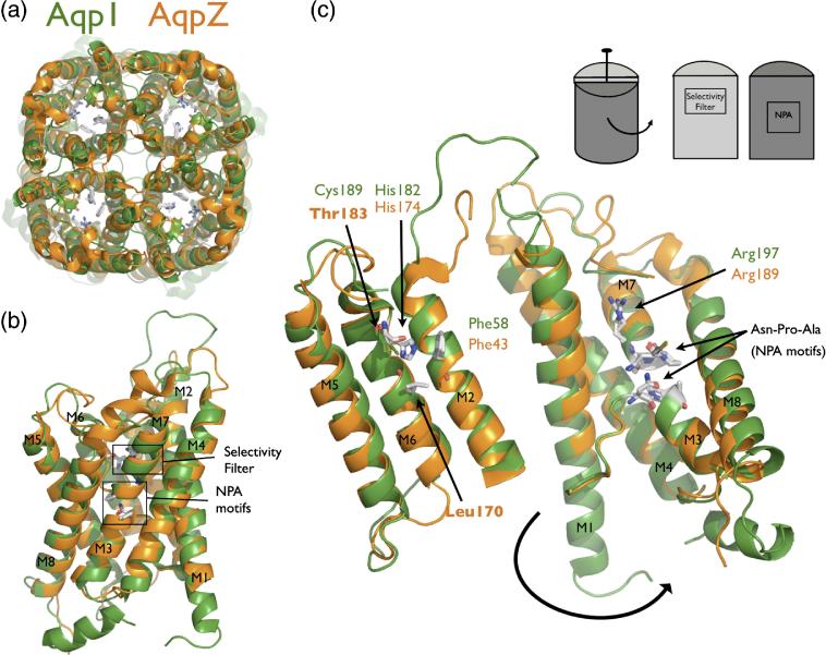
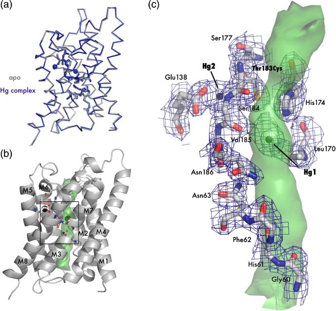
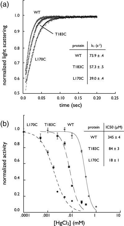
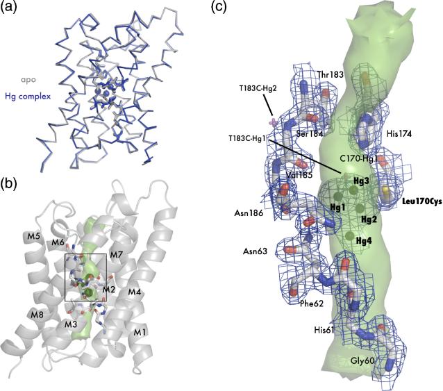
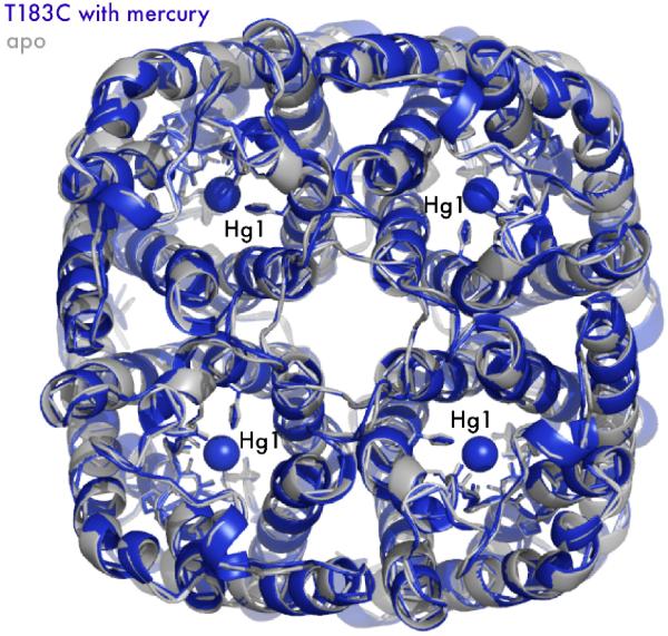
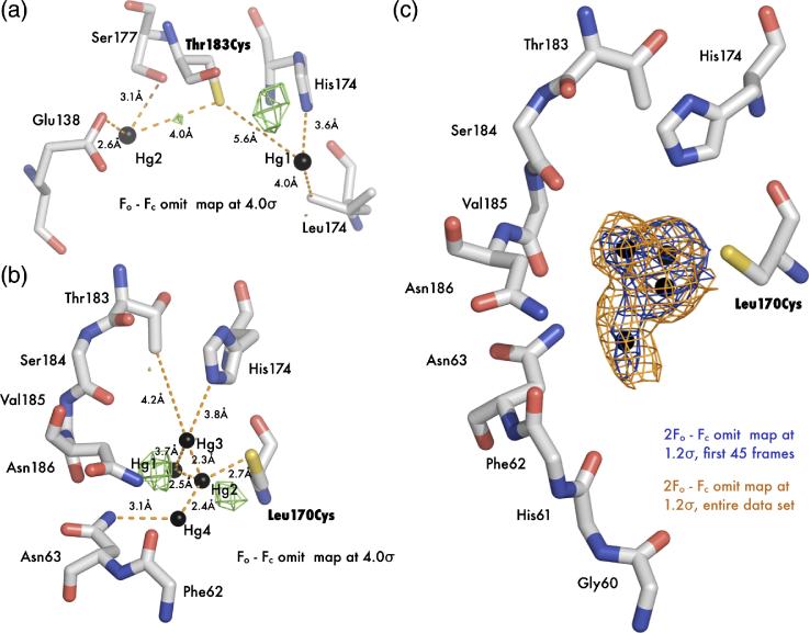
The aquaporin family of channels was defined based on the inhibition of water transport by mercurial compounds. Despite the important role of mercurials, little is known about the structural changes involved upon mercury binding leading to channel inhibition. To elucidate the mechanism we designed a mutant, T183C, of aquaporin Z (AqpZ) patterned after the known mercury-sensitive site of aquaporin 1 (AQP1) and determined the X-ray crystal structures of the unbound and mercury blocked states. Superposition of the two structures shows no conformational rearrangement upon mercury binding. In the blocked structure, there are two mercury sites, one bound to Cys183 and occluding the pore, and a second, also bound to the same cysteine but found buried in an interstitial cavity. To test the mechanism of blockade we designed a different mutant, L170C, to produce a more effective mercury block at the pore site. In a dose-response inhibition study, this mutant was 20 times more sensitive to mercury than wild-type AqpZ and four times more sensitive than T183C. The X-ray structure of L170C shows four mercury atoms at, or near, the pore site defined in the T183C structure and no structural change upon mercury binding. Thus, we elucidate a steric inhibition mechanism for this important class of channels by mercury.


Figures

Figure 1
Figure 1
AqpZ is the bacterial homolog of AQP1. (A) Cartoon representation of the AqpZ (orange) and AQP1 (green) tetramers. Note the presence of the four monomer channels and the hypothetical channel down the tetramer axis. (B) Cartoon representation of the AqpZ and AQP1 monomers. Helices are labeled M1 through M8, and the selectivity filter and NPA motifs are designated with boxes. (C) Monomer opened up showing conservation of the water-selective motif. In this cartoon representation, the monomer is peeled open as shown in the inset schematic. The conserved selectivity filter and NPA motif are shown in sticks. Thr183 and Leu170 in AqpZ are the positions of cysteine mutants in this study. All molecular structure figures were made in Pymol (Delano Scientific).
Figure 2
Figure 2
Crystal structure of apo T183C and mercury bound T183C mutants. (A) Main chain overlay of the apo (gray) and Hg-complex (blue) with an RMSD (Cα) of 0.27 Å. Bound Hg2+ atoms are displayed as spheres with a van der Waals radius of 1.10 Å. (B) Cartoon representation of T183C. Transmembrane helices are labeled M1-M8 and the interior surface of the channel is drawn as a green surface. The black square denotes the area of interest depicted in panel C. (C) Structure of the blocked channel. Amino acids involved with water binding in AQPs are shown as sticks and with 2Fo-Fc electron density mapped contoured at 1.2σ drawn in blue. Mercury atoms are shown as spheres. In this orientation it can be seen that T183C-Hg1 sterically blocks the pore (green surface).
Figure 3
Figure 3
Kinetic Studies of Aquaporin Z. (A) Water conduction of WT, T170C, and T183C. Proteins were reconstituted in liposomes, challenged with a higher osmotic gradient in a stopped-flow device, and liposome shrinkage measured by light scattering at 440 nm. Plots were fit to a single exponential and the resulting rates are shown in the inset table (n=5). (B) Dose-response curve of proteoliposomes incubated with HgCl2. After incubation with HgCl2 proteoliposomes were assayed as in panel A and the rates (normalized to maximum rate for each mutant, n=5) were fit to a sigmoid dose-response curve in Kaleidagraph (Synergy Software). IC50 values are shown in the inset table.
Figure 4
Figure 4
Crystal structure of apo L170C and mercury bound L170C. (A) Main chain overlay of the apo (gray) and Hg-complex (blue) with an RMSD of 0.27 Å. Bound mercury atoms are displayed as spheres with a van der Waals radius of 1.10 Å. (B) Cartoon representation of L170C. Transmembrane helices are labeled M1-M8 and the interior surface of the channel is drawn as a green surface. The black square denotes the area of interest depicted in panel C. (C) Structure of the blocked channel. Amino acids classically involved with water binding in AQPs are shown as sticks and with 2Fo-Fc - electron density mapped contoured at 1.2 σ drawn in blue. Mercury are shown as spheres. Superposition of mercury atoms from the T183C structure are shown as magenta crosses. In this orientation it can be seen that all three mercury atoms sterically block the pore (green surface).
Figure 5
Figure 5
Mercury blocks the monomer channel. While AQPs are tetramers in the membrane, the monomer is the functional unit. By imposing crystal symmetry on both the apo (grey) and complex structures (blue), T183C is drawn as a tetramer in cartoon representation. Mercury-Hg1, with its proper van der Waals radius, is drawn as a sphere blocking the channel. Note there is almost no structural change to the tetrameric axis.
Figure 6
Figure 6
Mercury disorder in electron density maps. (A) Fo-Fc electron density map (green) of mercury bound T183C structure contoured at 4 σ. (B) Fo-Fc electron density map (green) of mercury bound L170C structure contoured at 4 σ. (C) 2Fo-Fc omit electron density map solved with the first 45 frames of data (blue) and the entire dataset (orange). Both maps are contoured at 1.2 σ around the three mercury atoms.

References

    1. Borgnia M, Nielsen S, Engel A, Agre P. Cellular and molecular biology of the aquaporin water channels. Annu Rev Biochem. 1999;68:425–58. - PubMed
    1. Hille B. Ion channels of excitable membranes. 3rd edit Sinauer; Sunderland, Mass: 2001.
    1. Macey RI. Transport of water and urea in red blood cells. Am J Physiol. 1984;246:C195–203. - PubMed
    1. Zeidel ML, Ambudkar SV, Smith BL, Agre P. Reconstitution of functional water channels in liposomes containing purified red cell CHIP28 protein. Biochemistry. 1992;31:7436–40. - PubMed
    1. Preston GM, Carroll TP, Guggino WB, Agre P. Appearance of water channels in Xenopus oocytes expressing red cell CHIP28 protein. Science. 1992;256:385–7. - PubMed

Publication types

MeSH terms

LinkOut - more resources