. 2007 May;124(5):350-63.
doi: 10.1016/j.mod.2007.02.001. Epub 2007 Feb 21.

The maternally localized RNA fatvg is required for cortical rotation and germ cell formation

Affiliations

The maternally localized RNA fatvg is required for cortical rotation and germ cell formation

Agnes P Chan et al. Mech Dev. 2007 May.

Abstract

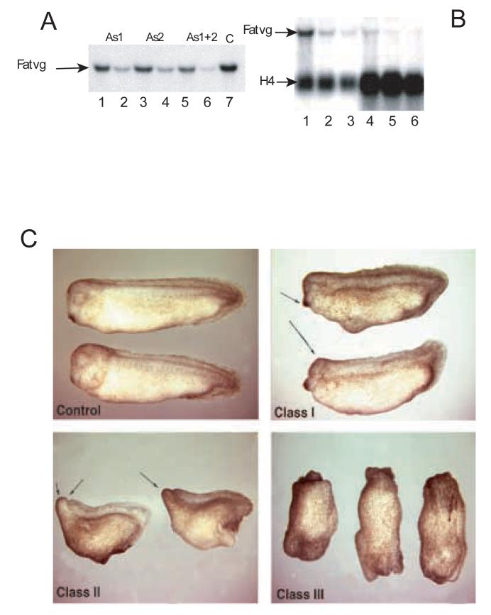
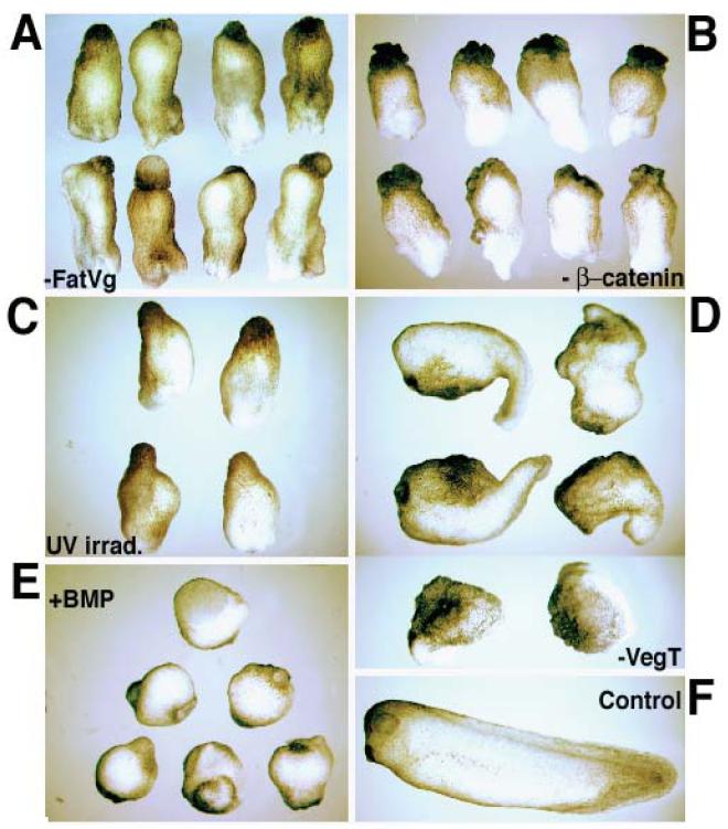
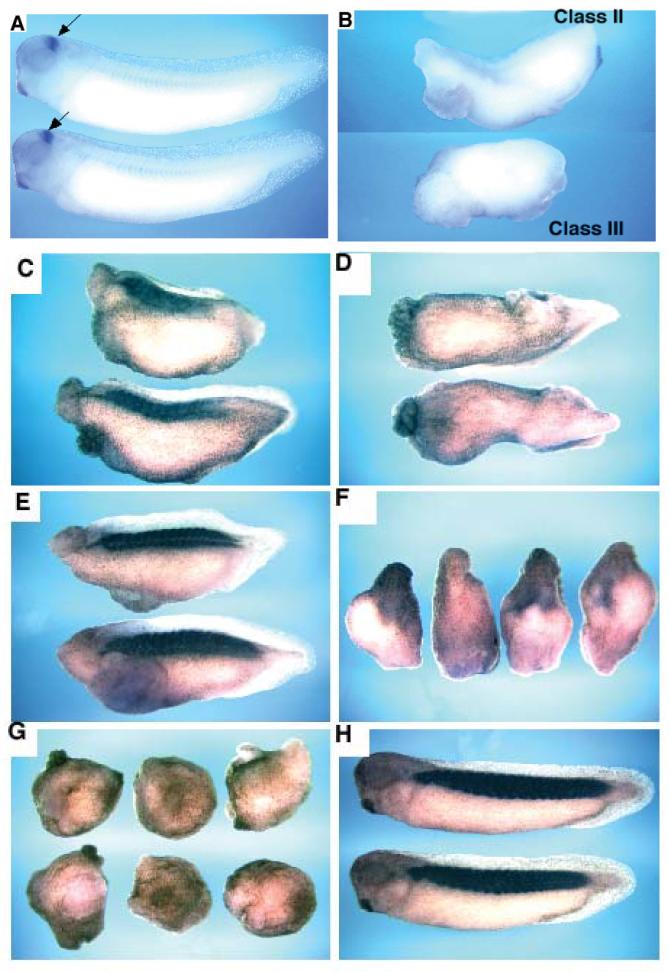
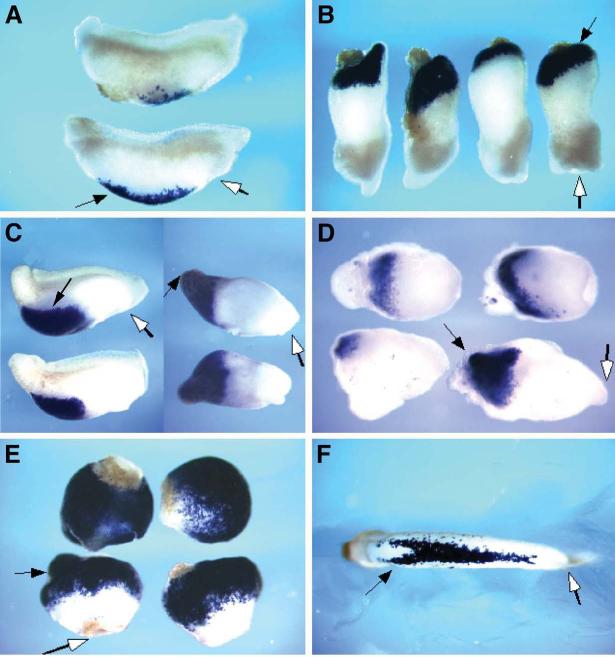
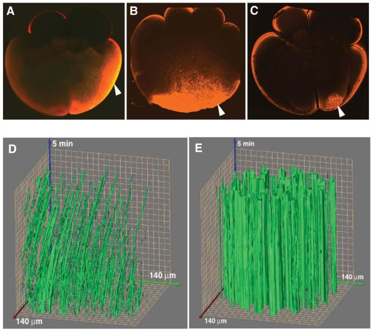
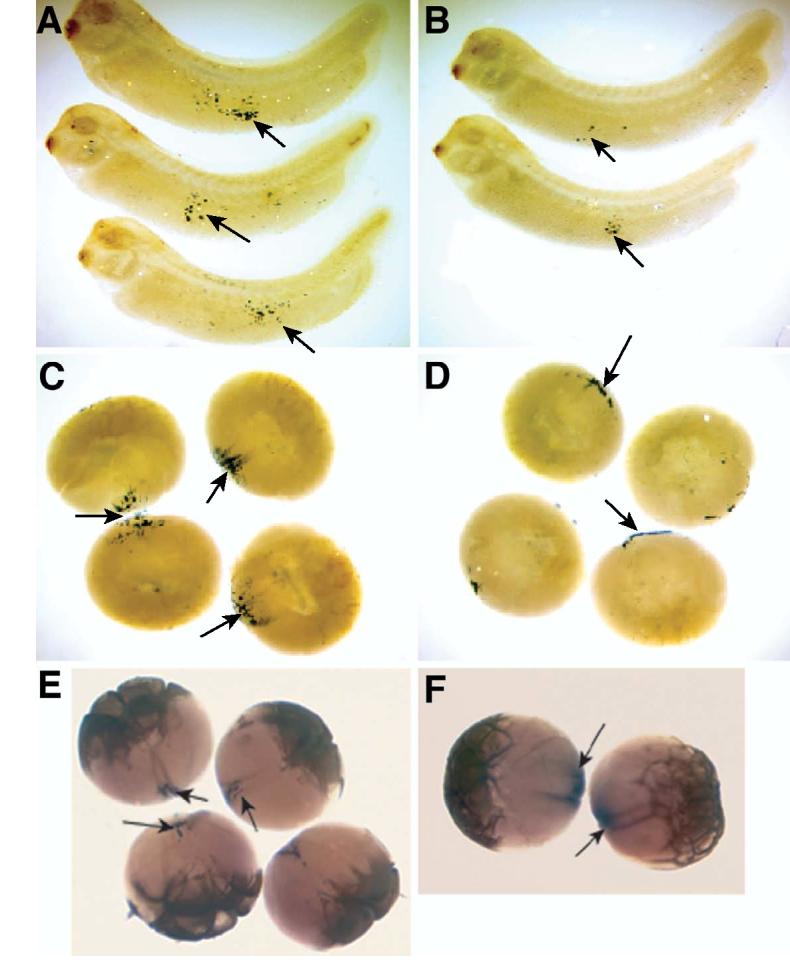
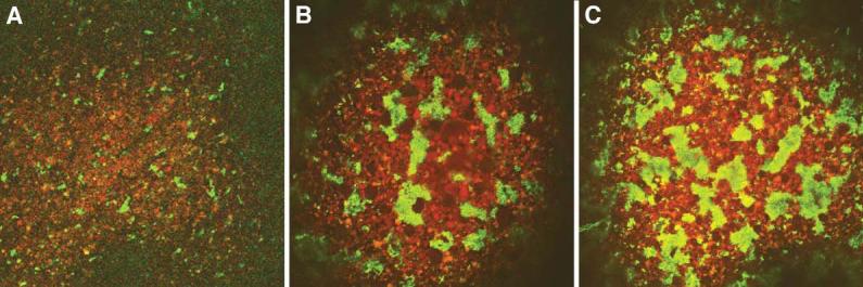
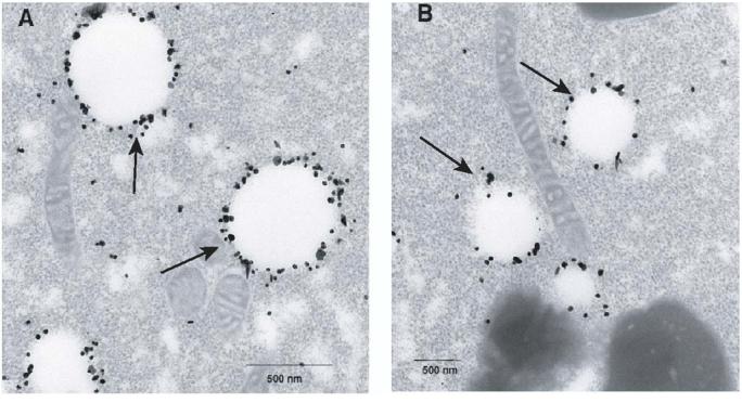
Fatvg is a localized maternal transcript that translocates to the vegetal cortex of Xenopus laevis oocytes through both the METRO and Late RNA localization pathways. It is a member of a gene family that functions in vesicular trafficking. Depletion of the maternal store of fatvg mRNA results in a dual phenotype in which embryos are ventralized and also lack primordial germ cells. This complex fatvg loss of function phenotype is the result of stabilization of the dorsalizing factor beta-catenin at the vegetal pole and the inability of the germ cell determinants to move to their proper locations. This is coincident with the inhibition of cortical rotation and the abnormal aggregation of the germ plasm. Fatvg protein is located at the periphery of vesicles in the oocyte and embryo, supporting its proposed role in vesicular trafficking in the embryo. These results point to a common fundamental mechanism that is regulated by fatvg through which germ cell determinants and dorsalizing factors segregate during early development.


Figures

Figure 1
Figure 1
Northern blot analysis of antisense oligo-injected oocytes. (A) The level of endogenous fatvg mRNA was analyzed after oocytes were injected, singly or with a mixture of two antisense ODNs, in two doses. Lane 1, 2.5 ng of fatvg antisense 1 (fatvgAS1); Lane 2, 5ng fatvgAS1; Lane 3, 2.5 ng fatvgAS2; Lane 4, 5ng fatvgAS2; Lane 5, total 2.5ng fatvgAS1 and 2; Lane 6, total 5ng fatvgAS1 and 2; Lane 7 Control. (B) Oocytes and gastrula stage embryos derived from oocytes injected with a total of 2.5 ng fatvg antisense ODN were analyzed by Northern analysis. Lane 1, levels of fatvg mRNA in uninjected stage VI oocytes; Lane 2 levels of fatvg mRNA in fatvgAS1 injected oocytes; Lane 3, level of fatvg mRNA in fatvgAS1 and2 injected oocytes; Lane 4, Level of fatvg mRNA in uninjected stage 10 embryos; Lane 5, Levels of fatvg mRNA in fatvgAS 1 injected embryos at stage 10; Lane 6, Levels of fatvg mRNA in fatvgAS1 and 2 injected embryos at stage 10. The level of fatvg mRNA in embryos derived from depleted oocytes did not increase, indicating that there was no detectable zygotic transcription of fatvg. The histone gene H4 was used as a loading control and was zygotically activated. Fatvg-depleted embryos display axial defects. (C) Axis formation in embryos derived from uninjected oocytes is normal. Class I embryos have reduced anterior development and a shortened axis, but the most anterior structure, the cement gland (arrows), is still formed. Class II embryos have anterior truncation of head structures (arrows) and are lacking the cement gland and the eyes. Dorsal-ventral polarity can be distinguished from the formation of the dorsal fin. Class III embryos show a complete loss of axial identity and these embryos appear as an elongated mass of tissue with a highly ruffled pigmented end and a blastopore formed at the other end.
Figure 2
Figure 2
Ventralization caused by fatvg-depletion is reminiscent of UV irradiation and β-catenin-depleted embryos but different than VegT depleted embryos. (A) fatvg-depleted embryos showing a class III phenotype with a complete loss of axial identity. (B) β-catenin-depleted embryos showing a similar phenotype. (C) Embryos that had been irradiated at the vegetal pole after fertilization. (D) VegT-depleted embryos are defective in endoderm and mesoderm formation. The two bottom embryos show the most severe phenotype of VegT depletion in which only epidermal development is apparent. (E) Embryos injected with 2ng BMP-4 at 2-cell stage. (F) Control embryo at stage 39.
Figure 3
Figure 3
The anterior marker engrailed is not expressed in fatvg-depleted embryos. (A) Embryos derived from uninjected oocytes express engrailed mRNA (arrows) in the midbrain-hindbrain boundary as analyzed by in situ hybridization. (B) The upper embryo is a class II fatvg-depleted embryo and the lower one is a class III embryo. Niether express engrailed. Muscle differentiation is deficient in fatvg-depleted embryos. (C) Class II fatvg-depleted embryos showed a reduced amount of 12/101 immunostaining which is a specific marker for muscle differentiation. (D) No 12/101 staining was detected in class III fatvg-depleted embryos. (E) A reduced amount of 12/101 staining is obtained in embryos exposed to a low dose of UV irradiation. (F) High dose of UV irradiation generated ventralized embryos that show no staining. Dark shadows are regions of the embryo concentrated with pigments. (G) No staining is detected in BMP-4-injected embryos. (H) Control embryos showing 12/101 staining in the somites.
Figure 4
Figure 4
The ventral mesodermal marker α-T4 globin expression is enhanced in fatvg-depleted class III embryos. (A) α-T4 globin is expressed at a normal level in class II embryos as shown by in situ hybridization. (B) Class III embryos have an increase amount of α-T4 globin expression close to one end of the embryo. (C) UV irradiated embryos have elevated levels of expression of α-T4 globin. Embryos on the left are treated with a low UV dose. A higher dose was used for the embryos on the right. (D) β-catenin depleted embryos have increased α-T4 globin expression. (E) α-T4 globin is highly expressed in BMP-4 injected embryos. (F) A control embryo showing α-T4 globin expression in the developing blood islands derived from the ventral mesoderm. Arrows point to the expression domain of α-T4 globin. Open arrows point to the position of the blastopore.
Figure 5
Figure 5
Confocal analysis of β-catenin protein distribution in 8-cell stage fatvg-depleted embryos showing abnormal accumulation of β-catenin at the vegetal pole of the embryos. Embryos were bisected along animal/vegetal axis into halves before the immunostaining procedure was carried out. (A) In embryos derived from uninjected oocytes that had undergone the host transfer procedure β-catenin protein is detected in an arc region in the outer region of the dorsal blastomeres. (B) In fatvg-depleted embryos an accumulation of β-catenin is seen at the vegetal pole region, rather than in the future dorsal blastomeres of the embryo (C) In some fatvg-depleted embryos, low levels of β-catenin are seen in a vegetal blastomere slightly displaced from the vegetal pole. Arrows point to β-catenin staining. Organelle transport and cortical rotation are inhibited in fatvg-depleted embryos. Displacement of DiOC(6)3-labeled organelles located in the egg periphery was monitored over time during cortical rotation. The DiOC(6)3-labeled organelles in an optical section (140 μm × 140 μm field of view) are plotted along the x-y axis. Images of this field of view were collected at 3-sec intervals for 5 min, and the movement of organelles across this field, from frame to frame, is plotted along the z-axis. Movement of the organelles in the control egg (D) is seen as an arcing of the green line to the right; discontinuous green lines indicate organelles that enter or leave the plane of focus over time. (E) In fatvg antisense ODN injected eggs in which β-catenin accumulated at the vegetal pole, the large clusters of DiOC(6)3-labeled organelles remain in the same position over time, as indicated by the perfectly straight green columns along the z-axis.
Figure 6
Figure 6
Fatvg-depleted embryos are deficient in primordial germ cells. Xpat mRNA is detected in the germ plasm and primordial germ cells as shown by in situ hybridization. Germ cells can be detected in the endoderm by Xpat in situ hybridization signal (arrows) in control embryos at stage 40 (A). The number of Xpat-expressing cells is reduced in fatvg-depleted embryos (B). Germ plasm islands as detected by Xpat mRNA signal can be observed in control embryos at early cleavage stage. Ingression of the germ plasm material into the interior of control early cleavage embryos is apparent (C, arrows). In fatvg-depleted embryos, Xpat mRNA did not ingress, but remained at the vegetal cortex (D, arrows). Xcat2 is associated with the germ plasm that has ingressed into the interior of the blastomeres. Xcat2 (Forristall et al., 1995) expression in control cleavage stage embryos (E, arrows). In fatvg-depleted embryos, Xcat2 mRNA remains close to the cortical region and does not undergo ingression (F, arrows).
Figure 7
Figure 7
Effects of fatvg depletion on germ plasm aggregation. Control and fatvg antisense ODN injected oocytes were matured in progesterone and prick activated with a glass needle. Eggs were stained with DiOC(6)3, and the behavior of the germ plasm was followed during the first 30 minutes. Full aggregation normally required several hours in prick activated eggs (Savage and Danilchik, 1993; Robb et al., 1999) (A) Control activated eggs showing the beginning stages of aggregation; (B) and (C) are examples of precocious aggregation of germ plasm in fatvg-depleted embryos.
Figure 8
Figure 8
Distribution of fatvg protein on vesicles at the vegetal cortex. Oocytes were fixed, sectioned, incubated with immunogold anti-fatvg antibody, and examined using electron microscopy. A and B are two examples of the localization of the fatvg protein on vesicles (arrows) located at the vegetal cortex. These structures are located among the mitochondria containing material that contains the germ plasm and dorsal determinants at the vegetal cortex.

Similar articles

Cited by

References

    1. al-Mukhtar KA, Webb AC. An ultrastructural study of primordial germ cells, oogonia and early oocytes in Xenopus laevis. J Embryol Exp Morphol. 1971;26:195–217. - PubMed
    1. Basyuk E, Galli T, Mougel M, Blanchard J-M, Sitbon M, Bertrand E. Retroviral Genomic RNAs Are Transported to the Plasma Membrane by Endosomal Vesicles. Developmental Cell. 2003;5:161–174. - PubMed
    1. Birsoy B, Kofron M, Schaible K, Wylie C, Heasman J. Vg 1 is an essential signaling molecule in Xenopus development. Development. 2006;133:15–20. - PubMed
    1. Blackler AW. Contribution to the study of germ-cells in the Anura. J Embryol Exp Morph. 1958;6:491–503. - PubMed
    1. Brasaemle DL, Barber T, Wolins NE, Serrero G, Blanchette-Mackie EJ, Londos C. Adipose differentiation-related protein is an ubiquitously expressed lipid storage droplet-associated protein. J Lipid Res. 1997;38:2249–2263. - PubMed

Publication types

MeSH terms

LinkOut - more resources