

nanos function is essential for development and regeneration of planarian germ cells
- PMID: 17376870
- PMCID: PMC1851589
- DOI: 10.1073/pnas.0609708104
nanos function is essential for development and regeneration of planarian germ cells
Abstract
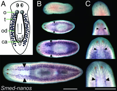
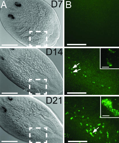
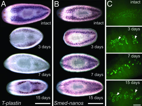
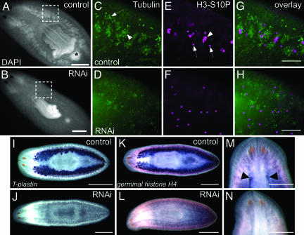
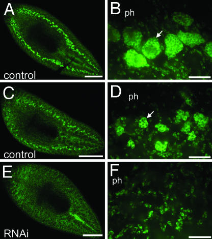
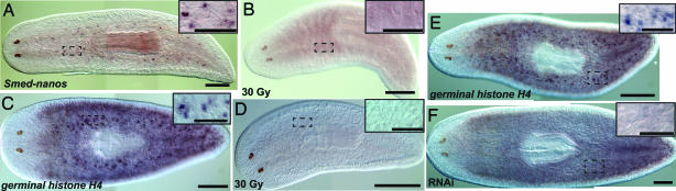
Germ cells are required for the successful propagation of sexually reproducing species. Understanding the mechanisms by which these cells are specified and how their totipotency is established and maintained has important biomedical and evolutionary implications. Freshwater planarians serve as fascinating models for studying these questions. They can regenerate germ cells from fragments of adult tissues that lack reproductive structures, suggesting that inductive signaling is involved in planarian germ cell specification. To study the development and regeneration of planarian germ cells, we have functionally characterized an ortholog of nanos, a gene required for germ cell development in diverse organisms, from Schmidtea mediterranea. In the hermaphroditic strain of this species, Smed-nanos mRNA is detected in developing, regenerating, and mature ovaries and testes. However, it is not detected in the vast majority of newly hatched planarians or in small tissue fragments that will ultimately regenerate germ cells, consistent with an epigenetic origin of germ cells. We show that Smed-nanos RNA interference (RNAi) results in failure to develop, regenerate, or maintain gonads in sexual planarians. Unexpectedly, Smed-nanos mRNA is also detected in presumptive testes primordia of asexual individuals that reproduce strictly by fission. These presumptive germ cells are lost after Smed-nanos RNAi, suggesting that asexual planarians specify germ cells, but their differentiation is blocked downstream of Smed-nanos function. Our results reveal a conserved function of nanos in germ cell development in planarians and suggest that these animals will serve as useful models for dissecting the molecular basis of epigenetic germ cell specification.
Conflict of interest statement
The authors declare no conflict of interest.
Figures
Similar articles
-
The Dr-nanos gene is essential for germ cell specification in the planarian Dugesia ryukyuensis.Int J Dev Biol. 2012;56(1-3):165-71. doi: 10.1387/ijdb.113433hn. Int J Dev Biol. 2012. PMID: 22451004
-
The planarian Schmidtea mediterranea as a model for epigenetic germ cell specification: analysis of ESTs from the hermaphroditic strain.Proc Natl Acad Sci U S A. 2005 Dec 20;102(51):18491-6. doi: 10.1073/pnas.0509507102. Epub 2005 Dec 12. Proc Natl Acad Sci U S A. 2005. PMID: 16344473 Free PMC article.
-
Germ cell specification and regeneration in planarians.Cold Spring Harb Symp Quant Biol. 2008;73:573-81. doi: 10.1101/sqb.2008.73.022. Epub 2008 Nov 6. Cold Spring Harb Symp Quant Biol. 2008. PMID: 19022767 Review.
-
The planarian nanos-like gene Smednos is expressed in germline and eye precursor cells during development and regeneration.Dev Genes Evol. 2007 May;217(5):403-11. doi: 10.1007/s00427-007-0146-3. Epub 2007 Mar 28. Dev Genes Evol. 2007. PMID: 17390146
-
From worm to germ: Germ cell development and regeneration in planarians.Curr Top Dev Biol. 2019;135:127-153. doi: 10.1016/bs.ctdb.2019.04.001. Epub 2019 May 2. Curr Top Dev Biol. 2019. PMID: 31155357 Review.
Cited by
-
Single-cell transcriptomics in planaria: new tools allow new insights into cellular and evolutionary features.Biochem Soc Trans. 2022 Oct 31;50(5):1237-1246. doi: 10.1042/BST20210825. Biochem Soc Trans. 2022. PMID: 36281987 Free PMC article. Review.
-
The history and enduring contributions of planarians to the study of animal regeneration.Wiley Interdiscip Rev Dev Biol. 2013 May-Jun;2(3):301-26. doi: 10.1002/wdev.82. Epub 2012 Jul 23. Wiley Interdiscip Rev Dev Biol. 2013. PMID: 23799578 Free PMC article. Review.
-
Boule-like genes regulate male and female gametogenesis in the flatworm Macrostomum lignano.Dev Biol. 2011 Sep 1;357(1):117-32. doi: 10.1016/j.ydbio.2011.06.030. Epub 2011 Jun 28. Dev Biol. 2011. PMID: 21740899 Free PMC article.
-
Genetic expansion of chaperonin-containing TCP-1 (CCT/TRiC) complex subunits yields testis-specific isoforms required for spermatogenesis in planarian flatworms.Mol Reprod Dev. 2017 Dec;84(12):1271-1284. doi: 10.1002/mrd.22925. Epub 2017 Nov 10. Mol Reprod Dev. 2017. PMID: 29095551 Free PMC article.
-
Stem cells (neoblasts) and positional information jointly dominate regeneration in planarians.Heliyon. 2025 Jan 9;11(2):e41833. doi: 10.1016/j.heliyon.2025.e41833. eCollection 2025 Jan 30. Heliyon. 2025. PMID: 39877626 Free PMC article. Review.
References
-
- Seydoux G, Braun RE. Cell. 2006;127:891–904. - PubMed
-
- Nieuwkoop PD, Sutasurya LA. Primordial Germ Cells in the Chordates. Cambridge, UK: Cambridge Univ Press; 1979.
-
- Nieuwkoop PD, Sutasurya LA. Primordial Germ Cells in the Invertebrates: From Epigenesis to Preformation. Cambridge, UK: Cambridge Univ Press; 1981.
-
- Extavour CG, Akam M. Development (Cambridge, UK) 2003;130:5869–5884. - PubMed
-
- Johnson AD, Drum M, Bachvarova RF, Masi T, White ME, Crother BI. Evol Dev. 2003;5:414–431. - PubMed
Publication types
MeSH terms
Substances
Associated data
- Actions
Grants and funding
LinkOut - more resources
Full Text Sources
Research Materials
