Comparative Study
. 2007 Jun;81(11):5594-606.
doi: 10.1128/JVI.00202-07. Epub 2007 Mar 21.

Unique pathology in simian immunodeficiency virus-infected rapid progressor macaques is consistent with a pathogenesis distinct from that of classical AIDS

Affiliations
Comparative Study

Unique pathology in simian immunodeficiency virus-infected rapid progressor macaques is consistent with a pathogenesis distinct from that of classical AIDS

Charles R Brown et al. J Virol. 2007 Jun.

Abstract

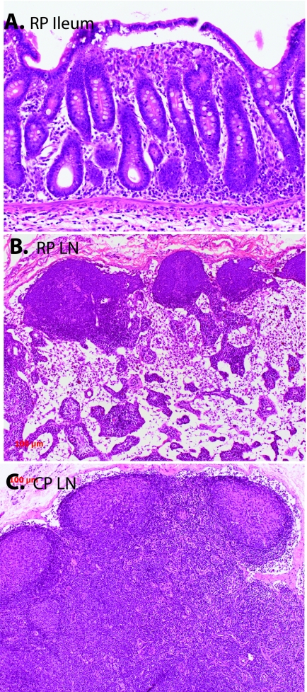
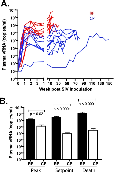
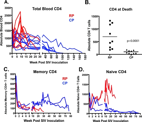
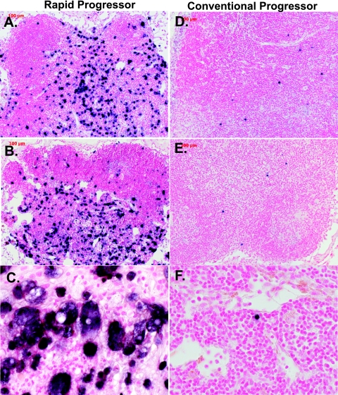
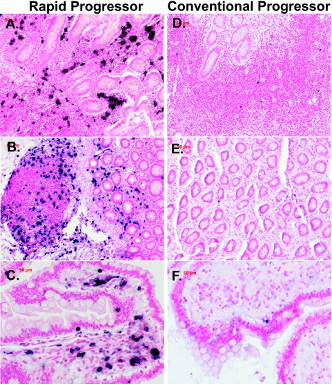
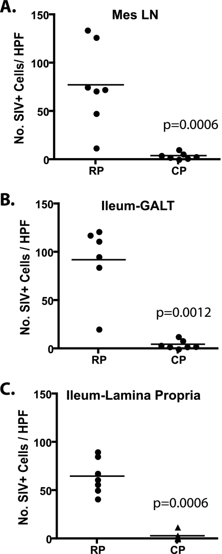
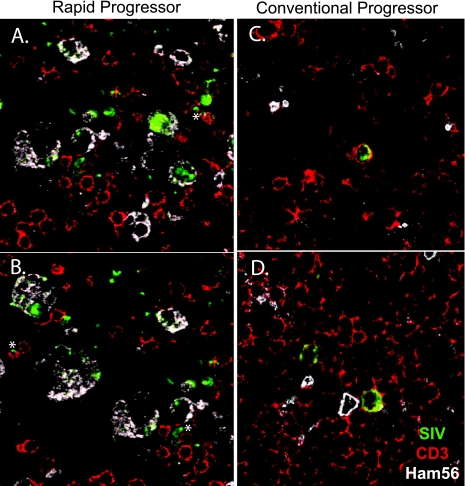
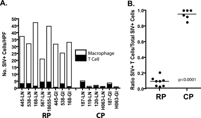
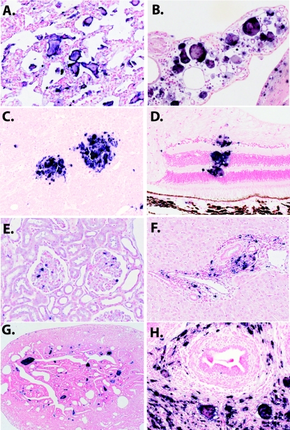
Simian immunodeficiency virus (SIV) infection of macaques and human immunodeficiency virus type 1 (HIV-1) infection of humans result in variable but generally fatal disease outcomes. Most SIV-infected macaques progress to AIDS over a period of 1 to 3 years, in the face of robust SIV-specific immune responses (conventional progressors [CP]). A small number of SIV-inoculated macaques mount transient immune responses and progress rapidly to AIDS (rapid progressors [RP]). We speculated that the underlying pathogenic mechanisms may differ between RP and CP macaques. We compared the pathological lesions, virus loads, and distribution of virus and target cells in SIVsmE660- or SIVsmE543-infected RP and CP rhesus macaques at terminal disease. RP macaques developed a wasting syndrome characterized by severe SIV enteropathy in the absence of opportunistic infections. In contrast, opportunistic infections were commonly observed in CP macaques. RP and CP macaques showed distinct patterns of CD4(+) T-cell depletion, with a selective loss of memory cells in RP macaques and a generalized (naive and memory) CD4 depletion in CP macaques. In situ hybridization demonstrated higher levels of virus expression in lymphoid tissues (P < 0.001) of RP macaques and a broader distribution to include many nonlymphoid tissues. Finally, SIV was preferentially expressed in macrophages in RP macaques whereas the primary target cells in CP macaques were T lymphocytes at end stage disease. These data suggest distinct pathogenic mechanisms leading to the deaths of these two groups of animals, with CP macaques being more representative of HIV-induced AIDS in humans.


Figures

FIG. 1.
FIG. 1.
Hematoxylin-and-eosin-stained sections of the ileum of an RP (top) and comparison of the lymph nodes (LN) of a representative RP macaque and a CP macaque. (A) Section of ileum of RP macaque H445 showing the blunting and fusion of intestinal villi that was characteristic of RP intestinal tissues. (B) Mesenteric lymph node of RP macaque H445 showing depletion of the paracortex and a lack of secondary germinal centers. (C) Inguinal lymph node of CP macaque H133 showing large germinal centers with irregular mantles and hyalinization indicative of involution.
FIG. 2.
FIG. 2.
Plasma viremia in RP and CP macaques is shown over the time course of infection. (A) Sequential plasma viral loads in RP (red) and CP (blue) macaques. (B) Comparison of peak, set point, and terminal plasma viral RNA levels in RP and CP macaques with statistically significant differences between the two groups at each of these time points (Mann-Whitney U test).
FIG. 3.
FIG. 3.
CD4+ T cells in the peripheral blood of RP and CP macaques over the time course of infection. (A) Total CD4+ T cells are compared in RP (red) and CP (blue) macaques. (B) Comparison of total CD4+ T cells in peripheral blood at the time of euthanasia shows significantly lower total CD4+ T-cell counts in CP versus RP macaques (Mann-Whitney U test). Comparison of memory (C) and naive (D) CD4+ T cells in the blood of two RP and two CP macaques inoculated with SIVsmE543-3 shows the selective depletion of memory CD4+ T cells that occurs in RP macaques, as determined by CD28 and CD95 expression. In contrast, CP macaques demonstrate early depletion of memory CD4+ T cells with partial replenishment but subsequent progressive loss of both memory and naive CD4+ T-cell subsets during long-term progression.
FIG. 4.
FIG. 4.
Representative fields of view of ISH for SIV viral RNA in the mesenteric lymph nodes of RP and CP macaques. (A, B, and C) RP lymph nodes showing numerous SIV-expressing cells (dark blue) within the paracortex, follicle, intrafollicular zone, and cortex of the lymph nodes of SIV-infected RP macaques H538 (A; magnification, ×10), H445 (B; magnification, ×10), and H168 (C; magnification, ×40) showing large numbers of SIV-expressing cells. Lower numbers of SIV-expressing cells were observed in the mesenteric lymph nodes of representative CP macaques H063 (D), H119 (E), and H460 (F). Magnification, ×40.
FIG. 5.
FIG. 5.
Representative fields of view of ISH for SIV viral RNA in the ilea of RP (left) and CP (right) macaques. (A and B) Representative sections of the ilea of RP macaques H147 and H538 showing large numbers of SIV-expressing cells in the GALT, crypt regions, and lamina propria (magnification, ×10). Panel C shows a higher magnification of the ileum of macaque 18655 (magnification, ×40). (D, E, and F) Representative sections of the ilea of CP macaques H187 and H063 showing a scattering of SIV-expressing cells in the GALT (magnifications: D and E, ×10; F, ×40).
FIG. 6.
FIG. 6.
Comparison of the numbers of SIV-expressing cells per high-power field (HPF) in the mesenteric lymph nodes and ilea of RP and CP macaques. Five random fields of view (magnification, ×40) of ISH-stained tissues from seven RP and seven CP macaques were photographed from each tissue, and the mean number of SIV+ cells per high-power field for each tissue was calculated. Scatter plots of mean numbers of SIV+ cells in the mesenteric lymph nodes (A), GALT (B), and lamina propria (C) of the ileum show that RP macaque tissues contained significantly more SIV+ cells per high-power field compared to CP macaques (Mann-Whitney U test).
FIG. 7.
FIG. 7.
Confocal microscopic analysis of mesenteric lymph nodes of representative RP and CP macaques. Triple-label confocal microscopy of lymph nodes from an RP macaque (A and B) and a CP macaque (C and D) to identify macrophages (HAM56, white), SIV RNA expression (ISH, green), and T cells (CD3, red). The majority of SIV-expressing cells in the tissues from the RP macaque coexpressed Ham56, a marker for macrophages (magnification, ×63). Rare CD3+ T cells coexpressing SIV are indicated by asterisks. In contrast (C and D), the majority of SIV-expressing cells in tissues of the CP macaques (H187 and H063) coexpressed CD3 (magnification, ×63).
FIG. 8.
FIG. 8.
Graphic representation of the proportions of SIV-expressing macrophages and T cells in the lymph nodes and ilea of RP and CP macaques. (A) The mean numbers of SIV+ cells per high-power field (HPF) of each type in various tissues of RP and CP macaques are shown by a bar graph where the filled bars represent T cells and the open bars represent macrophages. (B) Ratios of infected T cells to total infected cells in RP and CP tissues, demonstrating a significant difference between these two groups.
FIG. 9.
FIG. 9.
ISH for SIV RNA in a wide range of tissues of RP macaques. (A) A section of lung tissue from RP macaque H168 shows abundant SIV-expressing giant cells (magnification, ×10). (B) SIV-expressing giant cells in the choroid plexus of macaque H567 (magnification, ×20). (C) ISH of the brain of RP macaque H567 showing perivascular infiltration of SIV-expressing macrophages and giant cells (magnification, ×10). (D) SIV-expressing macrophages in the retina of RP macaque H538 (magnification, ×20). (E) SIV-expressing cells within glomeruli of RP macaque H538. (F) SIV-expressing cells in periportal infiltrates in the liver of RP macaque H538. (G) SIV+ giant cells in the interstitial areas of the fallopian tube of female macaque H538. (H) SIV-expressing cells adjacent to seminiferous tubules of male macaque H168.

Similar articles

Cited by

References

    1. Allan, J. S., M. Short, M. E. Taylor, S. Su, V. M. Hirsch, P. R. Johnson, G. M. Shaw, and B. H. Hahn. 1991. Species-specific diversity among simian immunodeficiency viruses from African green monkeys. J. Virol. 65:2816-2828. - PMC - PubMed
    1. Anderson, M. G., D. Hauer, D. P. Sharma, S. V. Joag, O. Narayan, M. C. Zink, and J. E. Clements. 1993. Analysis of envelope changes acquired by SIVmac239 during neuroadaption in rhesus macaques. Virology 195:616-626. - PubMed
    1. Baskin, G. B., C. M. Murphey, E. D. Roberts, P. J. Didier, and L. N. Martin. 1992. Correlates of SIV encephalitis in rhesus monkeys. J. Med. Primatol. 21:59-63. - PubMed
    1. Baskin, G. B., M. Murphey-Corb, L. N. Martin, K. F. Soike, F. S. Hu, and D. Kuebler. 1991. Lentivirus-induced pulmonary lesions in rhesus monkeys (Macaca mulatta) infected with simian immunodeficiency virus. Vet. Pathol. 28:506-513. - PubMed
    1. Baskin, G. B., M. Murphey-Corb, E. A. Watson, and L. N. Martin. 1988. Necropsy findings in rhesus monkeys experimentally infected with cultured simian immunodeficiency virus (SIV)/delta. Vet. Pathol. 25:456-467. - PubMed

Publication types

MeSH terms