Comparative Study
. 2007 Jun;81(11):5995-6006.
doi: 10.1128/JVI.01714-06. Epub 2007 Mar 21.

Nuclear and nucleolar targeting of influenza A virus NS1 protein: striking differences between different virus subtypes

Affiliations
Comparative Study

Nuclear and nucleolar targeting of influenza A virus NS1 protein: striking differences between different virus subtypes

Krister Melén et al. J Virol. 2007 Jun.

Abstract

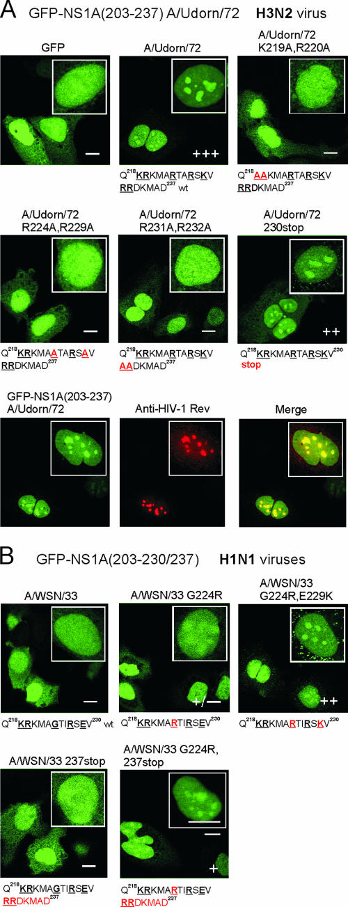
Influenza A virus nonstructural protein 1 (NS1A protein) is a virulence factor which is targeted into the nucleus. It is a multifunctional protein that inhibits host cell pre-mRNA processing and counteracts host cell antiviral responses. We show that the NS1A protein can interact with all six human importin alpha isoforms, indicating that the nuclear translocation of NS1A protein is mediated by the classical importin alpha/beta pathway. The NS1A protein of the H1N1 (WSN/33) virus has only one N-terminal arginine- or lysine-rich nuclear localization signal (NLS1), whereas the NS1A protein of the H3N2 subtype (Udorn/72) virus also has a second C-terminal NLS (NLS2). NLS1 is mapped to residues 35 to 41, which also function in the double-stranded RNA-binding activity of the NS1A protein. NLS2 was created by a 7-amino-acid C-terminal extension (residues 231 to 237) that became prevalent among human influenza A virus types isolated between the years 1950 to 1987. NLS2 includes basic amino acids at positions 219, 220, 224, 229, 231, and 232. Surprisingly, NLS2 also forms a functional nucleolar localization signal NoLS, a function that was retained in H3N2 type virus NS1A proteins even without the C-terminal extension. It is likely that the evolutionarily well-conserved nucleolar targeting function of NS1A protein plays a role in the pathogenesis of influenza A virus.


Figures

FIG. 1.
FIG. 1.
Influenza A virus NS1 binds to all importin α isoforms. (A) 35S-labeled and in vitro translated NS1A protein from A/Udorn/72 virus strain (H3N2) was allowed to bind to E. coli-expressed and Sepharose-immobilized GST-importin β, α1, α3, α5, α6, or α7 at +4°C for 1 h. After being washed twice with binding buffer, importin-bound NS1A was dissolved in Laemmli sample buffer, separated by 12% SDS-PAGE, and autoradiographed. A similar gel was also stained with Coomassie blue to visualize the amount of Sepharose-immobilized GST-importin β and α isoforms. C, control; M, Sepharose matrix-bound in vitro translated NS1A. (B) 35S-labeled and in vitro translated NS1A protein (A/Udorn/72) was allowed to bind to Sf9-expressed and Sepharose-immobilized GST and GST-importin α1, α3, α4, α5, or α7 as described above. 35S-labeled and in vitro translated NS1A protein from A/WSN/33 virus strain (H1N1) was allowed to bind to E. coli-expressed (C) or Sf9-expressed (D) and Sepharose-immobilized GST-importins as described above. C, the control of in vitro translated NS1. (E) Cell extracts of cultured A549 cells were prepared, and the proteins in cell extracts were allowed to bind to E. coli-expressed and Sepharose-immobilized GST and GST-NS1A (A/Udorn/72) at +4°C for 1 h. Sepharose-bound proteins were dissolved in Laemmli sample buffer followed by 8% SDS-PAGE and Western blotting with anti-importin α1, α3, and α7 antibodies. As a control, cell extracts of A549 cells containing 20 μg of protein were stained in lane C. A similar gel was also stained with Coomassie blue to visualize the amount of Sepharose-immobilized GST and GST-NS1A A/Udorn/72.
FIG. 2.
FIG. 2.
Variation of the amino acid sequence around NLS1 and NLS2 of influenza A virus NS1A protein. (A) Variation of NLS1 (amino acids 34 to 44). The number of human, avian, swine, and equine NS1A protein sequences from different virus strains is shown at right. The previously identified putative NLS1 (amino acids 34 to 38) (14) is underlined, and critical arginines (R) at positions 35 and 38 and lysine (K) at position 41, which regulate importin α binding, are shaded. (B) Variation of putative C-terminal NLS2 in selected influenza A virus NS1A proteins. Critical amino acids 219, 220, 224, 227, 229, 231, and 232 that regulate NS1A protein interaction with importin α (this study) are shaded.
FIG. 3.
FIG. 3.
Influenza virus A/Udorn/72 NS1A protein has two importin α binding sites, and A/WSN/33 NS1A protein has one importin α binding site. (A) Influenza virus A/Udorn/72 NS1A protein binds to importin α protein via two separate NLSs. 35S-labeled and in vitro translated A/Udorn/72 NS1A wt and point mutant proteins were allowed to bind to E. coli-expressed and Sepharose-immobilized GST, GST-importin β, and GST-importin α1 proteins as indicated in the figure. Importin-bound NS1A was dissolved in Laemmli sample buffer, separated by 12% SDS-PAGE, and autoradiographed. A similar gel was also stained with Coomassie blue to visualize the amount of Sepharose-immobilized GST and GST-importin β and α1 proteins. For the translation control 1 μl of 35S-labeled and in vitro translated NS1 wt and six point mutants were separated by 12% SDS-PAGE and autoradiographed. (B) 35S-labeled and in vitro translated A/Udorn/72 NS1A wt and seven point mutant proteins covering putative NLS1 and NLS2 as indicated in the figure. The binding experiment was carried out as described for panel A. (C) 35S-labeled and in vitro translated A/WSN/33 NS1A wt and four point mutant proteins were allowed to bind to E. coli-expressed and Sepharose-immobilized GST and GST-importin α1 proteins as indicated in the figure. The binding experiment was as described for panel A.
FIG. 4.
FIG. 4.
Importin α1 does not compete with dsRNA for binding to the NS1A protein. (A) Gel shift assay of complexes formed between radiolabeled 55-bp dsRNA and C-terminally deleted A/Udorn/72 NS1A(1-215) protein in the absence and presence of importin α1. The indicated polypeptides [40 or 400 nM of NS1A(1-215) and 4 to 1,600 nM of importin α1] were incubated with the 55-bp dsRNA (10,000 cpm; 1 nM), and the polypeptide-RNA complexes were separated from free RNA by nondenaturing gel electrophoresis (6% acrylamide; bis:acrylamide 1:100). (B) Gel shift assay of complexes formed between radiolabeled 55-bp dsRNA and importin α1. The last lane shows the complex formed between this dsRNA and the NS1A(1-215) protein.
FIG. 5.
FIG. 5.
Point mutations to NLSs regulate nuclear/cytoplasmic distribution of the influenza A virus NS1A protein. (A) HuH7 cells were transiently transfected with wt or NLS mutant (mt) influenza A/Udorn/72 NS1A gene constructs as indicated in the figure. NLS1, NLS1(R37A R38A); NLS2, NLS2(K219A R220A) plus NLS2(R231A R232A). (B) HuH7 cells were transiently transfected with wt or NLS mutant (mt) influenza A/WSN/33 NS1A gene constructs as indicated in the figure. The cells were stained with rabbit anti-NS1A protein and secondary rhodamine-labeled anti-rabbit antibodies. Bar, 5 μm.
FIG. 6.
FIG. 6.
C-terminal deletion mutant influenza A/Udorn/72 NS1AΔ231-237 protein binds to importin α6. (A) 35S-labeled and in vitro translated A/Udorn/72 NS1A wt and three mutant proteins were allowed to bind to E. coli-expressed and Sepharose-immobilized GST and GST-importin α6 proteins as indicated in the figure. Importin-bound NS1A was dissolved in Laemmli sample buffer, separated by 12% SDS-PAGE, and autoradiographed. A similar gel was also stained with Coomassie blue to visualize the amount of Sepharose-immobilized GST and GST-importin (GST-imp) α6 proteins. For the translation control, 1 μl of 35S-labeled and in vitro translated NS1 wt and three mutants were separated by 12% SDS-PAGE and autoradiographed. (B) HuH7 cells were transiently transfected with wt or C-terminal deletion mutant influenza A/Udorn/72 NS1A gene constructs as indicated in the figure. NLS1, NLS1(R37A R38A); NLS2, NLS2(K219A R220A). Bar, 5 μm.
FIG. 7.
FIG. 7.
Intracellular localization of NS1A protein during influenza A virus infection. (A) A549 cells grown directly on coverslips were infected with wt influenza A/Udorn/72, recombinant A/Udorn/72 NS1AΔ221-237 with a stop codon at position 221 of the NS1A protein gene, or wt A/WSN/33 viruses for 4 to 24 h as indicated in the figure. After fixation, the cells were stained with rabbit anti-NS1A and fluorescein isothiocyanate-labeled anti-rabbit antibodies, followed by analysis with confocal laser microscopy. Bar, 5 μm. (B) A549 cells were infected with different wt H3N2 or H1N1 influenza A virus strains as indicated in the figure. Nuclear and nucleolar localization of NS1A protein was detected at 6 and 12 h after infection by indirect immunofluorescence microscopy. For each virus the percentage of cells expressing NS1A protein in the nucleolus was calculated from 300 NS1A protein-expressing cells.
FIG. 8.
FIG. 8.
C-terminal end of NS1A protein in H3N2 type influenza A viruses encode a functional NoLS. C-terminal fragments of NS1A genes encoding amino acids 203 to 237 in A/Udorn/72 and amino acids 203 to 230 in A/WSN/33 were inserted into GFP expression vector pCMX-SAH/Y145F to express GFP-NS1A fusion proteins [GFP-NS1A(9203-230/237)]. (A) HuH7 cells were transiently transfected with GFP-wt, mutant, or deletion A/Udorn/72 NS1A gene constructs for 48 h as indicated in the figure. The intensity of nucleolar localization was scored by immunofluorescence microscopy as no nucleolar staining (−) or weak (+), moderate (++), or strong (+++) nucleolar staining. Critical basic amino acids involved in nuclear/nucleolar targeting are marked in boldface and underlined. Mutated amino acids are marked in red. To verify nucleolar localization, HuH7 cells were transiently transfected with GFP-wt NS1A(203-237) A/Udorn/72 and HIV-1 Rev gene constructs for 48 h as indicated in the figure. After fixation the cells were stained with anti-HIV-1 Rev antibodies, and colocalization with GFP-NS1A(203-237) protein was detected with confocal microscopy. (B) HuH7 cells were transiently transfected with GFP-wt and mutant A/WSN/33 NS1A gene constructs for 48 h as indicated in the figure. Critical and mutated amino acids are marked as above. Bars, 5 μm.
FIG. 9.
FIG. 9.
(A) Schematic representation of NS1A intracellular targeting signals. Both WSN and Udorn virus NS1A proteins have an NLS constituting basic residues 35, 38, and 41 (NLS1). The Udorn NS1A protein also has a 7-amino-acid C-terminal extension (residues 231 to 237) and a second NLS (NLS2) at the end of the molecule, constituting arginines or lysines at positions 219, 229, 224, 229, 231, and 323. The same residues form a functional NoLS. NLS and NoLS signals are shown in bold and underlined. (B) Mechanisms of nuclear import of Udorn virus NS1A protein. Newly synthesized dimeric Udorn NS1A protein interacts via its NLS1 or NLS2 with different cytoplasmic importin (Imp) α (all 6 isotypes)/importin β complex, followed by nuclear translocation of the complex via the NPC. Most cytoplasmic NS1A is likely bound to importin α interfering with dsRNA binding to NS1A in the cytoplasm. In the nucleus NS1A protein is released from the transport complex, and importin α and importin β are transported back into the cytoplasm through the NPC. After the release of importin α, the C-terminal NoLS of the NS1A protein is exposed, and the NS1A protein is targeted into the nucleolus. Simultaneously, the dsRNA-binding domain of the NS1A protein is also exposed, and the protein becomes competent to bind dsRNA. This leads to sequestration of dsRNA and lack of activation of the oligoadenylate synthetase/RNase L antiviral pathway. RBD, dsRNA-binding domain.

References

    1. Belmont, A. 2003. Dynamics of chromatin, proteins, and bodies within the cell nucleus. Curr. Opin. Cell Biol. 15:304-310. - PubMed
    1. Birbach, A., S. T. Bailey, S. Ghosh, and J. A. Schmid. 2004. Cytosolic, nuclear and nucleolar localization signals determine subcellular distribution and activity of the NF-κB inducing kinase NIK. J. Cell Sci. 117:3615-3624. - PubMed
    1. Bornholdt, Z. A., and B. V. Prasad. 2006. X-ray structure of influenza virus NS1 effector domain. Nat. Struct. Mol. Biol. 13:559-560. - PubMed
    1. Carmo-Fonseca, M. 2002. The contribution of nuclear compartmentalization to gene regulation. Cell 108:513-521. - PubMed
    1. Chen, Z., Y. Li, and R. M. Krug. 1999. Influenza A virus NS1 protein targets poly(A)-binding protein II of the cellular 3′-end processing machinery. EMBO J. 15:2273-2283. - PMC - PubMed

Publication types

MeSH terms

Substances

LinkOut - more resources