Comparative Study
. 2007 Jun;81(11):6043-56.
doi: 10.1128/JVI.02074-06. Epub 2007 Mar 21.

Sustained induction of NF-kappa B is required for efficient expression of latent human immunodeficiency virus type 1

Affiliations
Comparative Study

Sustained induction of NF-kappa B is required for efficient expression of latent human immunodeficiency virus type 1

Samuel A Williams et al. J Virol. 2007 Jun.

Abstract

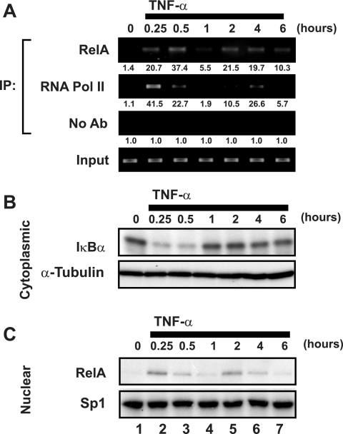
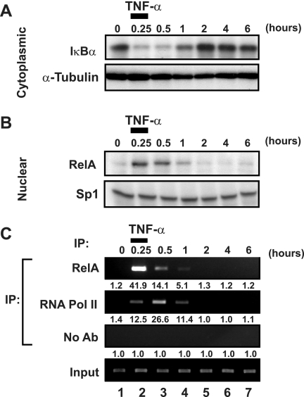
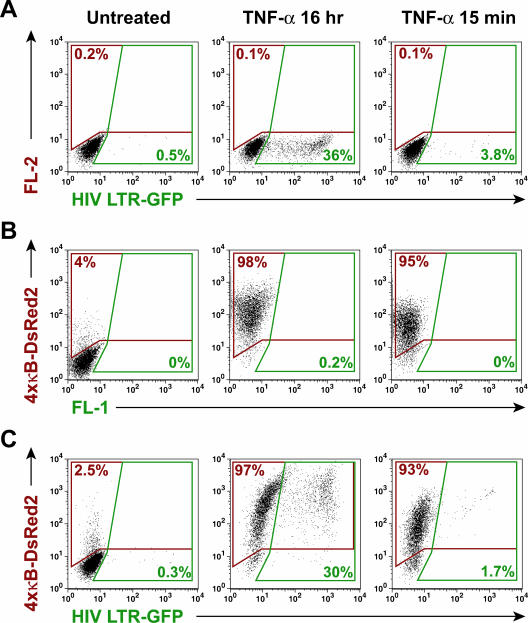
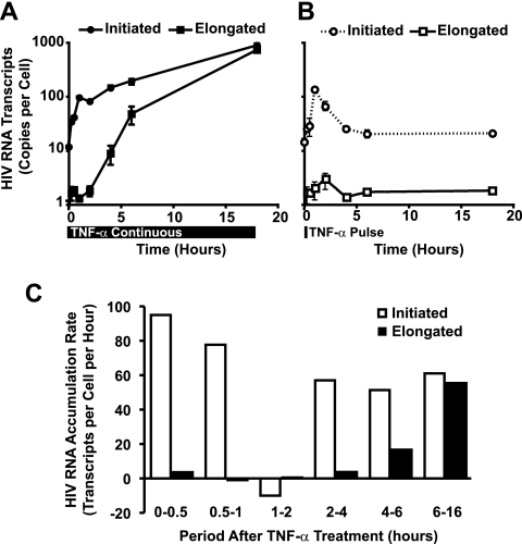
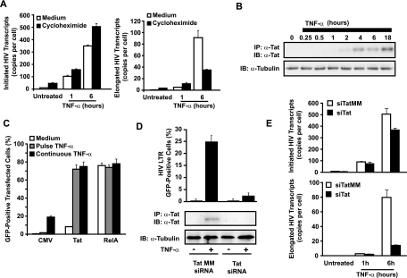
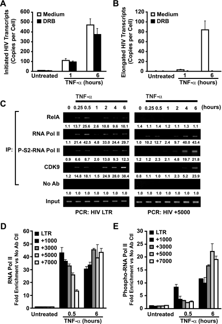
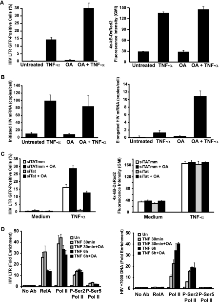
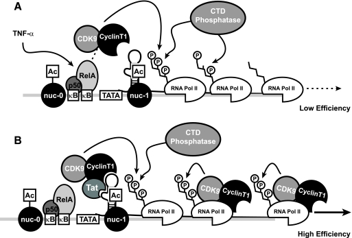
Cells harboring infectious, but transcriptionally latent, human immunodeficiency virus type 1 (HIV-1) proviruses currently pose an insurmountable barrier to viral eradication in infected patients. To better understand the molecular basis for HIV-1 latency, we used the J-Lat model of postintegration HIV-1 latency to assess the kinetic relationship between the induction of NF-kappaB and the activation of latent HIV-1 gene expression. Chromatin immunoprecipitation analyses revealed an oscillating pattern of RelA recruitment to the HIV-1 long terminal repeat (LTR) during continuous tumor necrosis factor alpha (TNF-alpha) stimulation. RNA polymerase II (Pol II) recruitment to the HIV-1 LTR closely mirrored RelA binding. Transient stimulation of cells with TNF-alpha for 15 min induced only a single round of RelA and RNA Pol II binding and failed to induce robust expression of latent HIV-1. Efficient formation of elongated HIV-1 transcripts required sustained induction by NF-kappaB, which promoted de novo synthesis of Tat. Cyclin-dependent kinase 9 (CDK9) and serine-2-phosphorylated RNA Pol II were rapidly recruited to the HIV-1 LTR after NF-kappaB induction; however, these elongating polymerase complexes were progressively dephosphorylated in the absence of Tat. Okadaic acid promoted sustained serine-2 phosphorylation of the C-terminal domain of RNA Pol II and stimulated efficient transcriptional elongation and HIV-1 expression in the absence of Tat. These findings underscore important differences between NF-kappaB and Tat stimulation of RNA Pol II elongation. While NF-kappaB binding to the HIV-1 LTR induces serial waves of efficient RNA Pol II initiation, elongation is impaired by the action of an okadaic acid-sensitive phosphatase that dephosphorylates the C-terminal domain of RNA Pol II. Conversely, the action of this phosphatase is overcome in the presence of Tat, promoting very efficient RNA Pol II elongation.


Figures

FIG. 1.
FIG. 1.
TNF-α stimulation induces an oscillating pattern of RelA and RNA Pol II binding to the latent HIV-1 LTR. (A) TNF-α stimulation leads to synchronous but oscillating recruitment of RelA and RNA Pol II to the latent HIV-1 LTR. Fixed chromatin extracts from J-Lat 6.3 cells either untreated or treated with 20 ng/ml TNF-α for various times were immunoprecipitated with antibodies specific for RelA, RNA Pol II, or no antibody as a control. Immunoprecipitates were assessed for enrichment in HIV-1 LTR DNA by UV visualization of PCR products in an ethidium bromide-stained gel. Data are representative of three independent experiments. Note the synchronicity of RelA and RNA Pol II recruitment to the HIV-1 LTR, as well as the nadir in binding 1 h after TNF-α stimulation. Quantitation of enrichment (fold increase) above the “no-antibody” control is indicated below each sample. (B) TNF-α-induced recruitment of RelA and RNA Pol II to the latent HIV-1 LTR coincides with IκBα degradation. Cytoplasmic extracts of samples treated as for panel A were prepared, and IκBα levels were assessed by immunoblotting. Cytoplasmic levels of α-tubulin were assessed to confirm equivalent loading of samples. (C) TNF-α induces bimodal nuclear enrichment of RelA. Nuclear extracts of samples treated as for panel A were prepared and analyzed for RelA enrichment by immunoblotting. Nuclear Sp1 levels were assessed to confirm equivalent loading. Note the largely overlapping patterns of nuclear enrichment of RelA and its recruitment to the latent HIV-1 LTR as assessed by ChIP (A).
FIG. 2.
FIG. 2.
Transient induction of NF-κB induces unimodal recruitment of RNA polymerase II to the latent HIV-1 LTR. (A) Pulsed administration of TNF-α induces IκBα degradation. J-Lat 6.3 cells were stimulated with 20 ng/ml TNF-α or left untreated for 15 min, washed twice in medium, and returned to culture for various times. Cytoplasmic extracts were prepared, and IκBα levels were assessed by immunoblotting. Cytoplasmic α-tubulin levels were assessed to confirm equivalent loading of samples. Note the similarity in depletion of IκBα in transiently and continuously TNF-α treated samples (Fig. 1B). (B) Pulsed administration of TNF-α induces transient activation of NF-κB. Nuclear extracts of samples treated as for panel A were prepared and analyzed for recruitment of RelA by immunoblotting. Nuclear Sp1 levels were assessed to confirm equivalent loading. (C) Pulsed TNF-α administration induces a unimodal pattern of RelA and RNA Pol II recruitment to the latent HIV-1 LTR. Fixed chromatin extracts of samples treated as for panel A were prepared and subjected to immunoprecipitation with antibodies specific to RelA or RNA Pol II or without antibody, as a nonspecific control. Immunoprecipitates were assessed for enrichment in HIV-1 LTR DNA by UV visualization of PCR products in a gel stained with ethidium bromide. Data are representative of three separate experiments. Note the absence of a second wave of RNA Pol II recruitment to the latent HIV-1 LTR. Quantitation of enrichment (fold increase) above the no-antibody control is indicated below each sample.
FIG. 3.
FIG. 3.
Transient induction of NF-κB is sufficient to induce robust general κB-dependent, but not latent HIV-1, gene expression. (A) Transient TNF-α administration induces poor expression of latent HIV-1. J-Lat 6.3 cells were left untreated or stimulated with 20 ng/ml TNF-α continuously or for 15 min, followed by washing and continued culture. HIV-LTR-dependent expression of GFP was assessed by flow cytometry. Note the overall lack of GFP expression in samples transiently treated with TNF-α. (B) Transient induction of NF-κB is sufficient to stimulate general κB-dependent gene expression. JκRed cells were treated as for panel A, and κB-dependent expression of DsRed2 was assessed by flow cytometry. Note the strong induction of κB-dependent gene expression by transient TNF-α stimulation. (C) Transient NF-κB induction induces robust expression of κB-dependent genes, but not latent HIV-1, in J-Lat 6.3 cells. J6.3κRed cells were treated as for panel A, and HIV-1 LTR-dependent expression of GFP and κB-dependent expression of DsRed2 were assessed by flow cytometry. Note the strong induction of κB-dependent gene expression and relative absence of HIV-1 gene expression induced by transient TNF-α stimulation.
FIG. 4.
FIG. 4.
Efficient elongation of HIV-1 mRNA transcripts is delayed in TNF-α-activated J-Lat cells. (A) TNF-α treatment of J-Lat cells induces rapid accumulation of initiated, but not elongated, HIV-1 mRNA transcripts. J-Lat 6.3 cells were treated with 20 ng/ml TNF-α for various times, and total RNA was extracted. Initiated and elongated HIV-1 mRNA transcripts were quantitated by real-time RT-PCR. Note the delayed emergence of elongated HIV-1 transcripts relative to the rapid increase in initiated transcripts. (B) Transient induction of NF-κB does not induce accumulation of elongated HIV-1 mRNA transcripts. J-Lat 6.3 cells were treated with 20 ng/ml TNF-α for 15 min, washed twice, and returned to culture for various times. Initiated and elongated HIV-1 mRNA transcript abundance were assessed as for panel A. (C) The kinetics of initiated and elongated HIV-1 transcript formation in TNF-α-induced J-Lat cells are dynamic. The rate of transcript formation in continuously TNF-α-stimulated J-Lat 6.3 cells was determined from the data in panel A. Note that the rate of initiated HIV-1 mRNA transcript formation is relatively constant across time, in contrast to the accelerating rate of elongated transcript formation.
FIG. 5.
FIG. 5.
TNF-α-induced expression of HIV-1 is dependent on de novo synthesis of Tat. (A) TNF-α-induced accumulation of elongated, but not initiated, HIV-1 mRNA transcripts is dependent on de novo protein synthesis. J-Lat 6.3 cells were preincubated with 10 μg/ml cycloheximide for 30 min or left in complete culture medium before a 15-min pulse or continuous stimulation with TNF-α for 1 or 6 h. Total RNA was extracted and initiated, and elongated HIV-1 mRNA transcripts were quantitated by real-time RT-PCR. Note the continued accumulation of initiated HIV-1 transcripts in cycloheximide-treated samples at both 1 and 6 h after TNF-α stimulation, whereas accumulation of elongated transcripts is blunted by cycloheximide at 6, but not 1, h after TNF-α treatment. Data are representative of three independent experiments. (B) Expression of Tat is delayed in TNF-α-stimulated J-Lat cells. Tat was immunoprecipitated from whole-cell lysates of J-Lat 6.3 cells treated with TNF-α for various times, and expression levels were assessed by immunoblotting. Note that efficient elongation of HIV-1 transcripts in Fig. 4A coincides with the kinetics of Tat expression. (C) Ectopic expression of HIV-1 Tat rescues HIV-1 gene expression in response to transient TNF-α stimulus. J-Lat 6.3 cells were cotransfected with control empty CMV, Tat, or RelA expression vectors and a plasmid expressing the cell surface H-2Kk marker to identify transfected cells. Transfected cells were stimulated with TNF-α for 15 min or continuously, and GFP expression was assessed in the H-2Kk-expressing cells. (D) Tat is required for efficient TNF-α-induced expression of latent HIV-1. J-Lat 6.3 cells were nucleofected with siRNA targeting Tat mRNA or a mismatched sequence, and knockdown of Tat expression was confirmed by immunoblotting (bottom panel). siRNA-treated cells were stimulated with TNF-α, and the percentage of cells expressing HIV-GFP was quantitated by flow cytometry (top). (E) Total RNA was extracted from cells treated as for panel D, and initiated (left) or elongated (right) transcripts were quantitated by RT-PCR.
FIG. 6.
FIG. 6.
NF-κB induces promoter-proximal, but not downstream, recruitment of CDK9 and serine-2 phosphorylation of RNA Pol II. (A) Early and late TNF-α-induced HIV-1 mRNA transcript elongation is DRB sensitive. J-Lat 6.3 cells were either untreated or pretreated with DRB for 30 min and then stimulated with 20 ng/ml TNF-α for 1 or 6 h or left unstimulated. Total RNA was extracted and initiated, or elongated HIV-1 mRNA transcripts were quantitated by real-time RT-PCR (B). (C) TNF-α stimulation induces rapid recruitment of CDK9 and serine-2-phosphorylated RNA Pol II to the HIV-1 LTR. Conversely, recruitment to downstream DNA is delayed. Fixed chromatin extracts prepared from J-Lat 6.3 cells stimulated with 20 ng/ml TNF-α continuously for various times were immunoprecipitated with antibodies specific for serine-2-phosphorylated RNA Pol II and assessed for enrichment in HIV-1 LTR DNA (left) or HIV+5000 DNA (right) by UV visualization of PCR products in a gel stained with ethidium bromide. Data are representative of three independent experiments. Note the presence of serine-2-phosphorylated RNA Pol II on the HIV-1 LTR in samples stimulated for 15 min; this association was markedly decreased when HIV+5000 DNA was analyzed. Quantitation of enrichment (fold increase) above the no-antibody control is indicated below each sample. (D) RNA Pol II is decreasingly associated with downstream regions of HIV-1 DNA in early, but not late, TNF-α-induced transcription of latent HIV-1. Samples stimulated with TNF-α for 30 min and immunoprecipitated with RNA Pol II or phospho-S2-RNA Pol II antibodies were subjected to real-time PCR quantitative analysis for the indicated HIV-1 DNA regions (E).
FIG. 7.
FIG. 7.
OA rescues NF-κB induction of efficient expression of latent HIV. (A) OA synergizes with TNF-α to promote expression of latent HIV. J6.3κRed cells were incubated with or without 30 nM OA for 1 h, and stimulated with 20 ng/ml TNF-α or left unstimulated, and HIV-GFP (left panel) and kB-DsRed2 expression (right) was quantitated 18 h later by flow cytometry. (B) OA promotes early TNF-α-induced transcriptional elongation. Total RNA was extracted from cells treated as for panel A, and initiated (left panel) and elongated (right panel) HIV-1 transcripts were quantitated by real-time RT-PCR. Note that OA does not affect initiated transcript abundance but effectively promotes elongated HIV-1 mRNA abundance. (C) OA promotes TNF-α-induced expression of latent HIV-1 in the absence of Tat. J6.3κRed cells were nucleofected with siRNA directed against Tat mRNA or a mismatch sequence, treated with 30 nM OA or left untreated, and stimulated with TNF-α or left unstimulated, and HIV-GFP (left) or κB-DsRed2 (right) expression was assessed by flow cytometry. Note the rescue of HIV-GFP expression in Tat siRNA-treated cells with OA and the absence of effect on κB-DsRed2 expression. (D) OA promotes downstream association of P-S-RNA Pol II in early transcription. J6.3κRed cells were pretreated for 1 h with 30 nM OA or left untreated and stimulated with TNF-α for 30 min, 1 h, or left untreated, and ChIP assessment for RelA, RNA Pol II, P-S2-Pol II, and P-S5-Pol II was conducted at the HIV-1 LTR and HIV+7000 DNA by quantitative PCR. Note the increase in Pol II and P-S2-Pol II enrichment in HIV+7000 DNA in samples treated for 30 min with TNF with OA versus without OA.
FIG. 8.
FIG. 8.
Model for events influencing early and late TNF-α-induced HIV-1 transcription. (A) TNF-α stimulation of J-Lat cells induces NF-κB-mediated recruitment of PTEF-b to the HIV LTR, which drives serine-2 phosphorylation of the CTD of coincidentally recruited RNA Pol II. This phosphorylated polymerase is progressively dephosphorylated during elongation, limiting processivity. (B) Early, inefficient elongation produces low levels of HIV-1 Tat, which recruits CDK9 to the HIV-1 LTR in a context capable of transiting with the elongating polymerase. This continued association permits reinforcement of serine-2 phosphorylation of the CTD and prevents transcriptional stalling due to the action of CTD phosphatases.

Similar articles

Cited by

References

    1. Andrews, N. C., and D. V. Faller. 1991. A rapid micropreparation technique for extraction of DNA-binding proteins from limiting numbers of mammalian cells. Nucleic Acids Res. 19:2499. - PMC - PubMed
    1. Barboric, M., R. M. Nissen, S. Kanazawa, N. Jabrane-Ferrat, and B. M. Peterlin. 2001. NF-κB binds P-TEFb to stimulate transcriptional elongation by RNA polymerase II. Mol. Cell 8:327-337. - PubMed
    1. Blankson, J. N., D. Persaud, and R. F. Siliciano. 2002. The challenge of viral reservoirs in HIV-1 infection. Annu. Rev. Med. 53:557-593. - PubMed
    1. Bohnlein, E., J. W. Lowenthal, M. Siekevitz, D. W. Ballard, B. R. Franza, and W. C. Greene. 1988. The same inducible nuclear proteins regulates mitogen activation of both the interleukin-2 receptor-alpha gene and type 1 HIV. Cell 53:827-836. - PubMed
    1. Bres, V., N. Gomes, L. Pickle, and K. A. Jones. 2005. A human splicing factor, SKIP, associates with P-TEFb and enhances transcription elongation by HIV-1 Tat. Genes Dev. 19:1211-1226. - PMC - PubMed

Publication types

MeSH terms