. 2007 Jun;81(11):5841-9.
doi: 10.1128/JVI.00096-07. Epub 2007 Mar 21.

Infectivity determinants of the hepatitis B virus pre-S domain are confined to the N-terminal 75 amino acid residues

Affiliations

Infectivity determinants of the hepatitis B virus pre-S domain are confined to the N-terminal 75 amino acid residues

Matthieu Blanchet et al. J Virol. 2007 Jun.

Abstract

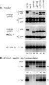
The N-terminal pre-S domain of the large hepatitis B virus (HBV) envelope protein plays a pivotal role at the initial step of the viral entry pathway. In the present study, the entire pre-S domain was mapped for infectivity determinants, following a reverse-genetics approach and using in vitro infection assays with hepatitis delta virus (HDV) or HBV particles. The results demonstrate that lesions created within the N-terminal 75 amino acids of the pre-S region abrogate infectivity, whereas mutations between amino acids 76 and 113, overlapping the matrix domain, had no effect. In contrast to the results of a recent study (L. Stoeckl, A. Funk, A. Kopitzki, B. Brandenburg, S. Oess, H. Will, H. Sirma, and E. Hildt, Proc. Natl. Acad. Sci. 103:6730-6734, 2006), the deletion of a cell membrane translocation motif (TLM) located between amino acids 148 and 161 at the C terminus of pre-S2 did not interfere with the infectivity of the resulting HDV or HBV mutants. Furthermore, a series of large deletions overlapping the pre-S2 domain were compatible with infectivity, although the efficiency of infection was reduced when the deletions extended to the pre-S1 domain. Overall, the results demonstrate that the activity of the pre-S domain at viral entry solely depends on the integrity of its first 75 amino acids and thus excludes any function of the matrix domain or TLM.


Figures

FIG. 1.
FIG. 1.
Schematic representation of wt and mutant L-HBsAg. (A) The L-HBsAg polypeptide is depicted by a horizontal thin line. The pre-S1, pre-S2, and S domains are indicated. The sequence of the wt preS-1 domain is indicated, and each deletion mutant is designated by the positions of the first and last deleted amino acids. The letters KL correspond to an insertion of a Lys-Leu dipeptide at the site of the deletion. (B) The sequences of the wt and mutant polypeptides overlapping part of the pre-S1 domain and the pre-S2 region are indicated. Deletion mutants are designated by the positions of the first and last deleted amino acids. ΔTLM corresponds to the deletion of amino acids 149 to 160 of L-HBsAg.
FIG. 2.
FIG. 2.
Characterization of HDV particles carrying preS-1 mutations. Production of wt and mutant HDV particles was achieved by transfection of 106 Huh-7 cells with 1 μg each of pSVLD3, p123, and p124 (or mutant derivatives) as described in Materials and Methods. (A) Particles from the culture fluids of transfected cells were concentrated and assayed for the presence of HBV envelope (Env.) proteins after SDS-PAGE, transfer to a PVDF membrane, and immunodetection with a mixture of rabbit anti-S and anti-preS2 antibodies (1:500 dilution each). Signals are from 1 ml of culture medium. The S-HBsAg (S) and L-HBsAg (L) HBV envelope proteins are indicated. The glycosylated (gp) and nonglycosylated (p) forms of S- and L-HBsAg are indicated. (B) RNA extracted from supernatants was subjected to electrophoresis through a 2.2 M formaldehyde, 1.2% agarose gel and analyzed by Northern blotting using a 32P-labeled RNA probe specific for detection of genomic HDV RNA. Signals are from 70 μl of culture medium. The size of genomic HDV RNA is indicated. Wt SL, HDV particles coated with wt S- and L-HBsAg. Wt S, HDV particles coated with the S-HBsAg only. Mutant HDV particles bearing KL insertion/deletions in L-HBsAg are designated by the positions of the first and last deleted amino acids.
FIG. 3.
FIG. 3.
Infectivity of pre-S1 HDV mutants in HepaRG cell cultures. HepaRG cells (3.3 × 10⁵ cells/20-mm-diameter well) were inoculated with 108 ge of wt or mutant HDV particles in the presence of 5% PEG. (A) (Left) Amount of HDV RNA in 70 μl of undiluted or serially diluted (twofold) wt HDV particles. (Right) HDV RNA signal from 70 μl of supernatants containing wt or mutant HDV particles after normalization to approximately 108 ge/ml. (B) At day 8 postinoculation, HepaRG cells were analyzed by Northern blot hybridization with 32P-labeled RNA probes specific for detection of antigenomic (AG) or genomic (G) HDV RNA. Signals are from 6.6 × 10⁴ cells. Signals of nonspecific hybridization of the antigenomic-specific HDV RNA probe to rRNA served as a loading control. Quantification of HDV RNA signals by phosphorimager is indicated as percentages of the wt value. The size of HDV RNA is indicated. Wt SL, HDV particles coated with wt S- and L-HBsAg. Wt S, HDV particles coated with the S-HBsAg only. Mutant HDV particles bearing KL insertion/deletions in L-HBsAg are designated by the positions of the first and last deleted amino acids.
FIG. 4.
FIG. 4.
Infectivity of ΔTLM HDV particles in HepaRG cell cultures. Production of wt or mutant HDV particles was achieved by transfection of 106 Huh-7 cells with 1 μg each of pSVLD3, p123, and p124 (or a ΔTLM derivative) plasmids. (A) Culture fluids from transfected cells were harvested, and viral particles were concentrated and assayed for the presence of HBV envelope proteins as described in the legend to Fig. 2. Signals are from 1 ml of culture medium. RNA was extracted from the clarified supernatant, subjected to electrophoresis, and analyzed by Northern blotting using a 32P-labeled RNA probe specific for genomic HDV RNA. Signals are from 70 μl of supernatant. (B) HepaRG cells (3.3 × 10⁵ cells/20 mm-diameter well) were exposed to 1 ml of inoculum containing approximately 108 ge in the presence of 5% PEG. At day 8 postinoculation, cells were harvested and cellular RNA was analyzed by Northern blot hybridization with 32P-labeled RNA probes specific for detection of antigenomic (AG) or genomic (G) HDV RNA. Signals are from 6.6 × 10⁴ cells. Quantification of HDV RNA signals by phosphorimager is indicated as percentages of the wt value. The size of HDV RNA is indicated. The glycosylated (gp) and nonglycosylated (p) forms of S-HBsAg (S) and L-HBsAg (L) proteins are indicated. Wt SL, HDV particles coated with wt S- and L-HBsAg. Wt S, HDV particles coated with the S-HBsAg only. ΔTLM, HDV particles coated with wt S-HBsAg and mutant L-HBsAg carrying a deletion of the TLM domain (residues 149 to 160).
FIG. 5.
FIG. 5.
Infectivity of ΔTLM HDV viral particles in HepaRG cell cultures in the absence of PEG. HepaRG cells (3.3 × 10⁵ cells/20 mm-diameter well) were exposed for 16 h to 1 ml of inoculum containing approximately 16, 8, 4, 2, 1, and 0.5 × 10⁸ ge, as indicated, in the absence of PEG. (A) Signals corresponding to viral RNA purified from 70 μl of inoculum after Northern blot hybridization using a 32P-labeled RNA probe specific for detection of genomic (G) HDV RNA. (B) At day 8 postinoculation, HepaRG cells were assayed for the presence of antigenomic (AG) or genomic (G) HDV RNA. Signals are from 6.6 × 10⁴ cells. The size of HDV RNA is indicated. Quantification of HDV RNA signals by phosphorimager is indicated below each panel as percentages of the wt value. Wt SL, HDV particles coated with wt S- and L-HBsAg. Wt S, HDV particles coated with the S-HBsAg only. ΔTLM, HDV particles coated with wt S-HBsAg and mutant L-HBsAg bearing a TLM deletion.
FIG. 6.
FIG. 6.
Infectivity of ΔTLM HDV particles in primary cultures of human hepatocytes. Two ml of each inoculum (2 × 10⁸ ge) described in Fig. 4 was added to primary cultures of human hepatocytes (2 × 10⁶ cells/12.5-cm2 flask) at day 3 postseeding and incubated with the cells for 16 h in the absence or the presence of 5% PEG. At day 8 postinoculation, cells were harvested and assayed for the presence of antigenomic (AG) or genomic (G) HDV RNA. Signals are from 2 × 10⁵ cells. Quantification of HDV RNA signals by phosphorimager is indicated as percentages of the wt value. The size of HDV RNA is indicated. Wt SL, HDV particles coated with wt L- and S-HBsAg. Wt S, HDV particles coated with the S-HBsAg only. ΔTLM, HDV particles coated with wt S-HBsAg and ΔTLM L-HBsAg mutant.
FIG. 7.
FIG. 7.
Infectivity of ΔTLM HBV particles in HepaRG cell cultures. Production of wt and mutant HBV particles was achieved by transfection of Huh-7 cells (106 cells) with a mixture of 1 μg each of pCIHBenv(−), p123, and p124 (or a ΔTLM mutant derivative) plasmids. (A) Culture fluids of transfected cells were assayed for the presence of HBV envelope proteins by SDS-PAGE and immunoblot analysis with a mixture of rabbit anti-S and anti-preS2 antibodies (1:500 dilution each). Signals are from 1 ml of supernatant. HBV virions were immunoprecipitated from the culture fluids of transfected cells using anti-pre-S1 antibodies, and viral DNA was extracted from the precipitate using the QIAamp DNA Mini Kit. Purified DNA was subjected to electrophoresis through a 1% agarose gel, transfer to a positively charged nylon membrane, and hybridization to a 32P-labeled RNA specific for detection of negative-strand HBV DNA. Signals are from 1 ml of supernatant. (B) HepaRG cells (3.3 × 10⁵ cells/20-mm-diameter well) were inoculated with 1 ml of inoculum containing approximately 1.5 × 10⁷ ge. Cells were harvested at day 12 postinoculation, and mRNAs were purified and assayed by Northern blot analysis using a 32P-labeled RNA specific for detection of HBV mRNA and a probe specific for detection of GAPDH mRNA (as a loading control). Signals are from 1.1 × 10⁵ cells. Quantification of HBV RNA signals by phosphorimager is indicated below each panel as percentages of the wt value. RC, relax circular; L, linear; SS, single strand. The size of HBV mRNAs is indicated. The glycosylated (gp) and nonglycosylated (p) forms of S-HBsAg (S) and L-HBsAg (L) proteins are indicated. Wt SL, HBV particles coated with wt S- and L-HBsAg. Δ26-30, HBV particles coated with wt S-HBsAg and L-HBsAg carrying a 26-to-30 deletion in the pre-S1 domain. ΔTLM, HBV particles coated with wt S-HBsAg and ΔTLM L-HBsAg mutant.
FIG. 8.
FIG. 8.
Effects of large deletions in the pre-S domain on HDV infectivity in HepaRG cell cultures. Production of wt and mutant HDV particles was achieved by transfection of 106 Huh-7 cells with 1 μg each of pSVLD3, p123, and p124 (or mutant derivatives) plasmids. (A) Culture fluids from transfected cells were harvested, and particles were concentrated and assayed for the presence of HBV envelope proteins before and after incubation with PNGase F as indicated. After SDS-PAGE and transfer to a PVDF membrane, the proteins were probed with a mixture of rabbit anti-S and anti-pre-S1 antibodies (1:500 dilution each). Signals are from 1 ml of culture medium. RNA was extracted from the clarified supernatant, subjected to electrophoresis, and analyzed by Northern blotting using a 32P-labeled RNA probe specific for detection of genomic HDV RNA. Signals are from 70 μl of supernatant. (B) Prior to infection assays, supernatants were normalized to approximately 108 ge/ml, and 1 ml of each preparation was inoculated into 3.3 × 10⁵ HepaRG cells in the presence of 5% PEG. At day 8 postinoculation, HepaRG cells were harvested and cellular RNA was analyzed by Northern blot hybridization with 32P-labeled RNA probes specific for antigenomic (AG) or genomic (G) HDV RNA. Signals are from 6.6 × 10⁴ cells. Quantification of HDV RNA signals by phosphorimager is indicated below each panel as percentages of the wt value. The size of HDV RNA is indicated. The glycosylated (gp) and nonglycosylated (p) forms of S-HBsAg (S) and L-HBsAg (L) proteins are indicated. Wt SL, HDV particles coated with wt L- and S-HBsAg. Wt S, HDV particles coated with the S-HBsAg only. Pre-S deletions mutants are designated by the positions of the first and last deleted amino acids.

Similar articles

Cited by

References

    1. Barrera, A., B. Guerra, H. Lee, and R. E. Lanford. 2004. Analysis of host range phenotypes of primate hepadnaviruses by in vitro infections of hepatitis D virus pseudotypes. J. Virol. 78:5233-5243. - PMC - PubMed
    1. Barrera, A., B. Guerra, L. Notvall, and R. E. Lanford. 2005. Mapping of the hepatitis B virus pre-S1 domain involved in receptor recognition. J. Virol. 79:9786-9798. - PMC - PubMed
    1. Blanchet, M., and C. Sureau. 2006. Analysis of the cytosolic domains of the hepatitis B virus envelope proteins for their function in viral particle assembly and infectivity. J. Virol. 80:11935-11945. - PMC - PubMed
    1. Bonino, F., K. H. Heermann, M. Rizzetto, and W. H. Gerlich. 1986. Hepatitis delta virus: protein composition of delta antigen and its hepatitis B virus-derived envelope. J. Virol. 58:945-950. - PMC - PubMed
    1. Bruss, V. 1997. A short linear sequence in the pre-S domain of the large hepatitis B virus envelope protein required for virion formation. J. Virol. 71:9350-9357. - PMC - PubMed

Publication types

MeSH terms

Substances

LinkOut - more resources