. 2007 Jun;81(11):5508-17.
doi: 10.1128/JVI.02796-06. Epub 2007 Mar 21.

Random screening for dominant-negative mutants of the cytomegalovirus nuclear egress protein M50

Affiliations

Random screening for dominant-negative mutants of the cytomegalovirus nuclear egress protein M50

Brigitte Rupp et al. J Virol. 2007 Jun.

Abstract

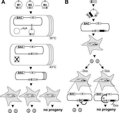
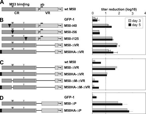
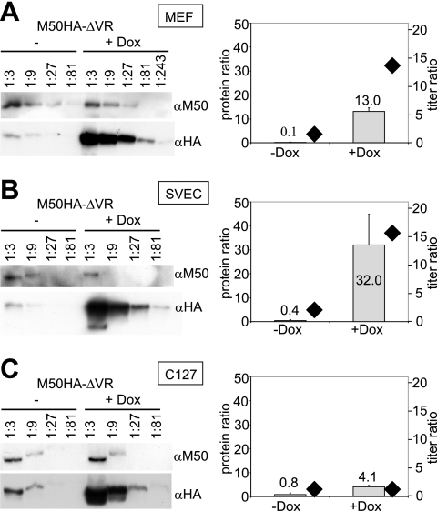
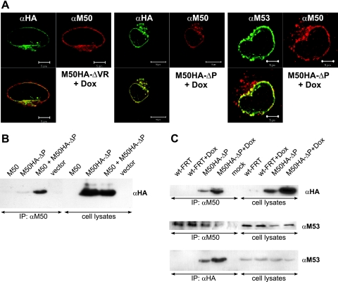
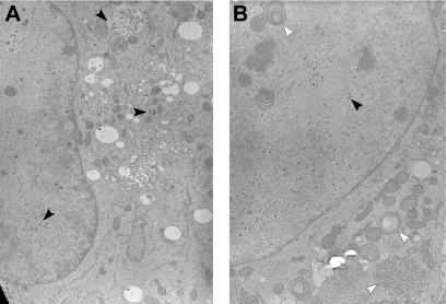
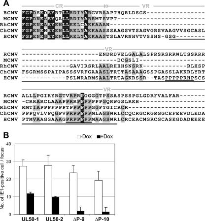
Inactivation of gene products by dominant-negative (DN) mutants is a powerful tool to assign functions to proteins. Here, we present a two-step procedure to establish a random screen for DN alleles, using the essential murine cytomegalovirus gene M50 as an example. First, loss-of-function mutants from a linker-scanning library were tested for inhibition of virus reconstitution with the help of FLP-mediated ectopic insertion of the mutants into the viral genome. Second, DN candidates were confirmed by conditional expression of the inhibitory proteins in the virus context. This allowed the quantification of the inhibitory effect, the identification of the morphogenesis block, and the construction of DN mutants with improved activity. Based on these observations a DN mutant of the homologous gene (UL50) in human cytomegalovirus was predicted and constructed. Our data suggest that a proline-rich sequence motif in the variable region of M50/UL50 represents a new functional site which is essential for nuclear egress of cytomegalovirus capsids.


Figures

FIG. 1.
FIG. 1.
Screening strategy for DN mutants of essential viral genes. (A) Screening for inhibitory mutants. An essential viral gene, the target gene (gray box, T), is subcloned and subjected to a random and comprehensive mutagenesis in vitro leading to a mutant library, M1, M2, … Mn (small black boxes indicate mutations). Mutated ORFs are placed under the control of a strong constitutive promoter into an insertion plasmid containing an FRT site (open box with gray triangle). The insertion plasmids can be maintained only in a special E. coli strain. Normal E. coli (open boxes) carrying an FRT-site-labeled viral BAC and a temperature-sensitive plasmid expressing FLP recombinase (FLP) are transformed with the insertion plasmids carrying different mutants one by one. The FLP recombinase mediates site-specific recombination between the FRT sites in the BAC and the insertion plasmids. This recombinant can then be isolated under combined antibiotic selection for both the BACs and the insertion plasmid. The FLP-expressing helper plasmid is removed by elevated temperature. Then, BAC DNA is prepared and permissive cells are transfected with each construct. The mutants which are able to inhibit the virus reconstitution can be selected on the basis of the inability to form plaques upon transfection. (B) Validation of DN mutants by conditional gene expression. The inhibitory mutants are subcloned under the control of a promoter regulated by TetR (black box) into an insertion plasmid with an FRT site. These constructs are delivered into the viral BAC as described above. Then, permissive cells are transfected with the recombinants in order to reconstitute viruses carrying the regulation cassettes for the inhibitory mutants. The inhibitory mutants are not expressed during reconstitution because in the absence of Dox (− Dox) the constitutively expressed TetR blocks their transcription. The inhibitory function of the mutants can be analyzed upon Dox administration (+ Dox), which leads to the expression of the inhibitory mutant by releasing the expression cassette from TetR regulation.
FIG. 2.
FIG. 2.
Results of the primary screen for inhibitory mutants of M50. The amino acid sequence encoded by the M50 gene of MCMV is displayed to show the positions of the different mutations which were analyzed. Transposon insertions resulting in the introduction of 5 aa (arrowheads) are indicated along the ORF. Mutants that caused a null phenotype after they were placed into the M50-deleted MCMV genome are indicated by filled symbols; mutants which did not affect the function are indicated by empty ones (6). Black arrowheads indicate the mutants that interfered with virus reconstitution after insertion into the wt genome, and gray arrowheads indicate mutants that did not interfere. The amino acid sequence deleted in M50-ΔVR is indicated by white lettering in a black box. The sequence which is deleted in the ΔP mutant of M50 is indicated by bold lettering. The underlined sequence in the M50 ORF shows the region which is essential for binding to the M53 protein.
FIG. 3.
FIG. 3.
The effect of conditional expression of the inhibitory M50 mutants on the virus growth. The schematic representation of the analyzed mutants is shown on the left. The wt M50 ORF is shown first. (A) The N-terminal region of the M50 (CR), which is conserved in alpha-, beta-, and gammaherpesvirus families, is indicated by a dark gray box, and the C-terminal variable region (VR) is indicated by a light gray box. The open box labeled with TM indicates the transmembrane domain, and “P” indicates the proline-rich sequence. The HA tag is indicated by a black box. The open arrowheads show the positions of 5-aa insertions affecting M53 protein binding (aa 53 to 57 and aa 114) (6). The sequence used to generate the anti-M50 antiserum (aa 201 to 213) is shown by a thick bar (ab). (B to D) The analyzed M50 mutants carry an insertion (black triangle) of 5 aa after position 40, 56, or 125 of M50 or lack sequences (represented by lines), i.e., a deletion of aa 179 to 276 in the ΔVR constructs (B), of aa 51 to 59 in the ΔM constructs (C), and of aa 179 to 207 in the ΔP constructs (D). The bar diagram on the right side shows the regulation of the virus growth in response to Dox in these mutants. MEFs were infected at an MOI of 0.1 in the absence or presence of 1 μg/ml Dox for 3 (gray bars) or 5 (black bars) days. Cell-free supernatants were taken on the indicated days and were titrated on MEFs to determine the number of the released infectious units. The titer reduction induced by Dox is shown as a ratio between the titer in the absence of Dox and the titer in the presence of Dox. The titer reduction was considered not significant if the ratio was less than 10. Each set was analyzed two times. Shown are means and standard deviations with the exception of the panel shown in the second diagram, which shows results from one experiment.
FIG. 4.
FIG. 4.
The inhibitory effect is defined by the ratio of DN to wt protein. MEFs (A) and SVEC (B) and C127 (C) cells were infected with M50HA-ΔVR at an MOI of 0.1 in the absence or presence of 1 μg/ml Dox for 3 days. The expression of the wt and the mutant protein was analyzed by Western blotting (left). Lysates of infected cells were diluted serially, and proteins were separated in an SDS-polyacrylamide gel, transferred to a membrane, and stained with specific antibodies to M50 (αM50) or HA (αHA). The M50 antiserum and the HA antibody have comparable affinities for their epitopes (data not shown), allowing the relative quantification of the signals. The bar diagrams (to the right of each blot) show the ratio of the expression of the HA-tagged M50HA-ΔVR protein to that of the wt M50 gene product. The protein ratios were determined in at least two independent experiments and are depicted as gray bars referring to the left ordinate. In parallel, the amounts of the released infectious units were determined in each supernatant by plaque assay. The ratios of the titers in response to Dox (division of the titers obtained in the absence of Dox by the titers in the presence of Dox) are shown as black rhombuses (to be read according to the right ordinate).
FIG. 5.
FIG. 5.
Intracellular distribution of the M50-M53 complex in the presence of the DN M50 mutants. (A) Indirect immunofluorescence images of NIH 3T3 cells infected at an MOI of 0.2 with M50HA-ΔVR or M50HA-ΔP virus for 1 day in the presence of 1 μg/ml Dox were taken. The M50 mutants were stained with an anti-HA antibody coupled to FITC. wt M53 protein was stained with an anti-M53 antibody followed by treatment with an FITC-labeled anti-rat secondary antiserum. The wt M50 protein was stained with an anti-M50 antiserum followed by treatment with Texas Red-labeled anti-rabbit secondary antiserum. (B and C) Coimmunoprecipitation of wt M50, M50HA-ΔP, and M53 proteins. The M50 protein was precipitated from cell lysates by anti-M50/protein A-Sepharose (B and top and middle in panel C), and the M50HA-ΔP protein was precipitated by anti-HA coupled to beads (bottom in panel C). Samples were separated in a 10% SDS-polyacrylamide gel, transferred to a membrane, and stained with antisera to HA or M53. (B) 293 cells were transfected for 1 day with pOriR6K-zeo-ie-M50, pM50HA-ΔP, or pOriR6K-zeo-ie, either alone or in combination. (C) NIH 3T3 cells were infected with wt-FRT-MCMV or M50HA-ΔP virus at an MOI of 10 in the absence or presence of 1 μg/ml Dox for 28 h. IP, immunoprecipitation.
FIG. 6.
FIG. 6.
DN mutants of M50 prevent viral capsid egress from the cell nucleus. NIH 3T3 cells were infected at an MOI of 0.5 with M50HA-ΔP virus for 48 h in the absence (A) or presence (B) of 1 μg/ml Dox. Cells were fixed by high-pressure freezing, freeze-substituted, plastic embedded, and analyzed by electron microscopy. Black arrowheads show viral capsids; white arrowheads show accumulated membranes in the nucleus and cytoplasm after induction of overexpression of the M50HA-ΔP protein.
FIG. 7.
FIG. 7.
The deletion of the proline-rich sequence from UL50 of HCMV creates a DN mutant. (A) N-terminal sequence comparison of the CMV UL34 homologues. The amino acid sequence of M50 was aligned with the UL34 family members from CMVs by use of the Vector NTI Align X program (Invitrogen) via the BLOSUM 62 similarity matrix. (RCMV, rat CMV; RhCMV, rhesus monkey CMV; ChCMV, chimpanzee CMV). The alignment of the end of the conserved region (CR) and the N-terminal half of the variable region (VR) is shown. The strictly conserved amino acids are shown by white letters in black boxes. Blocks of similar amino acids are indicated by gray boxes. The region deleted from the ΔP mutant of the HCMV UL50 is underlined. The two proline-rich motifs found in UL50 of HCMV are double underlined. (B) HFFs were transfected with two independent clones of the recombinant BACs, AD169FRT-SVT-UL50-1 and AD169FRT-SVT-UL50-2 expressing the wt UL50 and AD169FRT-SVT-UL50-ΔP9 and AD169FRT-SVT-UL50-ΔP10 expressing the mutant ORF. After the first plaques became apparent, the cells were replated onto 96-well plates together with noninfected HFFs either in the absence (white bars) or in the presence (black bars) of Dox. Five days later the numbers of IE1-positive cell nuclei in infected foci were determined. The mean values for 8 to 10 infected foci obtained in two independent experiments for clones UL50-1, UL50-2, and ΔP10 and three experiments for clone ΔP9 are plotted.

Similar articles

Cited by

References

    1. Adler, B., L. Scrivano, Z. Ruzsics, B. Rupp, C. Sinzger, and U. Koszinowski. 2006. Role of human cytomegalovirus UL131A in cell type-specific virus entry and release. J. Gen. Virol. 87:2451-2460. - PubMed
    1. Baltimore, D. 1988. Gene therapy. Intracellular immunization. Nature 335:395-396. - PubMed
    1. Biery, M. C., F. J. Stewart, A. E. Stellwagen, E. A. Raleigh, and N. L. Craig. 2000. A simple in vitro Tn7-based transposition system with low target site selectivity for genome and gene analysis. Nucleic Acids Res. 28:1067-1077. - PMC - PubMed
    1. Brune, W., C. Menard, J. Heesemann, and U. H. Koszinowski. 2001. A ribonucleotide reductase homolog of cytomegalovirus and endothelial cell tropism. Science 291:303-305. - PubMed
    1. Brune, W., C. Menard, U. Hobom, S. Odenbreit, M. Messerle, and U. H. Koszinowski. 1999. Rapid identification of essential and nonessential herpesvirus genes by direct transposon mutagenesis. Nat. Biotechnol. 17:360-364. - PubMed

Publication types

LinkOut - more resources