Comparative Study
. 2007 Mar 21;27(12):3057-63.
doi: 10.1523/JNEUROSCI.4371-06.2007.

Minocycline reduces microglial activation and improves behavioral deficits in a transgenic model of cerebral microvascular amyloid

Affiliations
Comparative Study

Minocycline reduces microglial activation and improves behavioral deficits in a transgenic model of cerebral microvascular amyloid

Rong Fan et al. J Neurosci. .

Abstract

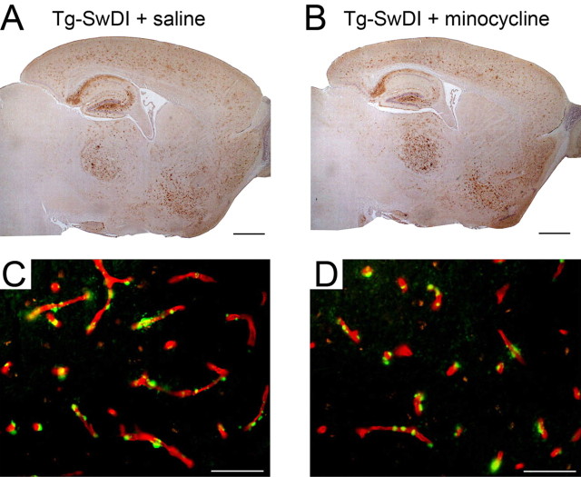
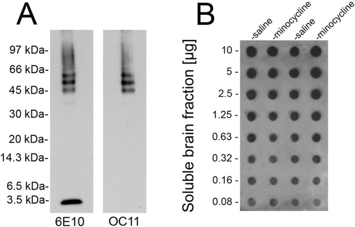
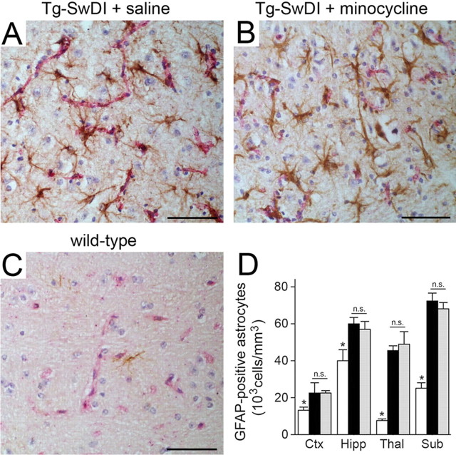
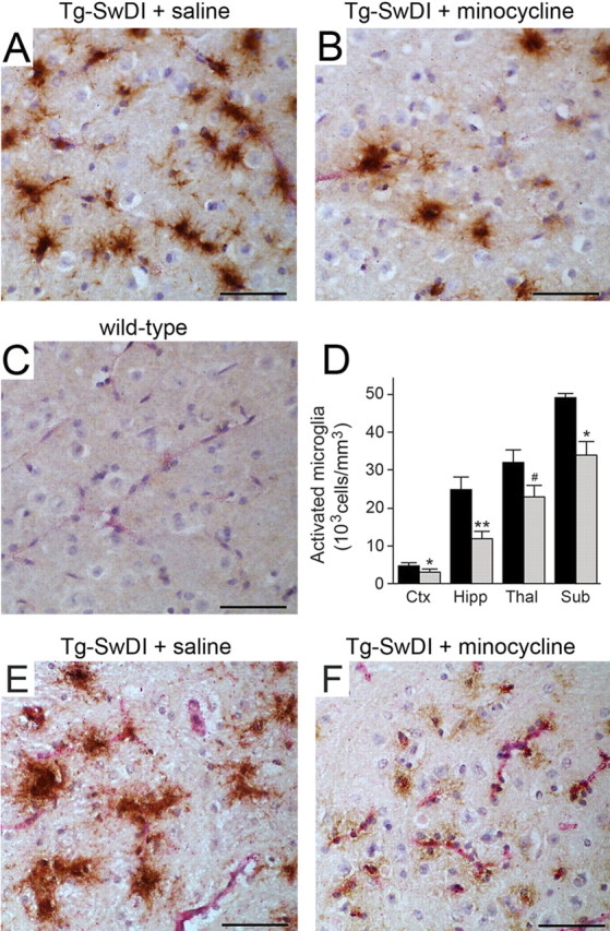
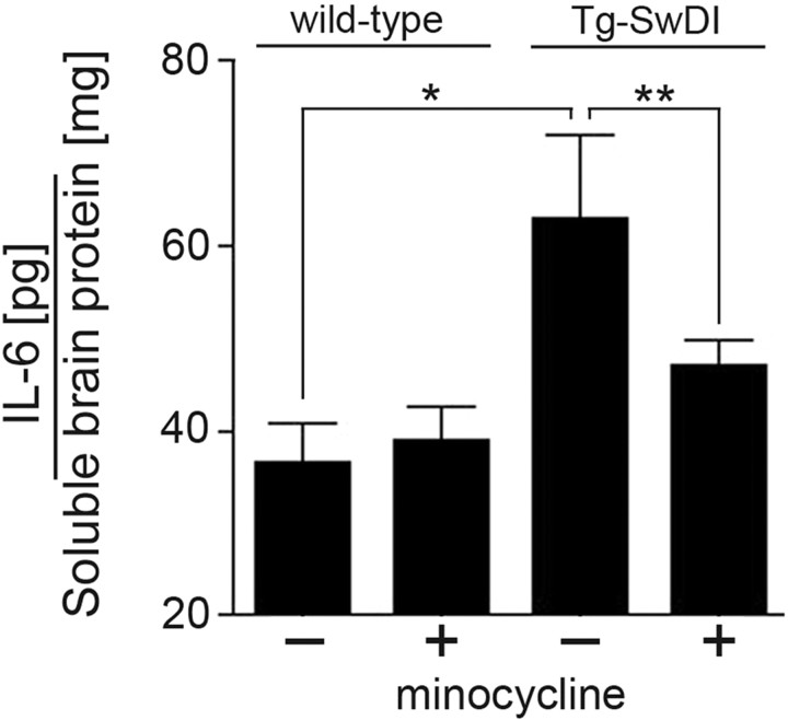
Cerebral microvascular amyloid beta protein (Abeta) deposition and associated neuroinflammation is increasingly recognized as an important component leading to cognitive impairment in Alzheimer's disease and related cerebral amyloid angiopathy disorders. Transgenic mice expressing the vasculotropic Dutch/Iowa (E693Q/D694N) mutant human Abeta precursor protein in brain (Tg-SwDI) accumulate abundant cerebral microvascular fibrillar amyloid deposits and exhibit robust neuroinflammation. In the present study, we investigated the effect of the anti-inflammatory drug minocycline on Abeta accumulation, neuroinflammation, and behavioral deficits in Tg-SwDI mice. Twelve-month-old mice were treated with saline or minocycline by intraperitoneal injection every other day for a total of 4 weeks. During the final week of treatment, the mice were tested for impaired learning and memory. Brains were then harvested for biochemical and immunohistochemical analysis. Minocycline treatment did not alter the cerebral deposition of Abeta or the restriction of fibrillar amyloid to the cerebral microvasculature. Similarly, minocycline-treated Tg-SwDI mice exhibited no change in the levels of total Abeta, the ratios of Abeta40 and Abeta42, or the amounts of soluble, insoluble, or oligomeric Abeta compared with the saline-treated control Tg-SwDI mice. In contrast, the numbers of activated microglia and levels of interleukin-6 were significantly reduced in minocycline-treated Tg-SwDI mice compared with saline-treated Tg-SwDI mice. In addition, there was a significant improvement in behavioral performance of the minocycline-treated Tg-SwDI mice. These finding suggest that anti-inflammatory treatment targeted for cerebral microvascular amyloid-induced microglial activation can improve cognitive deficits without altering the accumulation and distribution of Abeta.


Figures

Figure 1.
Figure 1.
Minocycline treatment does not alter the spatial accumulation of Aβ in Tg-SwDI mice. A, B, Aβ accumulation in the forebrain of 12-month-old Tg-SwDI mice treated with saline (A) or minocycline (B). Scale bars, 1 mm. C, D, Colocalization of vascular collagen IV immunostaining (red) and thioflavin-S amyloid staining (green) in the thalamus of 12-month-old Tg-SwDI mice treated with saline (C) or minocycline (D). Scale bars, 50 μm.
Figure 2.
Figure 2.
Minocycline treatment does not affect the levels of Aβ peptides in Tg-SwDI mouse forebrain. A, ELISA measurements of total Aβ40 (gray bars) and Aβ42 (black bars) in mouse forebrain tissue. B, ELISA measurements of soluble (Sol) Aβ (gray bars) and insoluble (Insol) Aβ (black bars) in mouse forebrain tissue. ELISA data shown are mean ± SD (n = 9 per group).
Figure 3.
Figure 3.
Minocycline treatment does not alter the levels of soluble Aβ oligomers in Tg-SwDI mouse forebrain. A, Immunoblot analysis of monomeric and oligomeric Dutch/Iowa Aβ40 using the anti-Aβ monoclonal antibody 6E10 (first lane) and anti-oligomeric Aβ polyclonal antibody OC11 (second lane). B, Representative dot blot analysis of Aβ oligomers in soluble mouse forebrain extracts using polyclonal antibody OC11.
Figure 4.
Figure 4.
Minocycline treatment does not affect the numbers of reactive astrocytes in Tg-SwDI mice. A–C, Microvascular-associated reactive astrocytes revealed by GFAP-positive immunostaining (brown) and collagen type IV (red). The thalamic regions of 12-month-old Tg-SwDI mice treated with saline (A) or minocycline (B) or 12-month-old wild-type mice (C) are shown. Scale bars, 50 μm. D, Quantitative stereological estimation of reactive astrocyte densities in different brain regions of wild-type mice (white bars), saline-treated Tg-SwDI mice (black bars), or minocycline-treated Tg-SwDI mice (gray bars). Brain regions measured include frontotemporal cortex (Ctx), hippocampus (Hipp), thalamus (Thal), and subiculum (Sub). Data shown are mean ± SD (n = 5). n.s., Not significant. *p < 0.001.
Figure 5.
Figure 5.
Minocycline treatment reduces microglial activation in Tg-SwDI mice. A–C, Microvascular-associated activated microglia revealed by MHCII-positive immunostaining (brown) and collagen type IV (red). The thalamic regions of 12-month-old Tg-SwDI mice treated with saline (A) or minocycline (B) or 12-month-old wild-type mice (C) are shown. Scale bars, 50 μm. D, Quantitative stereological estimation of activated microglial densities in different brain regions of saline-treated Tg-SwDI mice (black bars) or minocycline-treated Tg-SwDI mice (gray bars). Wild-type mice exhibited no activated microglia. Data shown are mean ± SD (n = 5). *p < 0.02; **p < 0.001; # p < 0.01. Brain regions measured include frontotemporal cortex (Ctx), hippocampus (Hipp), thalamus (Thal), and subiculum (Sub). E, F, Activated microglia revealed by 5D4-positive immunostaining in the thalamic region of 12-month-old Tg-SwDI mice treated with saline (E) or minocycline (F). Scale bars, 50 μm.
Figure 6.
Figure 6.
Minocycline reduces elevated levels of IL-6 in Tg-SwDI mice. The levels of IL-6 were measured in soluble forebrain extracts of 12-month-old wild-type and Tg-SwDI mice treated with saline or minocycline by ELISA analysis. Data shown are mean ± SD (n = 5). *p < 0.005; **p < 0.01.
Figure 7.
Figure 7.
Minocycline treatment improves spatial learning memory deficits in Tg-SwDI mice as assessed by performance in the Barnes maze task. Twelve-month-old wild-type mice (squares), saline-treated Tg-SwDI mice (open circles), or minocycline-treated Tg-SwDI mice (closed circles) were measured for their latencies to find the escape box over the course of acquisition. Data shown are mean ± SEM (n = 9 mice per group).

References

    1. Aisen PS. The potential of anti-inflammatory drugs for the treatment of Alzheimer's disease. Lancet Neurol. 2002;1:279–284. - PubMed
    1. Atterns J, Jellinger KA. Only cerebral capillary amyloid angiopathy correlates with Alzheimer pathology—a pilot study. Acta Neuropathol (Berl) 2004;107:83–90. - PubMed
    1. Bailey TL, Rivara CB, Rocher AB, Hof PR. The nature and effects of cortical microvascular pathology in aging and Alzheimer's disease. Neurol Res. 2004;26:573–578. - PubMed
    1. Barnes CA. Memory deficits associated with senescence: a neuropsychological and behavioral study in the rat. J Comp Physiol Psychol. 1979;93:74–104. - PubMed
    1. Combs CK, Karlo JC, Kao SC, Landreth GE. β-amyloid stimulation of microglia and monocytes results in TNFα-dependent expression of inducible nitric oxide synthase and neuronal apoptosis. J Neurosci. 2001;21:1179–1188. - PMC - PubMed

Publication types