Comparative Study
. 2007 Mar 21;27(12):3078-89.
doi: 10.1523/JNEUROSCI.3055-06.2007.

Lhx6 activity is required for the normal migration and specification of cortical interneuron subtypes

Affiliations
Comparative Study

Lhx6 activity is required for the normal migration and specification of cortical interneuron subtypes

Petros Liodis et al. J Neurosci. .

Abstract

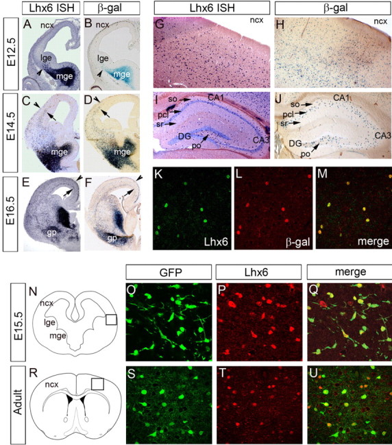
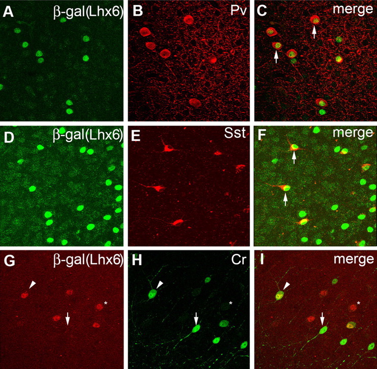
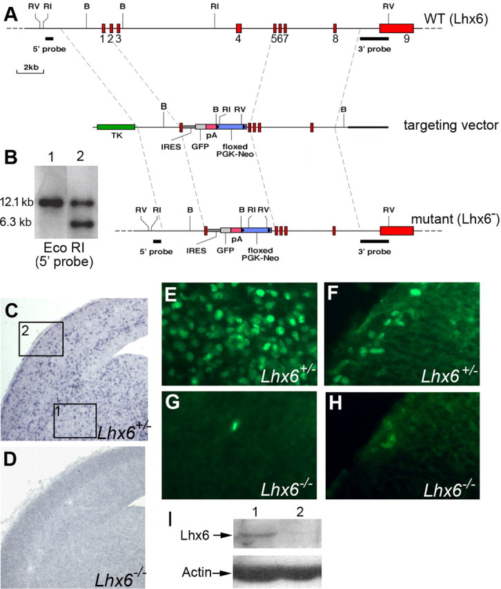
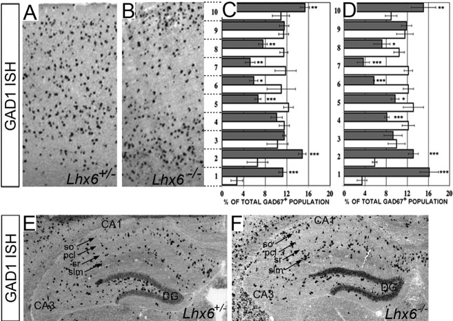
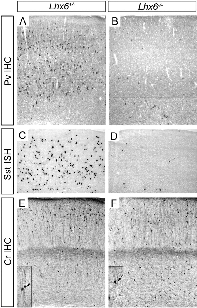
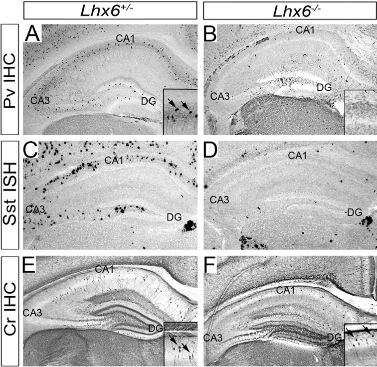
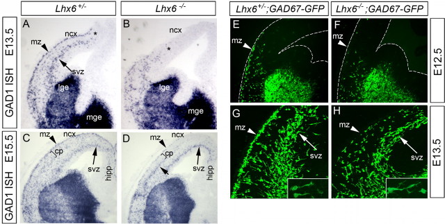
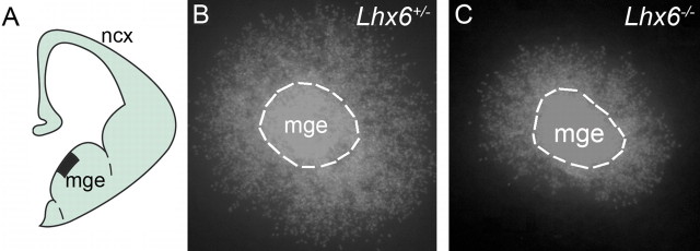
The cerebral cortex contains two main neuronal cell populations, the excitatory glutamatergic (pyramidal) neurons and the inhibitory interneurons, which synthesize GABA and constitute 20-30% of all cortical neurons. In contrast to the mostly homogeneous population of projection neurons, cortical interneurons are characterized by remarkable morphological, molecular, and functional diversity. Among the markers that have been used to classify cortical interneurons are the calcium-binding proteins parvalbumin and calretinin and the neuropeptide somatostatin, which in rodents identify mostly nonoverlapping interneuron subpopulations. Pyramidal neurons are born during embryogenesis in the ventricular zone of the dorsal telencephalon, whereas cortical interneurons are generated in the subpallium and reach the cortex by tangential migration. On completion of tangential migration, cortical interneurons switch to a radial mode of migration and enter the cortical plate. Although the mechanisms that control the generation of interneuron diversity are currently unknown, it has been proposed that their site of origin in the ventral forebrain determines their specification into defined neurochemical subgroups. Here, we show that Lhx6, a gene induced in the medial ganglionic eminence and maintained in parvalbumin- and somatostatin-positive interneurons, is required for the specification of these neuronal subtypes in the neocortex and the hippocampus. We also show that Lhx6 activity is required for the normal tangential and radial migration of GABAergic interneurons in the cortex.


Figures

Figure 1.
Figure 1.
A–J, Expression of the Lhx6LacZins allele recapitulates the pattern of expression of the wild-type Lhx6 locus. Coronal sections from the forebrain of E12.5 (A, B), E14.5 (C, D), and E16.5 (E, F) Lhx6+/LacZins embryos or the neocortex (G, H) and the hippocampus (I, J) of Lhx6+/LacZins adult animals were processed either for in situ hybridization with a Lhx6-specific riboprobe (A, C, E, G, I) or β-gal histochemistry (B, D, F, H, J). The arrowheads in A and B point to the early population of MGE-derived cells that migrate toward the cortex. The arrowheads and arrows in C–F indicate the MZ and IZ/SVZ, respectively. Note the similar distribution of cells identified by in situ hybridization and β-gal histochemistry. K–M, A cortical section from an Lhx6+/LacZins adult animal double immunostained with antibodies specific for Lhx6 (K) and β-gal (L). As shown by the merged image (M), all Lhx6+ cells coexpress β-gal. N–U, Brain sections from E15.5 (N–Q) and adult (R–U) GAD67GFP animals immunostained for GFP (O, S) and Lhx6 (P, T). The corresponding merged images are shown in Q and U. The areas shown in O–Q and S–U correspond to the boxes shown in N and K, respectively. β-gal, β-Galactosidase; gb, globus pallidus; ISH, in situ hybridization; lge, lateral ganglionic eminence; mge, medial ganglionic eminence; ncx, neocortex; poDG, polymorphic layer of the dentate gyrus; pcl, pyramidal cell layer; so, stratum oriens; sr, stratum radiatum.
Figure 2.
Figure 2.
The majority of Pv+ and Sst+ cortical interneurons express Lhx6. Confocal microscope images of cortical sections from Lhx6+/LacZins animals double immunostained for β-gal and either Pv (A–C), or Sst (D–F), or Cr (G–I). As is evident from the merged images, virtually all Pv- and Sst-expressing interneurons coexpress β-gal (C, F, arrows). In contrast, β-gal was generally excluded from neurons of bipolar morphology that expressed high levels of Cr (G–I, arrows). Colocalization of β-gal and Cr was observed in a subpopulation of neurons of variable morphology that expressed lower levels of Cr (G–I, arrowheads). The asterisk (in G–I) indicates a representative cell that is positive for β-gal but negative for Cr.
Figure 3.
Figure 3.
Targeting of the Lhx6 locus. A, Shown at the top is a diagrammatic representation of the Lhx6 locus. The middle diagram represents the targeting construct, whereas shown at the bottom is the mutant Lhx6 allele. B, Southern blot analysis of EcoRI-digested genomic DNA from a wild-type (lane 1) and a heterozygous (lane 2) clone using the 5′ external probe (indicated in A). C, D, In situ hybridization on forebrain sections from E15.5 heterozygous (C) and homozygous (D) Lhx6 embryos with an Lhx6-specific riboprobe. E–H, Immunostaining of forebrain sections from E15.5 heterozygous (E, F) and homozygous (G, H) embryos with an Lhx6-specific polyclonal antiserum. Boxes 1 and 2 in C show the areas represented in E, G, and F, H, respectively. I, Western blot analysis of protein extracts from the forebrain of wild-type (lane 1) and Lhx6−/− (lane 2) embryos. B, BamHI; RI, EcoRI; RV, EcoRV; TK, thymidine kinase; WT, wild type.
Figure 4.
Figure 4.
Normal number but abnormal distribution of GAD67+ cells in the cortex of Lhx6 mutants. Coronal brain sections from 2-week-old heterozygous (A) or homozygous (B) animals were hybridized with a Gad1-specific riboprobe. In contrast to wild-type sections, in which GAD67+ cells are distributed mostly uniformly, in mutant sections GAD67+ cells were mostly localized in the upper and deep cortical layers. The effect of the Lhx6 mutation on the distribution of GAD67+ cells along the ventricular–pial axis was quantified in the motor (C) and the somatosensory (D) cortex. For this, equivalent areas of cortex from heterozygous and homozygous Lhx6 animals were subdivided into 10 bins, and the proportion of GAD67+ cells present in each bin was determined. The white and gray bars represent the percentages of GAD67+ cells present in individual segments of the cortex of heterozygous and mutant animals, respectively. Error bars in C and D represent SDs (see Materials and Methods for details on statistical analysis) *, **, and *** correspond to p (t test) values of <0.05, <0.01, and <0.001, respectively. Coronal sections through the hippocampus of heterozygous (E) and mutant (F) animals hybridized with a Gad1-specific riboprobe. The number of GAD67+ cells is not reduced, but similar to the neocortex they appear to be redistributed in the hippocampal layers of the CA1 region. DG, Dentate gyrus; ISH, in situ hybridization; pcl, pyramidal cell layer; so, stratum oriens; slm, stratum lacunosum moleculare; sr, stratum radiatum.
Figure 5.
Figure 5.
Severe reduction in the number of Pv- and Sst-expressing interneurons in the neocortex of Lhx6 mutants. Equivalent sections from the neocortex of 2-week-old animals heterozygous (A, C, E) or homozygous (B, D, F) for Lhx6 were immunostained for Pv (A, B) or Cr (E, F) or hybridized with an Sst-specific riboprobe (C, D). The insets in E and F show high magnifications of representative Cr+ neurons (arrows) of the corresponding genotypes. IHC, Immunohistochemistry; ISH, in situ hybridization.
Figure 6.
Figure 6.
Severe reduction in the number of Pv- and Sst-expressing interneurons in the hippocampus of Lhx6 mutants. Equivalent sections from the hippocampus of 2-week-old animals heterozygous (A, C, E) or homozygous (B, D, F) for Lhx6 were immunostained for Pv (A, B) or Cr (E, F) or hybridized with an Sst-specific riboprobe (C, D). The insets in A, B, E, and F show high magnification of areas corresponding to the CA1 region of the hippocampus. Note the dramatic reduction in the number of Pv+ and Sst+ cells in the hippocampus of Lhx6-deficient animals. In contrast, the number of Cr+ cells in the CA1 region of mutant animals is unaffected. The arrows in the insets indicate positive cells. DG, Dentate gyrus; IHC, immunohistochemistry; ISH, in situ hybridization.
Figure 7.
Figure 7.
Delayed tangential migration and abnormal distribution of GABAergic interneurons in the cortex of Lhx6 mutant embryos. A–D, In situ hybridization of coronal brain sections from E13.5 (A, B) and E15.5 (C, D) embryos heterozygous (A, C) or homozygous (B, D) for the Lhx6 mutation. Note that, relative to controls, in E13.5 mutant embryos GAD67+ cells were restricted to the most ventrolateral cortex. In later-stage mutant embryos, GAD67+ cells were found in the IZ/SVZ of the neocortex and the hippocampus but were drastically reduced in the MZ and the CP. Generation of heterozygous and homozygous Lhx6 animals carrying the GAD67-GFP allele allowed us to use GFP as a reporter of tangentially migrating GABAergic interneurons. E–H, Immunostaining for GFP on coronal forebrain sections from E12.5 (E, F) or E13.5 (G, H) embryos heterozygous (E, G) or homozygous (F, H) for the Lhx6 mutation. Note the reduced number of GFP+ cells crossing the corticostriatal boundary in E12.5 mutant embryos (F) relative to controls (E). In E13.5 mutant embryos (H), the number of GFP+ cells present in the MZ was reduced relative to control embryos (G). No dramatic differences were observed in the morphology of tangentially migrating cells in the SVZ of control and Lhx6-deficient embryos (compare insets in G and H). The arrowheads indicate the MZ, whereas the arrows point to the IZ/SVZ. The asterisks indicate the front of tangentially migrating interneurons. cp, Cortical plate; hipp, hippocampus; ISH, in situ hybridization; lge, lateral ganglionic eminence, mge, medial ganglionic eminence; mz, marginal zone; ncx, neocortex; svz, subventricular zone.
Figure 8.
Figure 8.
Intrinsic migratory deficit of Lhx6-deficient MGE cells. A, Schematic presentation of the area of the subpallium used for the MGE explant cultures. MGE explants from E13.5 heterozygous (B) embryos show profuse migration into the collagen gel matrix. In contrast, reduced radial migration of MGE cells was observed in parallel cultures from Lhx6 mutant embryos (C). The black box in A shows the area used to generate the MGE explants. mge, Medial ganglionic eminence; ncx, neocortex.

Similar articles

Cited by

References

    1. Alcantara S, Soriano E, Ferrer I. Thalamic and basal forebrain afferents modulate the development of parvalbumin and calbindin D28k immunoreactivity in the barrel cortex of the rat. Eur J Neurosci. 1996;8:1522–1534. - PubMed
    1. Alifragis P, Liapi A, Parnavelas JG. Lhx6 regulates the migration of cortical interneurons from the ventral telencephalon but does not specify their GABA phenotype. J Neurosci. 2004;24:5643–5648. - PMC - PubMed
    1. Anderson SA, Eisenstat DD, Shi L, Rubenstein JL. Interneuron migration from basal forebrain to neocortex: dependence on Dlx genes. Science. 1997;278:474–476. - PubMed
    1. Anderson SA, Marin O, Horn C, Jennings K, Rubenstein JL. Distinct cortical migrations from the medial and lateral ganglionic eminences. Development. 2001;128:353–363. - PubMed
    1. Ang ES, Jr, Haydar TF, Gluncic V, Rakic P. Four-dimensional migratory coordinates of GABAergic interneurons in the developing mouse cortex. J Neurosci. 2003;23:5805–5815. - PMC - PubMed

Publication types

MeSH terms