Comparative Study
. 2007 Mar 21;27(12):3090-7.
doi: 10.1523/JNEUROSCI.4854-06.2007.

Anesthesia leads to tau hyperphosphorylation through inhibition of phosphatase activity by hypothermia

Affiliations
Comparative Study

Anesthesia leads to tau hyperphosphorylation through inhibition of phosphatase activity by hypothermia

Emmanuel Planel et al. J Neurosci. .

Abstract

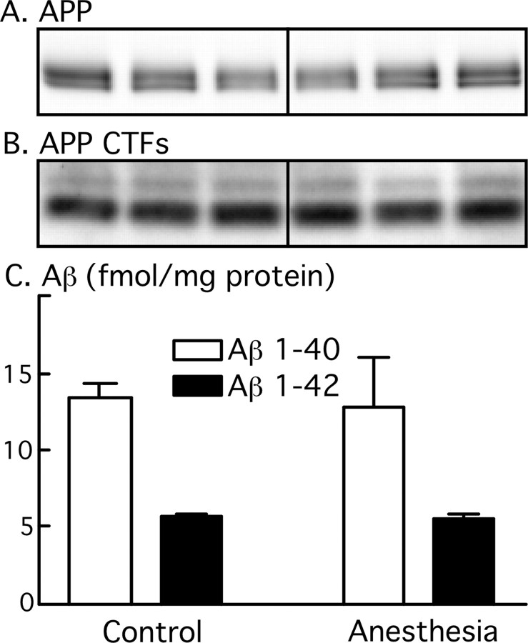
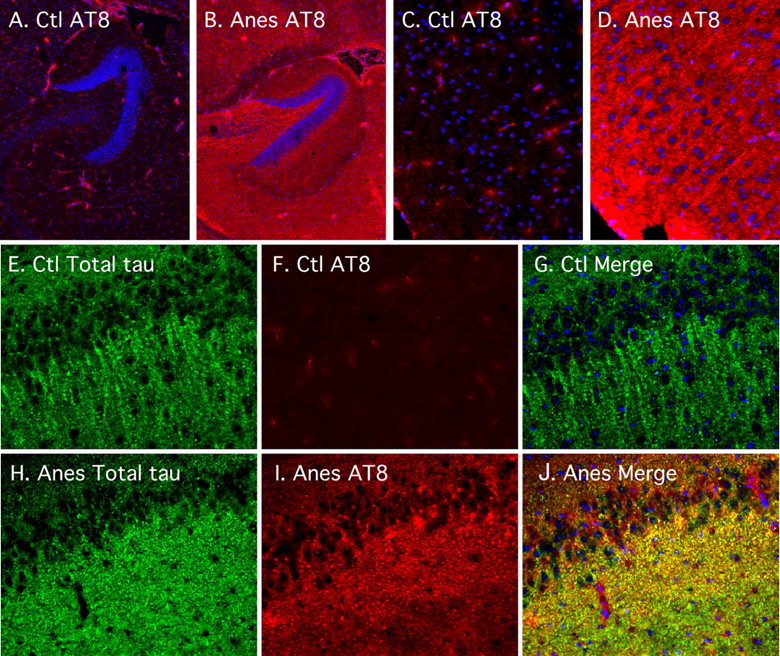
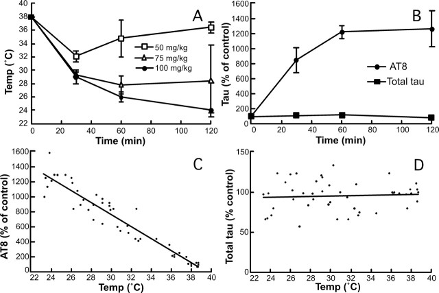
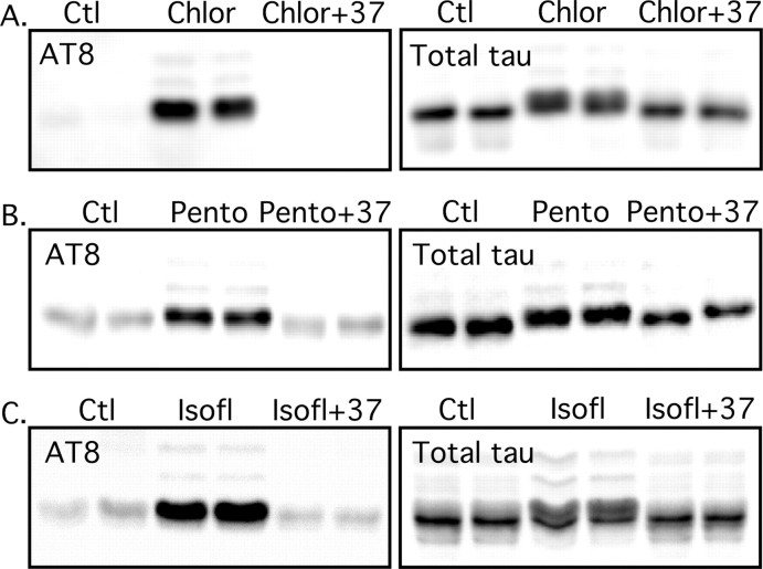
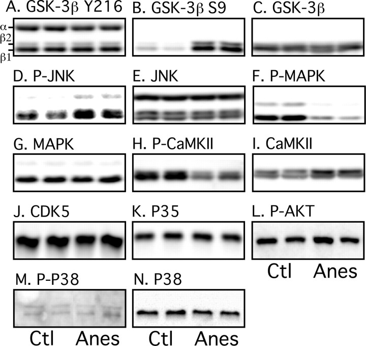
Postoperative cognitive dysfunction, confusion, and delirium are common after general anesthesia in the elderly, with symptoms persisting for months or years in some patients. Even middle-aged patients are likely to have postoperative cognitive dysfunction for months after surgery, and Alzheimer's disease (AD) patients appear to be particularly at risk of deterioration after anesthesia. Several investigators have thus examined whether general anesthesia is associated with AD, with some studies suggesting that exposure to anesthetics may increase the risk of AD. However, little is known on the biochemical consequences of anesthesia on pathogenic pathways in vivo. Here, we investigated the effect of anesthesia on tau phosphorylation and amyloid precursor protein (APP) metabolism in mouse brain. We found that, regardless of the anesthetic used, anesthesia induced rapid and massive hyperphosphorylation of tau, rapid and prolonged hypothermia, inhibition of Ser/Thr PP2A (protein phosphatase 2A), but no changes in APP metabolism or Abeta (beta-amyloid peptide) accumulation. Reestablishing normothermia during anesthesia completely rescued tau phosphorylation to normal levels. Our results indicate that changes in tau phosphorylation were not a result of anesthesia per se, but a consequence of anesthesia-induced hypothermia, which led to inhibition of phosphatase activity and subsequent hyperphosphorylation of tau. These findings call for careful monitoring of core temperature during anesthesia in laboratory animals to avoid artifactual elevation of protein phosphorylation. Furthermore, a thorough examination of the effect of anesthesia-induced hypothermia on the risk and progression of AD is warranted.


Figures

Figure 1.
Figure 1.
Effect of anesthesia on tau phosphorylation. Proteins from mouse brain hemispheres (hippocampus plus cortex) were separated by SDS-PAGE and identified with the antibodies indicated on top of the boxes. For each antibody, two representative data of four for each condition (Ctl, control; Anes, anesthesia by chloral hydrate) are displayed. Each lane represents an individual mouse. The bands were quantified and the immunoreactivity during anesthesia expressed as a percentage of the control treatment (100%): A, 187 ± 7**; B, 42 ± 3*; C, 1471 ± 177***; D, 326 ± 12*; E, 15683 ± 8059*; F, 475 ± 77**; G, 176 ± 40*; H, 230 ± 23***; I, 116 ± 2. Data are means ± SD. *, **, and *** indicate significant differences with p < 0.05, 0.01, and 0.001, respectively.
Figure 2.
Figure 2.
Effect of anesthesia on APP metabolism. Proteins from mouse brain hemispheres (hippocampus plus cortex) were separated by SDS-PAGE and identified with C1/6.1 antibody, which recognize APP (A) and CTFs (B). A and B display three representative data of five for each condition (Ctl, control; Anes, anesthesia by chloral hydrate). C shows the levels of Aβ1–40 (white bars) and Aβ1–42 (black bars) for control and anesthesia (n = 5), as detected by ELISA. Data are means ± SD.
Figure 3.
Figure 3.
Regional anatomical analysis of tau phosphorylation during anesthesia. Fluorescence photomicrographs of sagittal sections are shown. AT8 (in red) or total tau (in green) immunostaining was performed in control (Ctl) (A, C, E, F, G) and anesthetized (Anes) (pentobarbital; B, D, H, I, J) mice, in the hippocampus (A, B) (magnification, 50×), cortical regions (C, D) (magnification, 200×), or the CA1 (E–J) (magnification, 200×). DAPI nucleus-specific staining is visible in blue.
Figure 4.
Figure 4.
Effects of anesthesia on temperature and tau phosphorylation. A, Rectal temperatures of the mice were recorded after injection of 50, 75, or 100 mg/kg pentobarbital or vehicle (n = 3–5 for each point). B, AT8 and total tau levels were quantified from brains of animals anesthetized with 100 mg/kg pentobarbital, and displayed as percentage of the control animals at different times (n = 3–5 for each point). C, D, Immunoreactive signals of AT8 (C) and total tau (D) from animals anesthetized with 50, 75, or 100 mg/kg pentobarbital were expressed as a percentage of the control mice and plotted against the corresponding individual rectal temperature (n = 47). Data are means ± SD.
Figure 5.
Figure 5.
Effect of controlled temperature and different anesthetics on tau phosphorylation. Proteins from mouse brain hemispheres (hippocampus plus cortex) were separated by SDS-PAGE and identified with either AT8 or total tau antibodies. Anesthesia by chloral hydrate (A), pentobarbital (B), or isoflurane (C) induced a robust tau hyperphosphorylation (lanes 3 and 4) that could be totally rescued by returning the core temperature of the animals to normal (lanes 5 and 6). Total tau did not change during the experiment. Ctl, Control; Chlor, chloral hydrate; Pento, pentobarbital; Isofl, isoflurane.
Figure 6.
Figure 6.
Effect of anesthesia on tau kinase activation. Proteins from mouse brain hemispheres (hippocampus plus cortex) were separated by SDS-PAGE and identified with the antibodies indicated on top of the boxes. For each antibody, two representative data of four for each condition (Ctl, control; Anes, anesthesia by chloral hydrate) are displayed in the top part of the boxes. Each lane represents an individual mouse. The bands were quantified and the immunoreactivity during anesthesia expressed as a percentage of the control treatment (100%): A, 97 ± 13; B, 808 ± 51***; C, 112 ± 14; D, 128 ± 17*; E, 113 ± 4; F, 24 ± 8**; G, 125 ± 11; H, 72 ± 15*; I, 135 ± 8*; J, 91 ± 14; K, 106 ± 11; L, 119 ± 20; M, 127 ± 33; N, 101 ± 23. Data are means ± SD. *, **, and *** indicate significant differences with p < 0.05, 0.01, and 0.001, respectively.
Figure 7.
Figure 7.
Effect of anesthesia on phosphatase activity. A, Kinetic of tau dephosphorylation at PHF-1 epitope in brain extracts, as monitored by Western blotting, with or without 5 nm okadaic acid. Tau dephosphorylation was completely abolished by okadaic acid. B, Phosphatase activity was evaluated by endogenous tau dephosphorylation performed at the temperature of the animals before killing, followed by Western blot analysis. Tau from control brains incubated at 37°C was dephosphorylated faster (n = 3; p = 0.0017; two-way ANOVA followed by Bonferroni's posttests) than tau from animals anesthetized with pentobarbital and incubated at 26°C. C, To confirm these results, PP2A activity was evaluated with the PP2A assay system from Promega. Similarly to the endogenous assay, there was no significant difference in PP2A activity between control and anesthetized mice samples when incubated at 37°C, whereas ∼40% PP2A inhibition was detected when the reaction was performed at 26°C (n = 3; p < 0.05; ANOVA followed by Tukey–Kramer's posttests). Data are means ± SDs. * and ** indicate significant differences with p < 0.05 and 0.01, respectively.

References

    1. Alonso AC, Zaidi T, Novak M, Grundke-Iqbal I, Iqbal K. Hyperphosphorylation induces self-assembly of tau into tangles of paired helical filaments/straight filaments. Proc Natl Acad Sci USA. 2001;98:6923–6928. - PMC - PubMed
    1. Amaducci LA, Fratiglioni L, Rocca WA, Fieschi C, Livrea P, Pedone D, Bracco L, Lippi A, Gandolfo C, Bino G, Prencipe M, Bonatti ML, Girotti F, Carella F, Tavola B, Ferla S, Lenzi GL, Carolei A, Gambi A, Grigoletto F, et al. Risk factors for clinically diagnosed Alzheimer's disease: a case-control study of an Italian population. Neurology. 1986;36:922–931. - PubMed
    1. Ancelin ML, de Roquefeuil G, Ledesert B, Bonnel F, Cheminal JC, Ritchie K. Exposure to anaesthetic agents, cognitive functioning and depressive symptomatology in the elderly. Br J Psychiatry. 2001;178:360–366. - PubMed
    1. Avila J. Tau phosphorylation and aggregation in Alzheimer's disease pathology. FEBS Lett. 2006;580:2922–2927. - PubMed
    1. Avila J, Diaz-Nido J. Tangling with hypothermia. Nat Med. 2004;10:460–461. - PubMed

Publication types