Comparative Study
. 2007 Mar 21;27(12):3139-47.
doi: 10.1523/JNEUROSCI.5151-06.2007.

Asymmetric distribution of prickle-like 2 reveals an early underlying polarization of vestibular sensory epithelia in the inner ear

Affiliations
Comparative Study

Asymmetric distribution of prickle-like 2 reveals an early underlying polarization of vestibular sensory epithelia in the inner ear

Michael R Deans et al. J Neurosci. .

Abstract

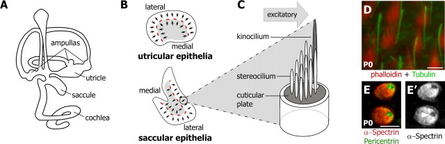
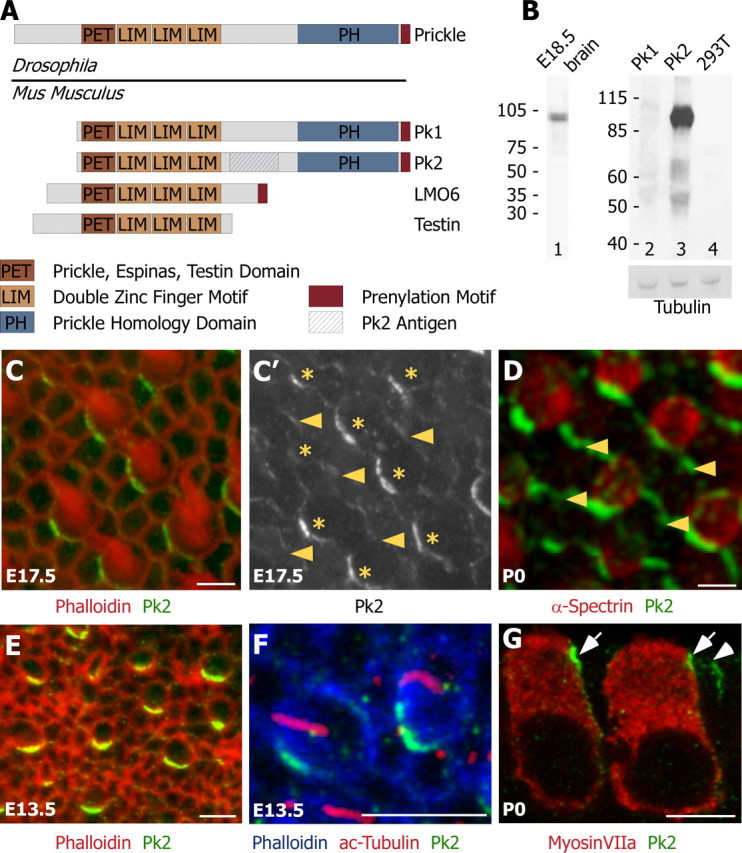
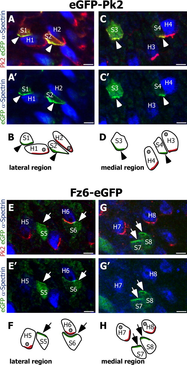
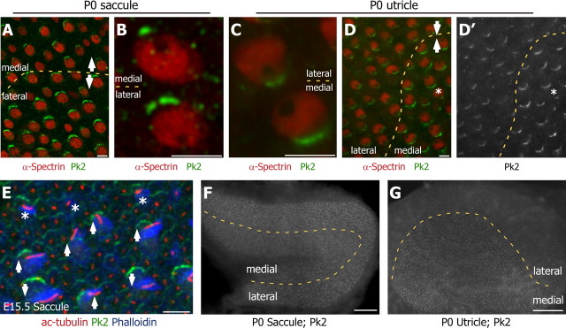
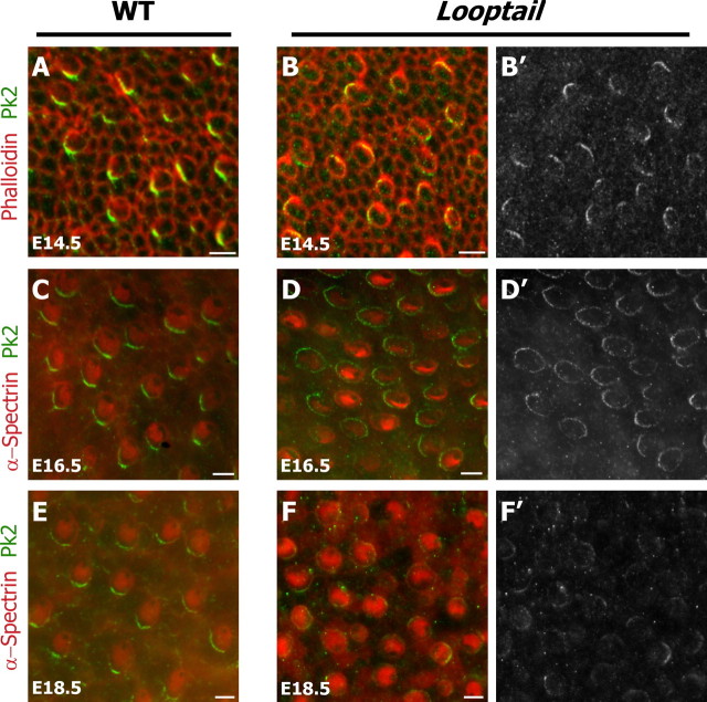
Vestibular hair cells have a distinct planar cell polarity (PCP) manifest in the morphology of their stereocilia bundles and the asymmetric localization of their kinocilia. In the utricle and saccule the hair cells are arranged in an orderly array about an abrupt line of reversal that separates fields of cells with opposite polarity. We report that the putative PCP protein Prickle-like 2 (Pk2) is distributed in crescents on the medial sides of vestibular epithelial cells before the morphological polarization of hair cells. Despite the presence of a line of polarity reversal, crescent position is not altered between hair cells of opposite polarity. Frizzled 6 (Fz6), a second PCP protein, is distributed opposite Pk2 along the lateral side of vestibular support cells. Similar to Pk2, the subcellular localization of Fz6 does not differ between cells located on opposite sides of the line of reversal. In addition, in Looptail/Van Gogh-like2 mutant mice Pk2 is distributed asymmetrically at embryonic day 14.5 (E14.5), but this localization is not coordinated between adjacent cells, and the crescents subsequently are lost by E18.5. Together, these results support the idea that a conserved PCP complex acts before stereocilia bundle development to provide an underlying polarity to all cells in the vestibular epithelia and that cells on either side of the line of reversal are programmed to direct the kinocilium in opposite directions with respect to the polarity axis defined by PCP protein distribution.


Figures

Figure 1.
Figure 1.
Vestibular hair cells of the utricle and saccule are organized within the inner ear to optimize the detection of linear accelerations in all directions. A, The vestibular hair cells within the utricle and saccule facilitate the detection of horizontal (utricle) and gravitational accelerations (saccule). In addition, hair cells within the ampullas of the semicircular canals detect rotational movements. Auditory hair cells detect sound and are located within the cochlea. B, In the utricular and saccular epithelia the adjacent hair cells share a similar polarity (small arrows) and are organized about a line of reversal (red dashed line) that separates lateral and medial domains (light gray shading). Arrows indicate the functional and morphological polarity of the bundle and are drawn from the shortest stereocilia to the kinocilium. C, The stereocilia bundle of an individual hair cell is arranged in a staircase manner, with the tallest stereocilia adjacent to the kinocilium. Deflections of the bundle toward the kinocilium are excitatory. D, Polarity can be visualized by using phalloidin (red) and an antibody against acetylated tubulin (green) to label the stereocilia and kinocilia, respectively. E, Alternatively, polarity can be visualized by using α-spectrin antibodies (red) to label the cuticular plate. Pericentrin immunolabeling (green) corresponds to the basal body beneath the kinocilium. E′, Via visualization with α-spectrin alone, the insertion point of the kinocilium and corresponding orientation of a hair cell can be determined by the position of a void of immunofluorescent labeling (arrowhead). Scale bars, 5 μm.
Figure 2.
Figure 2.
Pk2 is localized asymmetrically at cell boundaries throughout hair cell differentiation and polarization. A, Four Prickle-like proteins can be identified in mouse on the basis of the presence of a single PET and three LIM protein domains. Two of these, Pk1 and Pk2, also contain a Prickle homology domain and are the most similar to Drosophila Prickle. A region unique to Pk2 (hatched box) was used to generate Pk2-specific antiserum. B, On Western blots, the Pk2 antiserum recognizes a single band from E18.5 brain extract (lane 1) and HEK 293T cells expressing Pk2 (lane 3), but not Pk1 (lane 2) or nontransfected HEK 293T cell lysates (lane 4). Immunoblotting with a tubulin antibody was included as a loading control. C, In developing E17.5 utricles, Pk2 (green) is enriched at one edge of hair cells, labeled with phalloidin (red). C′, In the utricle, Pk2 is present at lower levels at boundaries between adjacent support cells than at boundaries between hair cells and support cells. Shown is a gray scale image of Pk2 labeling from C, with arrowheads indicating boundaries between adjacent supporting cells and asterisks marking the positions of hair cells. D, In the saccule, Pk2 labeling (green) is more prominent at boundaries between support cells (arrowheads) than in the utricle. Hair cells are labeled with α-spectrin (red). E, At E13.5, Pk2 (green) is enriched at one edge of hair cells that lack a stereocilia bundle but can be identified by their rounded shape and mosaic distribution via phalloidin stain (red). F, Pk2 accumulation (green) at E13.5 precedes the asymmetric localization of the kinocilium (red) to one edge of the cell (phalloidin; blue). G, In sections, Pk2 enrichment (green) is seen near the apical surface of the tissue at boundaries between supporting cells (arrowhead) and adjacent to hair cells (arrows), identified by the hair cell marker MyosinVIIa (red). Scale bars, 5 μm.
Figure 3.
Figure 3.
Pk2 and Fz6 are redistributed to opposite sides of utricular hair cells and support cells. A, Two support cells located within the lateral domain of the utricle (labeled S1 and S2) electroporated with eGFP-Pk2 (green) distribute eGFP-tagged Pk2 along their medial edges (arrowheads). Endogenous Pk2 (red) forms crescents along the medial cell boundary of two hair cells [identified by using α-spectrin (blue); labeled H1 and H2] and does not overlap with the distribution of eGFP-Pk2 in S1 or S2. The Pk2 antibody also labels exogenous eGFP-Pk2 in the support cells. Electroporated cells are located in the lateral domain of the utricle. B, Diagram illustrating the relative positions of support cells and hair cells from A and A′. In this and all subsequent diagrams the subcellular localizations of eGFP-tagged protein and endogenous Pk2 are indicated by green and red shading, respectively. The position of the hair cell kinocilia, based on α-spectrin labeling, is illustrated as a gray spot. C, C′, D, Support cells located on the opposite side of the line of reversal and within the medial region of the utricle (labeled S3 and S4) also distribute eGFP-tagged Pk2 (arrowheads) along their medial edges, similar to endogenous Pk2 in hair cells (labeled H3 and H4). E, E′, F, In contrast, support cells electroporated with Fz6-eGFP (labeled S5 and S6) redistribute Fz6-eGFP protein (green) to their lateral edge (arrows). In these cells Fz6-eGFP is enriched opposite of the endogenous Pk2 crescents (red) present in adjacent hair cells (H5 and H6). In E and F the electroporated cells are located in the lateral domain of the utricle. G, G′, H, Support cells located on the opposite side of the line of reversal and within the medial region of the utricle (labeled S7 and S8) also redistribute Fz6-eGFP to their lateral edge (arrows). This distribution is opposite from that of the endogenous Pk2 (red) in hair cells (labeled H7 and H8). For each utricle, the medial and lateral regions are identified by the position of the line of reversal, and images are oriented with the lateral edge of hair cells on top. Scale bars, 5 μm.
Figure 4.
Figure 4.
The relative distribution of Pk2 does not change across the line of reversal. A, D, The line of reversal (dashed yellow line) in P0 saccule A and utricle D is visualized easily by the position of the kinocilium void within the cuticular plate of hair cells that have been labeled with an antibody against α-spectrin (red). The polarities of pairs of cells on opposite sides of the line of reversal from both epithelia are indicated by arrows. Despite the change in hair cell polarity, the relative distribution of Pk2 (green) remains constant. B, C, Higher-magnification image containing two hair cells from saccule B and utricle C located on opposite sides of the line of reversal. In each the Pk2 (green) is located on the medial edge of the cell; however, there is a relative change in the amount of Pk2 protein between the two cells. D′, Gray scale image of Pk2 across the line of reversal in utricle reveals a correlation between the position of the kinocilium and relative levels of Pk2. An asterisk (D, D′) indicates a hair cell that does not share the polarity of its neighbors and has a corresponding decrease in Pk2 protein. E, The line of reversal is first apparent at E15.5 and can be visualized by the position of the kinocilium (acetylated tubulin; red) relative to the stereocilia bundle (phalloidin; blue). At this time, Pk2 (green) already is forming crescents along the medial edges of both mature and newly differentiated hair cells (asterisks). The polarity of developing hair cells is indicated by arrows. F, G, Low-magnification images of Pk2 immunofluorescence across the entire saccular (F) and utricular (G) epithelia emphasize the correlation between levels of Pk2 protein and the location of the line of reversal (dashed yellow line). Scale bars: A–E, 5 μm; F–G, 100 μm.
Figure 5.
Figure 5.
The Looptail mutation disrupts the maintenance, but not the early asymmetric localization, of Pk2. A, B, At E14.5, before maturation of the stereocilia bundle is evident with phalloidin (red), Pk2 (green) is localized to one edge of developing hair cells in wild-type and Looptail mutants. However, this localization is not coordinated between adjacent cells in mutant tissue (B). C, D, Similarly, at E16.5 Pk2 localization differs between wild-type (C) and Looptail mutant (D) hair cells labeled with α-spectrin (red). In Looptail, the distribution is symmetric and appears to surround individual hair cells. E, F, At E18.5 the Pk2 (green) is enriched at the medial edge of wild-type utricular hair cells (E). In contrast, the Pk2 crescents are disrupted in Looptail mutant hair cells (F), which also are misoriented relative to each other. B′, D′, F′, Gray scale images of the distribution of Pk2 corresponding to B, D, F. Scale bars, 5 μm.

References

    1. Adler PN. Planar signaling and morphogenesis in Drosophila. Dev Cell. 2002;2:525–535. - PubMed
    1. Amonlirdviman K, Khare NA, Tree DR, Chen WS, Axelrod JD, Tomlin CJ. Mathematical modeling of planar cell polarity to understand domineering nonautonomy. Science. 2005;307:423–426. - PubMed
    1. Axelrod JD. Unipolar membrane association of Dishevelled mediates Frizzled planar cell polarity signaling. Genes Dev. 2001;15:1182–1187. - PMC - PubMed
    1. Bastock R, Strutt H, Strutt D. Strabismus is asymmetrically localized and binds to Prickle and Dishevelled during Drosophila planar polarity patterning. Development. 2003;130:3007–3014. - PubMed
    1. Bekman E, Henrique D. Embryonic expression of three mouse genes with homology to the Drosophila melanogaster prickle gene. Mech Dev. 2002;119(Suppl 1):S77–S81. - PubMed

Publication types

MeSH terms