Comparative Study
. 2007 Mar 21;27(12):3157-62.
doi: 10.1523/JNEUROSCI.4969-06.2007.

Neuroblast division during migration toward the ischemic striatum: a study of dynamic migratory and proliferative characteristics of neuroblasts from the subventricular zone

Affiliations
Comparative Study

Neuroblast division during migration toward the ischemic striatum: a study of dynamic migratory and proliferative characteristics of neuroblasts from the subventricular zone

Rui Lan Zhang et al. J Neurosci. .

Abstract

Ischemic stroke induces neurogenesis in the subventricular zone (SVZ), and newly generated neurons in the SVZ migrate toward the ischemic boundary. However, the characteristics of migrating SVZ cells have not been investigated after stroke. Using time-lapse imaging in both SVZ cells and organotypic brain slice cultures, we measured the dynamics of SVZ cell division and migration of adult rats subjected to stroke. In normal brain slices, SVZ cells primarily migrated dorsally and ventrally along the lateral ventricular surface. However, in stroke brain slices, SVZ cells migrated laterally toward the striatal ischemic boundary. Cultured stroke-derived SVZ cells exhibited a significant (p < 0.01) increase in the migration distance (212 +/- 21 microm) compared with the nonstroke-derived SVZ cells (97 +/- 12 microm). Migrating stroke-derived SVZ cells spent significantly (p = 0.01) less time in cytokinesis (0.63 +/- 0.04 h) compared with the time (1.09 +/- 0.09 h) for nonstroke-derived SVZ cells. Newborn cells with a single leading process exhibited fast migration (7.2 +/- 0.8 microm/h), and cells with multiple processes showed stationary migration (3.6 +/- 0.8 microm/h). Stroke SVZ daughter cells further divided during their migration. The morphology of doublecortin (DCX)-positive cells in fixed brain sections resembled those observed in cultured newborn cells, and the DCX-positive cells proliferated in the ischemic striatum. Collectively, the present study suggests that stroke promotes cytokinesis of migrating neuroblasts, and these cells migrate toward the ischemic striatum with distinct migratory behaviors and retain the capacity for cell division during migration.


Figures
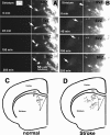
Figure 1.
Figure 1.
Time-lapse imaging of DiI-labeled cells in living brain coronal slices. The inset in A indicates coronal slices obtained at the striatal level of the coronal section. A, B, Arrows indicate migration of the same DiI-labeled SVZ cells over time from a representative nonstroke (A) or stroke (B) brain slice. DiI-labeled SVZ cells were localized to the SVZ at time 0 (A, B). In a nonstroke-derived brain slice, labeled cells within the SVZ migrated dorsally and ventrally (A, arrows; C), whereas labeled SVZ cells migrated laterally toward the striatum in a stroke-derived brain slice (B, arrows; D) during the 24 h experimental period. A line in A or B demarcates the striatal boundary of the SVZ. C, D, Migration paths of individual SVZ cells measured by tracking lines with different colors in nonstroke and stroke living brain slices, respectively. Each color in C and D represents one SVZ cell, and data are a summation of three individual observations from nonstroke (n = 3) and stroke (n = 3) rats. CC, Corpus callosum; LV, lateral ventricle.
Figure 2.
Figure 2.
Migration of SVZ cells out of neurospheres. A single neurosphere derived from nonstroke (A, B) or stroke (C, D) SVZ cells was placed in the Matrigel. After 24 h in the Matrigel, SVZ cells migrated out of the neurosphere (B, D; t = 24 h) compared with cells from time 0 (A, C; t = 0 h). More SVZ cells migrated out of the stroke-derived neurospheres (D) than the cells out of the nonstroke-derived neurospheres (B), and stroke-derived SVZ cells exhibited distant migration (D). Scale bars, 10 μm.
Figure 3.
Figure 3.
Division of migrating SVZ cells. A, B, An individual cell migrated out of the stroke neurosphere had leading (A, B, arrows) and tailing processes (B, arrowhead). C, D, This cell became round (C) and then divided into two daughter cells (D). E–G, A cell (E, arrow) within a process extended from the nonstroke neurosphere divided into two daughter cells (F, G, arrows). H, Distribution of total dividing cells during a 48 h experimental period, and each point represents a cell division. Scale bars, 10 μm.
Figure 4.
Figure 4.
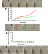
Time-lapse imaging of daughter cell migration. A, Fast migration of daughter cells from a representative stroke neurosphere. After cell division, one daughter cell had a bipolar shape (green arrow) and the other had a unipolar shape (red arrowhead). The unipolar daughter cell (red arrowhead) rapidly migrated away from the neurosphere. The bipolar daughter cell (green arrow) paused before resuming migration away from the neurosphere. B, Quantitative data of two daughter cell migrations, which were derived from the total of nonstroke- and stroke-derived daughter cells. Red and green lines represent unipolar and bipolar daughters, respectively. C, Stationary migration of daughter cells from a representative stroke neurosphere. Both daughter cells paused after division, then later migrated a very short distance. D, Quantitative data of two daughter cell migrations, which were derived from total of nonstroke- and stroke-derived daughter cells. Red and green lines represent each daughter cell marked in C. E, Reverse migration direction of daughter cells. The bipolar daughter cell (green arrow) reversed its polarity and migrated toward the neurosphere (N). Numbers in A, C, and E are minutes.
Figure 5.
Figure 5.
Dividing DCX-positive cells. A–H, In vitro morphology of daughter cells (A, C, E, G), which were mirrored by in vivo morphology of DCX-positive cells (B, D, F, H). I, Confocal images show that a DCX-positive cell (merged, green) in ischemic striatum was BrdU positive (merged, red). Z-stacks of DCX (green) and BrdU (red) immunoreactivity, at 1 μm intervals, are shown. Scale bars, 10 μm.

Similar articles

Cited by

References

    1. Arvidsson A, Collin T, Kirik D, Kokaia Z, Lindvall O. Neuronal replacement from endogenous precursors in the adult brain after stroke. Nat Med. 2002;8:963–970. - PubMed
    1. Chiasson BJ, Tropepe V, Morshead CM, van der Kooy D. Adult mammalian forebrain ependymal and subependymal cells demonstrate proliferative potential, but only subependymal cells have neural stem cell characteristics. J Neurosci. 1999;19:4462–4471. - PMC - PubMed
    1. Doetsch F, Garcia-Verdugo JM, Alvarez-Buylla A. Cellular composition and three-dimensional organization of the subventricular germinal zone in the adult mammalian brain. J Neurosci. 1997;17:5046–5061. - PMC - PubMed
    1. Edmondson JC, Hatten ME. Glial-guided granule neuron migration in vitro: a high-resolution time-lapse video microscopic study. J Neurosci. 1987;7:1928–1934. - PMC - PubMed
    1. Feng Y, Walsh CA. Protein-protein interactions, cytoskeletal regulation and neuronal migration. Nat Rev Neurosci. 2001;2:408–416. - PubMed

Publication types

MeSH terms

LinkOut - more resources