Comparative Study
. 2007 Mar 21;27(12):3230-43.
doi: 10.1523/JNEUROSCI.5265-06.2007.

Dlx-dependent and -independent regulation of olfactory bulb interneuron differentiation

Affiliations
Comparative Study

Dlx-dependent and -independent regulation of olfactory bulb interneuron differentiation

Jason E Long et al. J Neurosci. .

Abstract

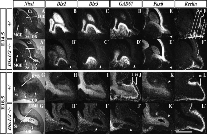
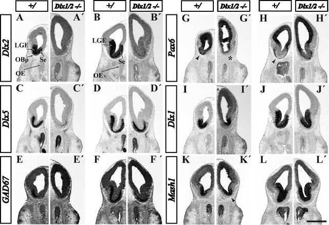
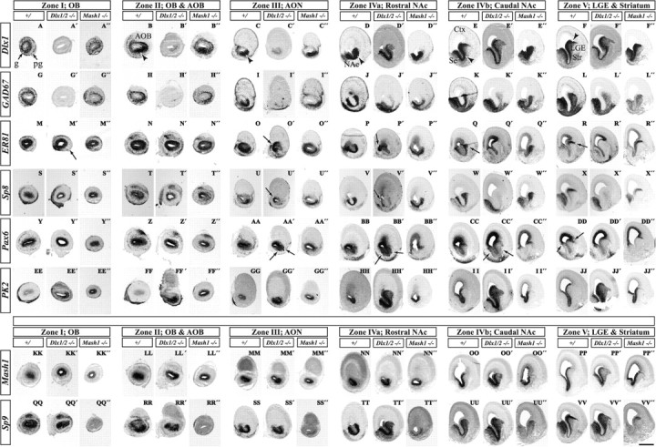
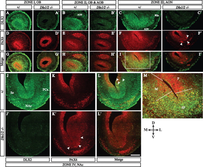
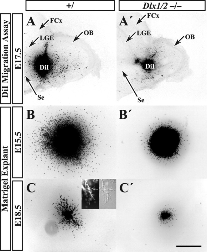
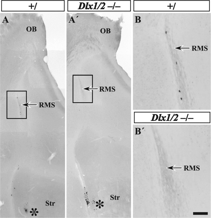
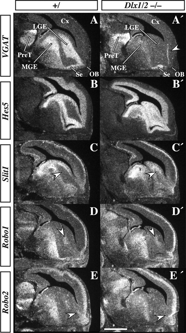
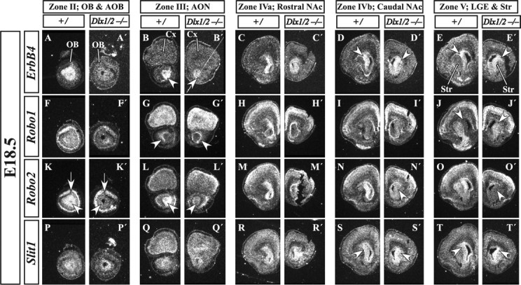
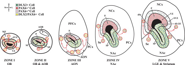
Olfactory bulb interneuron development is a complex multistep process that involves cell specification in the ventral telencephalon, tangential migration into the olfactory bulb, and local neuronal maturation. Although several transcription factors have been implicated in this process, how or when they act remains to be elucidated. Here we explore the mechanisms that result in olfactory bulb interneuron defects in Dlx1&2-/- (distal-less homeobox 1 and 2) and Mash1-/- (mammalian achaete-schute homolog 1) mutants. We provide evidence that Dlx1&2 and Mash1 regulate parallel molecular pathways that are required for the generation of these cells, thereby providing new insights into the mechanisms underlying olfactory bulb development. The analysis also defined distinct anatomical zones related to olfactory bulb development. Finally we show that Dlx1&2 are required for promoting tangential migration to the olfactory bulb, potentially via regulating the expression of ErbB4 (v-erb-a erythroblastic leukemia viral oncogene homolog 4), Robo2 (roundabout homolog 2), Slit1 (slit homolog 1), and PK2 (prokineticin 2), which have all been shown to play essential roles in this migration.


Figures

Figure 1.
Figure 1.
E14.5 and E18.5 Dlx1&2−/− mutant embryos show an enlarged OB ventricle and reduced OB gene expression. At E14.5, Nissl staining of sagittal sections reveals an enlarged OB ventricle in the Dlx1&2−/− mutants (A, A′), which is further expanded at E18.5 (G, G′). Radioactive in situ RNA hybridization analysis of Dlx2m (a Dlx2 transcript that is expressed in the Dlx1&2−/− mutant) shows a great reduction in the mutant OB at both E14.5 (arrowhead, B, B′) and E18.5 (arrowhead, H, H′). The same is true for Dlx5 (C, C′, I, I′) and GAD67 (D, D′, J, J′). Expression of Pax6 is reduced in the Dlx1&2−/− mantle zone of the OB at E14.5 (arrowhead, E, E′) and E18.5 (arrowhead, K, K′). An arrowhead points to the Pax6+ periglomerular layer (K, K′). Reelin expression (a marker of OB projection neurons) appears normal in Dlx1&2−/− mutants (F, F′, L, L′). Lines in F demarcate numbered coronal section planes (zones I–V) in Figures 3 and 8. Cx, Cortex; g, granule cell layer; m, mitral cell layer; OBv: olfactory bulb ventricle; pg, periglomerular cell layer; *RMS, defective rostral migratory stream. Scale bar: A–L′, 720 μm.
Figure 2.
Figure 2.
E12.5 Dlx1&2−/− mutant embryos show an early reduction of OB markers of differentiation despite maintenance of some early specification markers. Two coronal planes near the rostral pole of the telencephalon are shown. Hemisections (rostralmost in left panels) show nonradioactive in situ RNA hybridization analysis of Dlx2 in control (A, B) and Dlx1&2−/− mutant (A′, B′), Dlx5 in control (C, D) and Dlx1&2−/− mutant (C′, D′), GAD67 in control (E, F) and Dlx1&2−/− mutant (E′, F′), and Pax6 in control (G, H) and Dlx1&2−/− mutant (G′, H′) show a reduction in gene expression. Dlx1 in control (I, J) and Dlx1&2−/− mutant (I′, J′) and Mash1 in control (K, L) and Dlx1&2−/− mutant (K′, L′) embryos show maintenance of expression in the OB primordium. Arrowhead in I′, Increased Mash1 expression in the SVZ; arrowhead in K, L, arch of Pax6+ cells around rostral subpallium; star; loss of Pax6 expression in olfactory bulb primordium. OBp, Olfactory bulb primordium; OE, olfactory epithelium. Scale bar: A–L′, 1000 μm.
Figure 3.
Figure 3.
Nonradioactive in situ RNA hybridization analysis of control, Dlx1&2−/− mutant, and Mash1−/− mutant E15.5 embryos demonstrate Dlx1&2- and Mash1-dependent genes in OB development. Six coronal planes of the rostral telencephalon are shown; we conceptually organized the six planes into five zones (I–V) shown in Figure 1F (for an explanation of the five zones, see Results and Fig. 9; we subdivided zone IV into IVa and IVb). Hemisections show the expression of various genes in three types of mice: control (left column), Dlx1&2−/− mutant (middle column), and Mash1−/− mutant (right column) for each telencephalic zone. In general, the Mash1−/− mutation shows the opposite effect of the Dlx1&2−/− mutation. The following genes show decreased expression in the Dlx1&2−/− mutants (for details, see Results, because each gene shows variations on this general theme): Dlx1 (A–F), GAD67 (G–L), ER81 (M–R), Sp8 (S–X), and Pax6 (Y–DD). PK2 expression in the SVZ is elevated in the Dlx1&2−/− mutant and increased in the VZ of the Mash1−/− mutant (EE′–JJ′, EE″–JJ″). Mash1 expression is elevated in the subpallial SVZ of the Dlx1&2−/− mutants (KK′–PP′). Sp9 expression (QQ′–VV′) is reduced in the OB of both the Dlx1&2−/− and Mash1−/− mutants (QQ′, QQ″); more caudally, Sp9 expression is increased in the Dlx1&2−/− mutants and decreased in the Mash1−/− mutants (TT′–VV′, TT″–VV″). g, Granule cell layer; pg, periglomerular cell layer; Str, striatum. Scale bar: A–B″, G–H″, M–N″, S–T″, Y–Z″, EE–FF″, KK–LL″, QQ–RR″, 1000 μm; C–C″, I–I″, O–O″, U–U″, AA–AA″, GG–HH″, MM–NN″, SS–TT″, 750 μm; D–F″, J–L″, P–R″, V–X″, BB–DD″, II–JJ″, OO–PP″, UU–VV″, 500 μm.
Figure 4.
Figure 4.
PAX6 (red immunofluorescence) and DLX2 (green immunofluorescence) protein expression define zones and cell types related to the E15.5 olfactory bulb. Four coronal planes of the rostral telencephalon are shown; we conceptually organized these four planes into five zones (I–IV) shown in Figure 1F (for an explanation of these zones, see the Results and Fig. 9). In each zone, hemisections of each telencephalon show protein expression in the control (left column) and the Dlx1&2−/− mutant (right column) (except in zone IV: top is control and bottom is Dlx1&2−/− mutant). M is a higher-magnification view of the boxed region of zone III (I), showing the approximate boundaries between the pallial (P), mixed (M), and subpallial (SP) progenitor domains. Arrowheads in F′ and K′ show ectopic clusters of PAX6+ cells in the Dlx1&2−/− mutant. Scale bar: A–L′, 250 μm; M, 100 μm. PCx, Piriform cortex; D, dorsal, L, lateral; M, medial; V, ventral.
Figure 5.
Figure 5.
In vitro migration analysis of Dlx1&2−/− mutant embryos reveals reduced migratory capacity of the RMS. A DiI crystal was placed into the RMS of E17.5 Dlx1&2−/− mutant (A′) and control (A) embryos, and the sections were allowed to survive for 72 h. Immunofluorescence analysis (converted to black and white) shows that Dlx1&2−/− mutants have greatly reduced numbers of labeled cells migrating into the OB (A′). Small explants of approximately the same size of SVZ cells taken from the LGE of Dlx1&2−/− mutant and control embryos were placed into Matrigel and cultured for 72 h (B–C′). At E15.5, Dlx1&2−/− mutant explants lacked migration of Hoechst-stained cells away from the explant when compared with controls (C, C′). At E18.5, not only were the Dlx1&2−/− mutant cells unable to migrate away from the explant (D, D′), but they were also unable to form chains similar to controls (inset in D). FCx, Frontal cortex.. Scale bar: A, A′, 450 μm; B, B′, 100 μm; C, C′, 350 μm; inset, 50 μm.
Figure 6.
Figure 6.
In vivo transplantation of Dlx1&2−/− mutant cells into anterior SVZ of adult mice demonstrates a cell-autonomous defect in migration. CellTracker dye-labeled SVZ explants were taken from the striatal progenitor zone of E18.5 Dlx1&2−/− mutant and control embryos and injected into the SVZ of adult mice [adjacent to the rostral striatum (Str)] with survival for 6 d. Control cells integrated into and along the host RMS and migrated away from the injection site (asterisk) toward the OB (A). A higher-magnification view of the boxed area in A is provided in B. Dlx1&2−/− mutant cells were not found anywhere outside of a very short distance away from the injection site (asterisk, A′). A higher-magnification view of the boxed area in A′ is provided in B′. Scale bar: A, A′, 400 μm; B, B′, 100 μm.
Figure 7.
Figure 7.
Radioactive in situ RNA hybridization of VGAT, Hes5, Slit1, Robo1, and Robo2 in E14.5 parasagittal Dlx1&2−/− mutant and control embryos. Expression of VGAT marks a corridor from the LGE to the OB; this expression is nearly lost from the LGE and MGE of the Dlx1&2−/− mutant embryo but is maintained in part of the septum (Se; A, A′). Vertical stripe (arrowhead) in cortex of A′ is a tissue fold and is not VGAT expression. Hes5 expression, which is primarily expressed in the VZ, is increased in the SVZ of the LGE and MGE of the Dlx1&2−/− mutant embryo (B, B′). Slit1 is also ectopically expressed in the SVZ (C, C′). Expression of Robo1 in the LGE is slightly increased (compare arrowheads in D and D′), whereas Robo2 expression is lost from the rostral extension of the LGE near the OB (arrowhead in E, E′). Cx, Cortex; PreT, prethalamus. Scale bar: A–E′, 300 μm.
Figure 8.
Figure 8.
Radioactive in situ RNA hybridization in E18.5 coronal Dlx1&2−/− mutant embryos shows disruption of regulatory molecule expression. ErbB4 expression in the migrating immature OB interneurons is lost from the OB of the Dlx1&2−/− mutant embryos (A–B′). Additional caudal sections show an increase and expansion of ErbB4 expression in the striatum of Dlx1&2−/− mutant embryos (arrowheads in D–E′). Robo1 expression is increased in the SVZ of the striatum of the Dlx1&2−/− mutant embryo (arrowheads in J, J′). Robo2 expression is severely reduced in the LGE, RMS, and OB SVZ and granule cell layers (arrowheads in K′, L′, N′, O′), whereas its expression in the mitral cell (projection neuron) layer appears normal (arrows in K, K′). Slit1 expression is expanded in the SVZ and mantle of the striatum (arrowheads in S–T′). Cx, Cortex; Se, septum; Str, striatum. Scale bars: A–T′, 150 μm.
Figure 9.
Figure 9.
Schemas representing five zones of the embryonic telencephalon related to OB development. Five zones (I–V) have distinct anatomical and molecular characteristics. In each zone, only hemisections of each telencephalon is shown. The color code (top left) describes four types of DLX2 and PAX6 expression in individual nuclei. The olfactory bulb (zone I) appears to have a single progenitor domain consisting of a mixture of molecularly distinct cell types that can be identified by DLX2 and PAX6 expression. The caudal part of the OB (zone II) is defined by the presence of the AOB. Zone III corresponds to a region that includes the prefrontal cortex (PFCx) and AON. Zones IV and V are regions with prominent parts of the SE, NAc, and LGE. Unlike the OB, zones II–V have multiple progenitor domains and exhibit migrations emanating from the pallial/subpallial boundaries containing PAX6+, DLX2+, and PAX6+/DLX2+ cells. In zones II and III, PAX6 and DLX2 expression shows three types of VZ domains: pallial (P), subpallial (SP), and mixed (M) (the mixed domain is on the medial or septal side). The mixed domain most closely resembles the molecular profile of the OB VZ and forms at the medial intersection of pallial and subpallial progenitor domains. In zones IV and V, PAX6 and DLX2 labeling shows two types of VZ domains: pallial and subpallial. Zones III and IV are distinguished by the clear U-shape of the subpallial progenitor domains. We suggest that this SVZ domain corresponds to the embryonic rostral migratory stream. In Dlx1&2−/− mutants, differentiation of subpallial SVZ cells in domains II–V is defective, particularly in the embryonic RMS, resulting in loss of expression of several regulators of cell migration. NCx, Neocortex; PCx, piriform cortex; Se, septum.

Similar articles

Cited by

References

    1. Alcantara S, Ruiz M, D'Arcangelo G, Ezan F, de Lecea L, Curran T, Sotelo C, Soriano E. Regional and cellular patterns of reelin mRNA expression in the forebrain of the developing and adult mouse. J Neurosci. 1998;18:7779–7799. - PMC - PubMed
    1. Anchan RM, Drake DP, Haines CF, Gerwe EA, LaMantia AS. Disruption of local retinoid-mediated gene expression accompanies abnormal development in the mammalian olfactory pathway. J Comp Neurol. 1997;379:171–184. - PubMed
    1. Anderson SA, Eisenstat DD, Shi L, Rubenstein JL. Interneuron migration from basal forebrain to neocortex: dependence on Dlx genes. Science. 1997a;278:474–476. - PubMed
    1. Anderson SA, Qiu M, Bulfone A, Eisenstat DD, Meneses J, Pedersen R, Rubenstein JL. Mutations of the homeobox genes Dlx-1 and Dlx-2 disrupt the striatal subventricular zone and differentiation of late born striatal neurons. Neuron. 1997b;19:27–37. - PubMed
    1. Anderson SA, Marin O, Horn C, Jennings K, Rubenstein JL. Distinct cortical migrations from the medial and lateral ganglionic eminences. Development. 2001;128:353–363. - PubMed

Publication types

Substances

LinkOut - more resources