Comparative Study
. 2007 Mar 21;27(12):3260-7.
doi: 10.1523/JNEUROSCI.4033-06.2007.

Basic fibroblast growth factor modulates density of blood vessels and preserves tight junctions in organotypic cortical cultures of mice: a new in vitro model of the blood-brain barrier

Affiliations
Comparative Study

Basic fibroblast growth factor modulates density of blood vessels and preserves tight junctions in organotypic cortical cultures of mice: a new in vitro model of the blood-brain barrier

Kerstin Bendfeldt et al. J Neurosci. .

Abstract

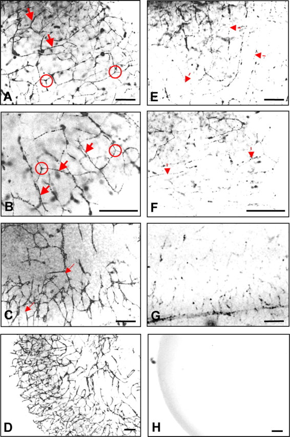
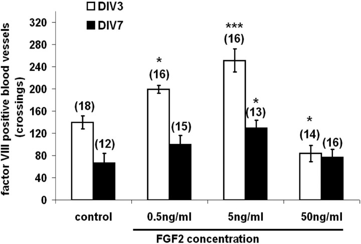
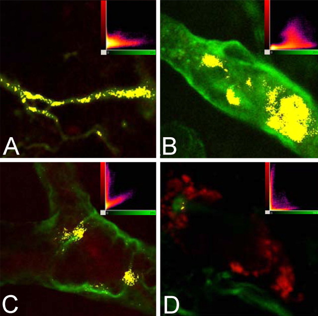
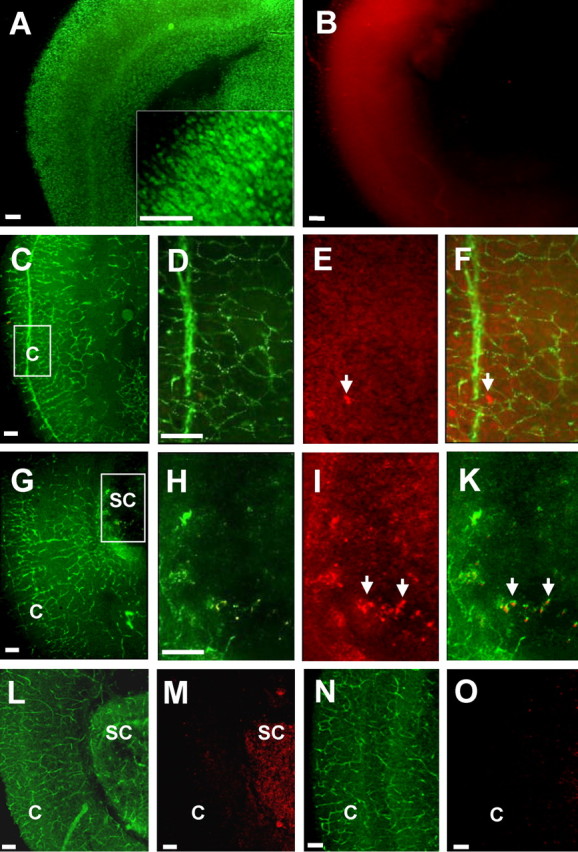
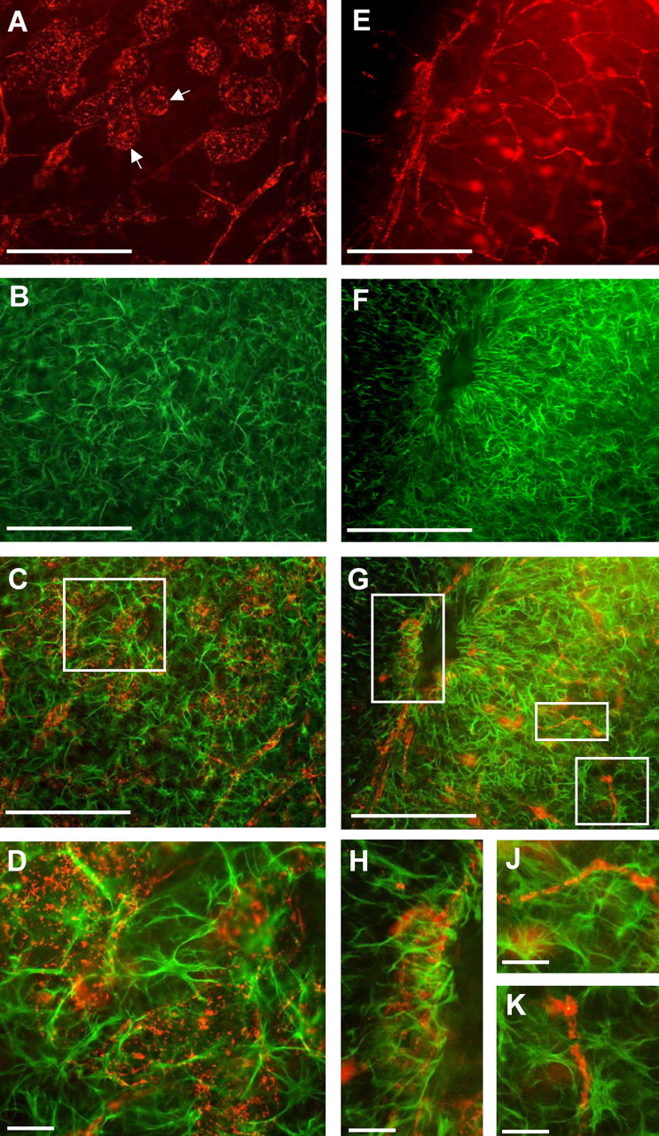
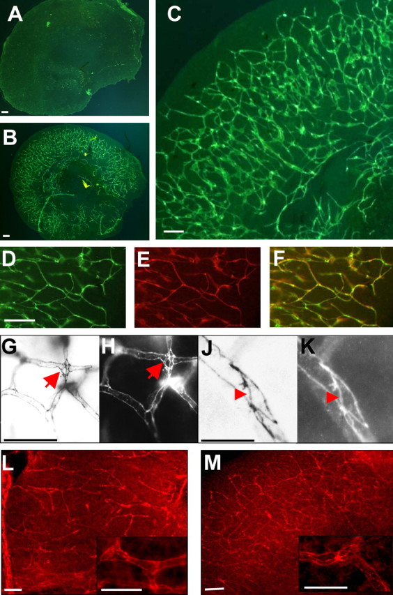
This study was performed to examine the maintenance of blood vessels in vitro in cortical organotypic slice cultures of mice with special emphasis on basic fibroblast growth factor (FGF-2), which is known to promote angiogenesis and to preserve the integrity of the blood-brain barrier. Slices of neonatal day 3 or 4 mouse brain were maintained for 3, 7, or 10 d in vitro (DIV) under standard culture conditions or in the presence of FGF-2. Immunohistochemistry for factor VIII-related antigen or laminin revealed a relative low number of blood vessels under standard conditions. In contrast, moderate FGF-2 concentrations increased the number of vessels: with 0.5 ng/ml FGF-2 it was 1.4-fold higher after DIV 3 or 1.5-fold after DIV 7 compared with controls; with 5 ng/ml it was almost doubled in both cases. With an excess of 50 ng/ml, FGF-2 vessels were reduced after DIV 3 or similar to controls after DIV 7. FGF receptor 1 was preferentially found on endothelial cells; its immunolabeling was reduced in the presence of the ligand. Cell death detected by an ethidium bromide analog or the apoptosis marker caspase-3 was barely detectable during the 10 d culture period. Immunolabeling of the tight junction proteins ZO-1 (zonula occludens protein 1), occludin, claudin-5, and claudin-3 revealed evidence for structural integrity of the blood-brain barrier in the presence of moderate FGF-2 concentrations. In conclusion, FGF-2 maintains blood vessels in vitro and preserves the composition of the tight junction. Hence, we propose FGF-2-treated organotypic cortical slices as a new tool for mechanistic studies of the blood-brain barrier.


Figures

Figure 1.
Figure 1.
Demonstration of microvessels in neonatal cortical slices under FGF-2 treatments. A–C, Immunofluorescence images labeled for FVIII-rAg revealed the presence of microvessels in COSCs after incubation with 5 ng/ml FGF-2 at DIV 3 (A), DIV 7 (B), and DIV 10 (C). Vessels appear as intact and frequently branching structures with continuous lines (arrows) and intersections (circles). D, Immunostaining for laminin also shows the complexity of the vascular network at DIV 10 after 5 ng/ml FGF-2. E, F, Control explants without FGF-2 displayed less labeling for FVIII-rAg at DIV 3 (E) and DIV 7 (F). Note discontinuous arrows marking few discontinuous vascular structures. G, In presence of 50 ng/ml FGF-2 at DIV 7 virtually no immunolabeling was observed. H, Control slice omitting the first antibody. Scale bars, 100 μm.
Figure 2.
Figure 2.
Quantification of vascular segments. Counting of microvessels revealed a relative low number of vessels in controls, which increased in number in a concentration dependent way up to 5 ng/ml FGF-2, independent from the duration of culture (e.g., ∼1.5-fold at 0.5 ng/ml and 2-fold at 5 ng/ml). Excessive amounts of 50 ng/ml FGF-2 were ineffective in preventing loss of vessels compared with controls. The absolute number of microvessels was halved at DIV 7 compared with DIV 3. Significances are given versus controls (*p ≤ 0.05; ***p ≤ 0.01). Error bars indicate SEM.
Figure 3.
Figure 3.
Localization of FGFR1. A–D, Colocalization of FGFR1 and the vascular marker laminin (A–C) or the astrocyte marker GFAP (D) is demonstrated by double immunofluorescence laser confocal microscopy. FGFR1 immunoreactivity is shown in red and laminin or GFAP immunoreactivity are shown in green in these images. Yellow indicates colocalization. Degree of colocalization is also indicated in the diagrams shown as insets in A–D. Laminin staining reveals the outline of typical capillaries (A) and larger microvessels (B, C) at DIV 7. FGFR1 staining localizes to subregions of the vessels; only little and weak expression of FGFR1 immunoreactivity is found outside of the vasculature. Note the more extensive expression of FGFR1 in controls (A, B) compared with FGF-2-treated slices (C). Virtually no colocalization of FGFR1 was seen with astrocytes stained for GFAP (D). All images were recorded with an apochromatic 100× NA 1.4 objective lens.
Figure 4.
Figure 4.
Survival of the slice culture. A, B, The organotypic cytoarchitecture of the cortex remained well preserved (A) and the overall survival of the tissue was superior in the presence of 5 ng/ml FGF-2 (B). A, Inset, NeuN labeling of neurons revealed well differentiated cells. B, Cell death as shown by EthD-1 staining was barely observed at DIV 7. C–K, Images of cortical slices labeled for FVIII-rAg antibody (green) and EthD-1 (red) at DIV 7 (C–F) and DIV 10 (G–K). C, Inset, D–F, Cell death of vascular endothelial cells was not observed in the presence of 5 ng/ml FGF-2 at DIV 7. Just a few dead cells occurred in the adjacent nervous tissue (E, arrows, F, overlay). G, Inset, H–K, At DIV 10, degeneration of the nervous tissue was apparent, but it occurred mainly in the center of the slices (I, arrows, K, overlay), indicating the degeneration of neurons and astrocytes rather than endothelial cells of cortical vessels. L–O, Apoptotic cell death as shown by caspase-3 staining (red) was nearly absent in the cortical regions of the cultures including cortical blood vessels (laminin staining, green) in the presence of 5 ng/ml FGF-2 at DIV 10. Apoptotic degeneration of the nervous tissue was observed sometimes in the center of the slices in the subcortical regions. c, Cortical; s.c., subcortical. Scale bars, 100 μm.
Figure 5.
Figure 5.
Astrocytes in untreated and FGF-2-treated slices. A–D, Untreated controls. E–K, FGF-2-treated slices (5 ng/ml, DIV 7). A, E, FVIII-rAg labeling of vascular structures (red). B, F, GFAP labeling of astrocytes (green). Insets in C and G are shown in D and H–K. A, In the absence of FGF-2, the disintegration of cerebral vessels is apparent (note arrows showing dilated vessels). E, In the presence of FGF-2, vascular structures appear intact. B, F, GFAP staining shows star-shaped astrocytes, which try to maintain their endfeet around the dilated vascular structures (C, inset, D). G, Insets, H–K, In the FGF-2-treated slices, the astrocytic endfeet are in close contact with vessels of different size and shape and surround the blood vessels in a regular manner. Scale bars: A–C, F, G, 100 μm; D, H–K, 25 μm.
Figure 6.
Figure 6.
Tight junctions of the BBB appear as intact structures. A, In the absence of FGF-2, the loss of tight junction proteins (here exemplarily, claudin-5 labeling) indicates the loss of the integrity of the BBB. B–M, Immunostaining revealed a complex network of tight junctions in the slices treated with moderate FGF-2 concentrations (here, 0.5 ng/ml) indicating the maintenance of the BBB. Note continuous large bands of claudin-5 (B–D), claudin-3 (G–K), ZO-1 (L), and occludin (M) delineating vascular walls, intersections between different vessels (G, H, arrows) and adjacent endothelial cells (J, K, arrowheads). E, Laminin labeling of vessels. F, Overlay of laminin and claudin-5. Scale bars, 100 μm.

Similar articles

Cited by

References

    1. Abbott NJ. Astrocyte-endothelial interactions and the blood–brain barrier permeability. J Anat. 2002;200:629–638. - PMC - PubMed
    1. Anderson JM, van Itallie CM. Tight junctions and the molecular basis for regulation of paracellular permeability. Am J Physiol. 1995;269:G467–G475. - PubMed
    1. Balda MS, Matter K. Tight junctions. J Cell Sci. 1998;111:541–547. - PubMed
    1. Bauer HC, Bauer H. The blood–brain barrier: Still an enigma? Cell Mol Neurobiol. 2000;20:13–29. - PMC - PubMed
    1. Brown RC, Mark KS, Egleton RD, Huber JD, Burroughs AR, Davis TP. Protection against hypoxia-induced increase in blood–brain barrier permeability: role of tight junction proteins and NFκB. J Cell Sci. 2003;116:693–700. - PubMed

Publication types

MeSH terms

Substances