Comparative Study
. 2007 Mar 21;27(12):3295-304.
doi: 10.1523/JNEUROSCI.4765-06.2007.

Amygdala gene expression correlates of social behavior in monkeys experiencing maternal separation

Affiliations
Comparative Study

Amygdala gene expression correlates of social behavior in monkeys experiencing maternal separation

Michael J Sabatini et al. J Neurosci. .

Abstract

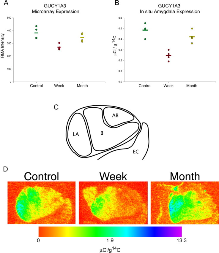
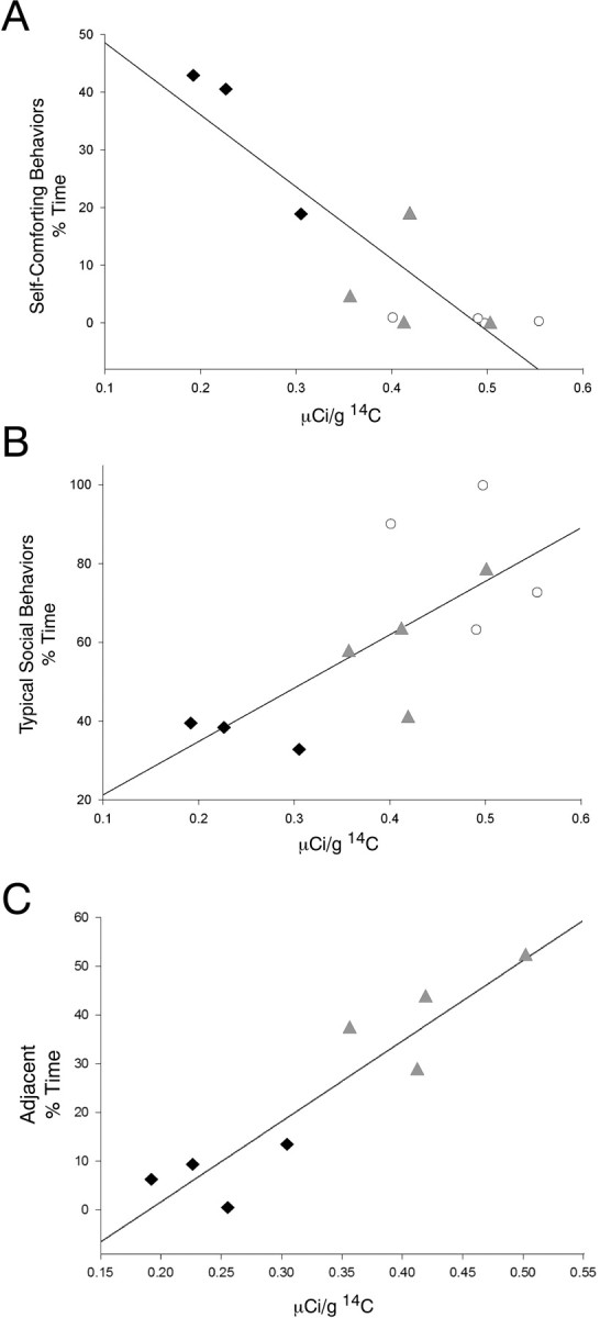
Children exposed to early parental loss from death or separation carry a greater risk for developing future psychiatric illnesses, such as major depression and anxiety. Monkeys experiencing maternal separation at 1 week of age show fewer social behaviors and an increase in self-comforting behaviors (e.g., thumb sucking) over development, whereas in contrast, monkeys experiencing maternal separation at 1 month of age show increased seeking of social comfort later in life. We sought to identify neural systems that may underlie these stress-induced behavioral changes by examining changes in mRNA content in amygdala tissue collected from 1 week separated, 1 month separated, and maternally reared infants at 3 months of age. mRNA from the right medial temporal lobe, primarily the amygdala, was analyzed using Affymetrix U133A 2.0 arrays. One gene, guanylate cyclase 1 alpha 3 (GUCY1A3), showed differential expression between the 1 week and maternally reared groups and the 1 week and 1 month groups; these changes were confirmed by in situ hybridization. The expression of this gene was positively correlated with acute social-comforting behavior (r = 0.923; p = 0.001) and longer-term close social behavior (r = 0.708; p = 0.015) and negatively correlated with self-comforting behaviors (r = -0.88; p < 0.001). Additional in situ hybridization studies of GUCY1A3 in normal monkeys showed that this gene is expressed at adult levels by 1 week of age and that its expression is greater in the amygdala than all other brain areas examined. We conclude that GUCY1A3 may contribute to the altered behavioral phenotypes that are differentially displayed depending on the age at which macaque infants experience an early-life stress.


Figures

Figure 1.
Figure 1.
GUCY1A3 mRNA expression. A, GUCY1A3 microarray gene expression level in the rostral medial temporal lobe. Circles represent the RMA normalized expression levels from each individual monkey; bars represent group mean. One week separated monkeys show significant decreases of 29.1% compared with controls and 22.0% compared with 1 month separated monkeys (p < 0.01). B, Quantification of radiographs of GUCY1A3 35S-labeled ribroprobe around the extent of the amygdala. Optical density levels were converted to known activities of 14C standards and expressed in microcuries per gram of 14C. Circles represent the average expression levels from three sections in each individual monkey; bars represent the group mean. Data were analyzed using ANOVA followed by least significant difference post hoc tests. Significant differences existed in comparisons of 1 week separated versus control monkeys and 1 week separated versus 1 month separated monkeys (p < 0.05). C, Graphic depiction of the region of the amygdala assessed in both the microarray and in situ hybridization studies. LA, Lateral nucleus of the amygdala; B, basal nucleus of the amygdala; AB, accessory basal nucleus of the amygdala; EC, entorhinal cortex. D, Representative pseudocolored image depicting regional amygdala expression in one animal per group expressed in a log scale. Each panel depicts the right amygdala at the coronal level immediately rostral to the lateral ventricle. Subnuclei evident in these panels from lateral (left) to medial (right) are as follows: lateral nucleus, basal nucleus, and accessory basal nucleus. Medial and adjacent to the amygdala is the periamygdalar cortex; inferomedial to the amygdala is the entorhinal cortex. Control, maternally raised; Week, 1 week separated; Month, 1 month separated. Color indicates the level of expression compared with 14C standard slides and is expressed in microcuries per gram of 14C.
Figure 2.
Figure 2.
Cellular and regional distribution of GUCY1A3. Representative photomicrograph from the lateral nucleus of the amygdala from one maternally raised control monkey. 35S-labeled riboprobes for GUCY1A3 appear as punctuate labeling of silver grains clustered around cell bodies counterstained with cresyl violet. The solid arrow depicts a typically labeled cell. The dashed arrow depicts a typical cell devoid of GUCY1A3 mRNA labeling. Scale bar, 10 μm.
Figure 3.
Figure 3.
Regional survey of GUCY1A3 expression in the monkey brain. Representative photomicrographs in a control monkey from: multiple coronal planes through forebrain (A–E) and two sagittal planes through the cerebellum (F). Labeled GUCY1A3 mRNA appears as graded levels of gray. Note the intense labeling in and around the amygdala, especially the lateral nucleus, and the periamygdalar cortex. Scale bar, 1 cm.
Figure 4.
Figure 4.
Anatomical expression of the development of GUCY1A3 mRNA expression. Images of representative scans of the radiographic films from one animal per age are as follows: A, 1 week; B, 4 weeks; C, 12 weeks; D, 63 weeks; and E, 176 weeks. Each image depicts the amygdala and surrounding cortical areas. Darker shades of gray represent high levels of GUCY1A3 mRNA expression, and lighter shades of gray represent low levels of GUCY1A3 mRNA expression. Intense labeling is present in the amygdala at all ages examined. F, Expression level in the amygdala was quantified from the radiographic OD and converted to known a known quantity of 14C standard. Each bar represents the average quantified GUCY1A3 mRNA level from three to six animals per age. Error bars represent 1 SD. There was no significant difference between any of the ages examined (F (4,15) = 0.937; p = 0.469). Scale bar, 10 mm.
Figure 5.
Figure 5.
Gene-behavior correlations. Black filled diamonds represent individual 1 week separated monkeys, gray filled triangles represent individual 1 month separated monkeys, and white open circles represent individual maternally reared monkeys. A, Level of self-comforting behavior during the third month of life is significantly correlated with GUCY1A3 mRNA levels in the amygdala (r = −0.878; p < 0.001). B, Level of typical social behavior during the third month of life is significantly correlated with GUCY1A3 mRNA levels in the amygdala (r = 0.708; p = 0.015). C, Level of social-comforting in the acute separation period is correlated with GUCY1A3 mRNA level at 3 months of age (r = 0.923; p = 0.001).

References

    1. Agid O, Shapira B, Zislin J, Ritsner M, Hanin B, Murad H, Troudart T, Bloch M, Heresco-Levy U, Lerer B. Environment and vulnerability to major psychiatric illness: a case control study of early parental loss in major depression, bipolar disorder and schizophrenia. Mol Psychiatry. 1999;4:163–172. - PubMed
    1. Ahern GP, Klyachko VA, Jackson MB. cGMP and S-nitrosylation: two routes for modulation of neuronal excitability by NO. Trends Neurosci. 2002;25:510–517. - PubMed
    1. Amaral DG. The amygdala, social behavior, and danger detection. Ann NY Acad Sci. 2003;1000:337–347. - PubMed
    1. Baron-Cohen S, Ring HA, Bullmore ET, Wheelwright S, Ashwin C, Williams SC. The amygdala theory of autism. Neurosci Biobehav Rev. 2000;24:355–364. - PubMed
    1. Bauman MD, Lavenex P, Mason WA, Capitanio JP, Amaral DG. The development of social behavior following neonatal amygdala lesions in rhesus monkeys. J Cogn Neurosci. 2004;16:1388–1411. - PubMed

Publication types

Substances

LinkOut - more resources