. 2007 Jun;18(6):1979-91.
doi: 10.1091/mbc.e06-12-1080. Epub 2007 Mar 21.

5-hydroxytryptamine 4 receptor activation of the extracellular signal-regulated kinase pathway depends on Src activation but not on G protein or beta-arrestin signaling

Affiliations

5-hydroxytryptamine 4 receptor activation of the extracellular signal-regulated kinase pathway depends on Src activation but not on G protein or beta-arrestin signaling

Gaël Barthet et al. Mol Biol Cell. 2007 Jun.

Abstract

The 5-hydroxytryptamine(4) (5-HT(4)) receptors have recently emerged as key modulators of learning, memory, and cognitive processes. In neurons, 5-hydroxytryptamine(4) receptors (5-HT(4)Rs) activate cAMP production and protein kinase A (PKA); however, nothing is known about their ability to activate another key signaling pathway involved in learning and memory: the extracellular signal-regulated kinase (ERK) pathway. Here, we show that 5-HT(4)R stimulation, in primary neurons, produced a potent but transient activation of the ERK pathway. Surprisingly, this activation was mostly PKA independent. Similarly, using pharmacological, genetic, and molecular tools, we observed that 5-HT(4)Rs in human embryonic kidney 293 cells, activated the ERK pathway in a G(s)/cAMP/PKA-independent manner. We also demonstrated that other classical G proteins (G(q)/G(i)/G(o)) and associated downstream messengers were not implicated in the 5-HT(4)R-activated ERK pathway. The 5-HT(4)R-mediated ERK activation seemed to be dependent on Src tyrosine kinase and yet totally independent of beta-arrestin. Immunocytofluorescence revealed that ERK activation by 5-HT(4)R was restrained to the plasma membrane, whereas p-Src colocalized with the receptor and carried on even after endocytosis. This phenomenon may result from a tight interaction between 5-HT(4)R and p-Src detected by coimmunoprecipitation. Finally, we confirmed that the main route by which 5-HT(4)Rs activate ERKs in neurons was Src dependent. Thus, in addition to classical cAMP/PKA signaling pathways, 5-HT(4)Rs may use ERK pathways to control memory process.


Figures
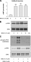
Figure 1.
Figure 1.
Stimulation of 5-HT4Rs coupled to Gαs in colliculi neurons leads to the activation of ERK, independently of PKA. (A) After 7 d of culture and differentiation, colliculi neurons were incubated in a medium containing 1 mM isobutylmethylxanthine (a phosphodiesterase inhibitor) and 10 μM BIMU8. cAMP accumulation was measured for 2, 5, and 15 min of stimulation at 37°C as indicated in Materials and Methods and expressed as picomoles per well. Results are the mean ± SEM of four independent experiments. **p < 0.01, significantly different from the corresponding cells before BIMU8 treatment. (B) Colliculi neurons were stimulated with 10 μM BIMU8 for the same periods as mentioned in A, and then they were lysed. The phosphorylation state of ERK1/2 of whole cell extract was analyzed by immunoblotting with the antibody to p-ERK1/2. Membranes were then stripped and analyzed with antibody to total ERK. The Western blot shown is representative of five independent experiments. (C) Inhibition of PKA does not lead to the inhibition of p-ERK1/2 in colliculi neurons. Colliculi neurons expressing endogenous 5-HT4R were pretreated with vehicle or 3 μM CMIQ for 30 min before treatment with 10 μM BIMU8 for 5 min. Cell lysates were successively analyzed by immunoblotting with the antibodies to phospho-PKA substrates, to p-ERK1/2, and to total ERK as described in B. The Western blot shown is representative of three independent experiments.
Figure 2.
Figure 2.
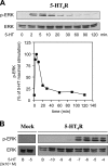
Time course of agonist-stimulated ERK1/2 phosphorylation. (A) HEK293 cells were transfected with 100 ng of 5-HT4R. Twenty-four hours later, the cells were starved in serum-free medium overnight and stimulated with 10 μM 5-HT for the indicated periods at 37°C. Phosphorylated (p-ERK1/2) and total ERK1/2 of the same cell lysate were determined by immunoblotting with the antibody to p-ERK1/2 or to total ERK. p-ERK1/2 bands were quantified by densitometry and normalized to ERK level and expressed as a percentage of the maximal p-ERK1/2 (obtained at 5 min). Data from four independent experiments are represented as the mean ± SEM and plotted in the graph below. (B) HEK293 cells expressing the 5-HT4R were starved in serum-free medium overnight and stimulated with increasing concentrations of 5-HT (3 × 10⁻¹⁰ – 3× 10⁻⁴ M). Representative blots of p-ERK1/2 and ERK are shown in A and B.
Figure 3.
Figure 3.
5-HT-stimulated second messengers (cAMP and IP) and phosphorylation of ERK1/2 in HEK293 cells. Comparison between the effects of 5-HT4R WT and defective mutants. HEK293 cells transiently expressing either 5-HT4R WT or mutants (D66N or W272A) were treated with increasing concentrations of 5-HT for cAMP production (5 min), IP production (30 min), and p-ERK1/2 (5 min). Receptor expression levels were similar for all the receptors as revealed by enzyme-linked immunosorbent assay that allowed quantifying relative cell surface receptor expression before agonist stimulation (data not shown). Basal and 5-HT–stimulated cAMP values were 1 ± 0.5 and 9 ± 2 pmol/well, respectively. Basal and 5-HT-stimulated IP values were 0.15 ± 0.5 and 10 ± 3 pmol/well, respectively. For WT, data are expressed as a percentage of the maximum second messenger (cAMP or IP) or p-ERK produced after stimulation with 5-HT 10 μM for 5 min (cAMP and p-ERK) or for 30 min (IP) (top). For mutants, data are expressed as a percentage of the maximum cAMP, IP, or p-ERK1/2 produced by the WT stimulated with 10 μM 5-HT, as indicated above. Analysis of p-ERK1/2 and total ERK content was performed by immunoblotting and quantified by densitometry. The amounts of p-ERK1/2 were always normalized to the total ERK signal obtained by stripping the same immunoblot and determined with the antibody to ERK42/44. Curve fitting was performed with the GraphPad Prism software. For all the curves, the data are the mean ± SEM of at least four independent experiments.
Figure 4.
Figure 4.
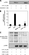
cAMP and downstream effector PKA do not participate in 5-HT4R–mediated ERK activation. (A) Inhibition of cAMP formation does not prevent ERK1/2 activation. HEK293 cells expressing 100 ng of 5-HT4R WT plasmid were serum starved overnight and exposed or not to 1 mM SQ 22536 for 30 min (an inhibitor of cAMP accumulation) before the stimulation by 10 μM 5-HT for 5 min. The cell lysates were analyzed by immunoblotting with antibody to p-ERK1/2. A representative immunoblot is shown. (B) In parallel, HEK293 cells coming from the same transfection as described in A for each condition were seeded into 24-well plates, stimulated with 10 μM 5-HT plus Ro-20-1724 (a phosphodiesterase inhibitor), and lysed in HBS plus Triton 0.1% as indicated in Materials and Methods. cAMP levels accumulated during the stimulation period of 5 min are expressed as the percentage of maximum cAMP response to 10 μM 5-HT, and they are reported in the absence or presence of pretreatment with SQ 22536. Values are the mean ± SEM of four independent experiments. **p < 0.01, significantly different from the corresponding cells stimulated with 5-HT before SQ 22536 treatment. (C) ERK phosphorylation does not depend on PKA activation. HEK293 cells expressing 5-HT4R were pretreated with either vehicle or 3 μM CMIQ or 1 μM H-89 for 30 min before treatment with 10 μM 5-HT for 5 min. Cell lysates were analyzed by immunoblotting successively with the antibodies to phospho-PKA substrates and to p-ERK1/2. The Western blot shown is representative of three independent experiments.
Figure 5.
Figure 5.
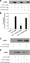
5-HT4R-mediated activation of p-ERK1/2 is independent of PLC, Gi/Go, and EGF receptors. (A) 5-HT4R–mediated ERK activation does not require PLC activation. Serum-starved HEK293 cells expressing 5-HT4R were pretreated with 10 μM U 73122, a PLC inhibitor, or by its inactive analogue U 73343 for 30 min before stimulation with 10 μM 5-HT for 5 min. Total lysates were analyzed by immunoblotting with antibody to p-ERK1/2. (B) In parallel, HEK293 cells coming from the same transfection as described in A for each condition were seeded into 24-well plates, pretreated by LiCl 10 min before a 30-min stimulation by 10 μM 5-HT. Quantification of IP production was performed by HTRF by using the IP-One assay as described in Materials and Methods. IP levels accumulated during the stimulation time are expressed as the percentage of maximum IP response to 10 μM 5-HT, and they are reported in the absence or presence of pretreatment with U 73122 and U 73343. Values are the mean ± SEM of four independent experiments. **p < 0.01, significantly different from the corresponding cells before U 73122-treatment. (C) 5-HT4R–mediated ERK activation does not require G protein PTX sensitive (Gi/Go). HEK293 cells expressing 5-HT4R were pretreated with 100 ng/ml PTX overnight before treatment with 10 μM 5-HT for 5 min. Total lysates were analyzed by immunoblotting with antibody to p-ERK1/2. (D) 5-HT4R–mediated ERK activation does not require transactivation of EGF-R tyrosine kinase. HEK293 cells expressing 5-HT4R were pretreated with 250 nM tyrphostin/AG1478 for 30 min before stimulation with 10 μM 5-HT. Total lysates were analyzed by immunoblotting with antibody to p-ERK 1/2. A representative blot of each experiment is shown in A, C, and D.
Figure 6.
Figure 6.
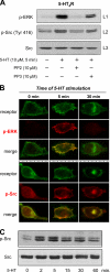
5-HT4R–mediated ERK1/2 activation depends on Src tyrosine kinase activation. (A) Stimulation of 5-HT4R activates Src. Inhibition of Src prevents ERK activation. Serum-starved HEK293 cells expressing 5-HT4R were pretreated with the Src kinase inhibitor PP2 at 10 μM or with the inactive analogue PP3 at 10 μM 30 min before a 5-min stimulation with 10 μM 5-HT. The cell lysates were analyzed by immunoblotting. The blots were probed sequentially to detect p-ERK1/2 with antibody against p-ERK1/2 (line 1), active Src with antibody to p-Src (Tyr 416) (line 2), and total Src with Pan antibody to Src inactive (line 3). (B) Colocalization of p-ERK1/2 and p-Src (Tyr 416) after stimulation of 5-HT4R WT. HEK293 cells transfected with 500 ng of Rho-tagged 5-HT4R were seeded onto coverslips. Twenty-four hours after transfection, cells were serum starved overnight. Cells were incubated 90 min with antibody against Rho-tagged 5-HT4R at 4°C before a 5- or 30-min stimulation with 10 μM 5-HT. After fixation and permeabilization, cells were sequentially incubated with primary antibody against p-ERK1/2 or p-Src (Tyr 416) and with fluorochrome-labeled secondary antibody. Fluorescence microscopy was then used to visualize the distribution of antibody-labeled receptors (green channel) and the appearance of phosphorylated form of ERK (p-ERK1/2) and Src (p-Src) (red channel). Immunofluorescence microscopy was performed using a Zeiss Axiophot2 microscope with Zeiss 63× NA 1.4 oil immersion lenses. Representative images from several independent experiments are shown. Top, distribution of Rho-tagged 5-HT4R before (basal) or after 5- and 30-min addition of 10 μM 5-HT to the culture medium at 37°C. The left column shows the distribution of the receptor and the absence of p-ERK1/2 before activation. An increase in phosphorylation state of ERK1/2 at the plasma membrane is visualized after 5-min treatment with 5-HT. Merged images were magnified to show colocalization of 5-HT4R with p-ERK1/2 at the plasma membrane after 5 min of stimulation. Bottom, phosphorylation state of p-Src (Tyr 416) before and after 5-HT4R stimulation. Phosphorylation state and colocalization with 5-HT4R is longer for p-Src than for p-ERK1/2. (C) HEK293 cells were transfected with 5-HT4R. Twenty-four hours later, the cells were starved in serum-free medium overnight and stimulated with 10 μM 5-HT for the indicated periods at 37°C. Phosphorylated Src (Tyr 416) of all the cell lysates was determined by immunoblotting with the antibody to active p-Src (Tyr 416) and to total Src.
Figure 7.
Figure 7.
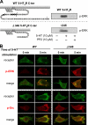
5-HT4R–mediated ERK phosphorylation through Src activation does not involve the C-terminal domain of 5-HT4R. (A) The C terminus is not implicated in Src-dependent 5-HT4R–mediated ERK activation. Topology of the C-terminal domain of 5-HT4R WT and Δ346 is represented on the left. Putative Ser/Thr phosphate acceptor sites are represented by black circles. HEK293 cells were transfected with 100 ng of Rho-tagged WT and Δ346 5-HT4R. Serum-starved HEK293 cells expressing the WT and the mutant were pretreated or not with the Src kinase inhibitor PP2 at 10 μM for 30 min before the stimulation with 10 μM 5-HT for 5 min. Whole cell lysates were prepared and analyzed by immunoblotting with antibody to p-ERK1/2. A representative blot of each experiment is shown. Both WT and truncated mutant Δ346 phosphorylate ERK1/2 in the same manner, dependent on Src kinase. (B) Colocalization of p-ERK1/2 and active p-Src after WT and Δ346 stimulation. HEK293 transfected with 500 ng of Rho-tagged 5-HT4R or Rho-tagged Δ346 were seeded onto coverslips. Twenty-four hours after transfection, cells were serum starved overnight. Cells were incubated 90 min with antibody against Rho-tagged 5-HT4R at 4°C before a 5- or 30-min stimulation period with 10 μM 5-HT. After fixation and permeabilization, the cells were sequentially incubated with primary antibody against p-ERK1/2 or p-Src (Tyr 416) and with fluorochrome-labeled secondary antibody. Fluorescence microscopy was then used to visualize distribution of antibody-labeled receptors (green channel) and appearance of the phosphorylated form of active ERK1/2 and active Src (red channel). Immunofluorescence microscopy was performed using a Zeiss Axiophot2 microscope with Zeiss 63× NA 1.4 oil immersion lenses. Representative images from several independent experiments are shown. Top, distribution of Rho-tagged 5-HT4R or Rho-tagged Δ346 before (basal) or after 5-min treatment with 10 μM 5-HT at 37°C. An increase in phosphorylation of p-ERK1/2 is visualized at the plasma membrane after 5-min stimulation with 5-HT of both WT and Δ346. Merged images were magnified to show colocalization of both receptors (WT and Δ346) with p-ERK1/2 at the plasma membrane after stimulation. Bottom, phosphorylation state of active Src before (basal) and after (5 min) 5-HT4R stimulation. Phosphorylation states were enhanced after stimulation with 5-HT and localization of p-ERK and p-Src do not differ between WT and Δ346 after 5-min stimulation.
Figure 8.
Figure 8.
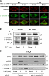
Arrestins are not implicated in Src-dependent 5-HT4R–mediated ERK1/2 activation. (A) Δ346 does not promote β-arrestin 2 redistribution to the plasma membrane after stimulation. HEK293 transfected with 500 ng of Rho-tagged 5-HT4R or Rho-tagged Δ346 were seeded onto coverslips. Twenty-four hours after transfection, cells were serum starved overnight. Cells were incubated 90 min with antibody against Rho-tagged 5-HT4R at 4°C before a 5-min stimulation with 10 μM 5-HT. After fixation and permeabilization, cells were incubated with Alexa Fluor 594-labeled secondary antibody. Fluorescence microscopy was then used to visualize the distribution of antibody-labeled receptors (red channel) and the redistribution of YFP-β-arrestin 2 (green channel). Immunofluorescence microscopy was performed using a Zeiss Axiophot2 microscope with Zeiss 63× NA 1.4 oil immersion lenses. Representative images from several independent experiments are shown. Left, distribution of Rho-tagged WT and β-arrestin 2-YFP. WT-5-HT4R is expressed at the plasma membrane under basal conditions, whereas β-arrestin 2-YFP is homogenously present in the cytosol. After 5-min stimulation with 10 μM 5-HT at 37°C, β-arrestin 2-YFP is redistributed to the plasma membrane where the stimulated 5-HT4Rs are localized. Merged images were magnified to show colocalization of wt 5-HT4R with β-arrestin 2-YFP at the plasma membrane. Right, distribution of Rho-tagged Δ346 and β-arrestin 2-YFP. Δ346, initially characterized to lack the ability to traffic in endosomes with β-arrestin 2 after a long-term stimulation, also lack the ability to promote the translocation of β-arrestin to the plasma membrane after a 5-min period of stimulation. (B) Δ346 activates Src despite its inability to interact with β-arrestins. Cells transfected with Rho-tagged 5-HT4R WT or Rho-tagged Δ346 (800 ng) were incubated with 10 μM 5-HT for 5 min. 5-HT4Rs were purified from proteins extracts by immunoprecipitation by using anti-Rho-tag antibody. Coprecipitated proteins (p-Src and β-arrestins 1 and 2) were analyzed by Western blotting by using the antibody to p-Src (Tyr 416) (line 1) and the antibody to β-arrestins 1 and 2 (line 2). The presence of the receptor was revealed by using the antibody anti-Rho-tag (line 3). (C) Down-regulation of β-arrestin expression by siRNA does not inhibit ERK phosphorylation mediated by 5-HT4R. Early passage HEK293 were transfected with 5-HT4R and with control or β-arrestin 1- and 2-specific siRNA. Seventy-two hours after transfection, cells were stimulated 5 or 30 min before lysis. p-Src, p-ERK, and β-arrestin contents were analyzed by Western blotting. The same blot was probed and stripped, sequentially, for p-Src (Tyr 416) and p-ERK1/2 (lines 1 and 2) and β-arrestins (lines 3 and 4). A representative illustration of three experiments is shown in A–C.
Figure 9.
Figure 9.
Src-dependent 5-HT4R–mediated ERK1/2 activation also occurs in neurons. (A) Colliculi neurons endogenously expressing 5-HT4R were stimulated with vehicle or BIMU8 (a 5-HT4R agonist). Total neuron lysates were analyzed by immunoblotting with the indicated antibodies. The same immunoblot was probed sequentially for p-Src with antibody to active p-Src (Tyr 416), for total inactive Src with antibody against pan Src, for p-ERK1/2 with the antibody to activate ERK1/2, and for total ERK1/2 with the antibody to ERK42/44. Quantification of p-Src and p-ERK1/2 was performed by densitometric analysis using NIH Image software. Data are means ± SEM of results obtained in four independent experiments. *p < 0.05 versus corresponding values measured in untreated neurons. (B) Colliculi neurons were pretreated with the Src kinase inhibitors PP2 at 10 μM for 30 min before the stimulation with 10 μM BIMU8 for 5 min. Neuron lysates were analyzed by immunoblotting with antibodies against p-Src and p-ERK1/2 as indicated in A. (C) 5-HT4R–mediated ERK activation does not require transactivation of EGF-R tyrosine kinase. Colliculi neurons were pretreated with 250 nM tyrphostin/AG1478 for 30 min before stimulation with 10 μM BIMU8 or 10 ng/ml EGF during 5 min. Total lysates were analyzed by immunoblotting with antibody to p-ERK1/2. (D) 5-HT4R–mediated ERK activation does not require G protein PTX sensitive (Gi/Go). Colliculi were pretreated with PTX at 50 or 100 ng/ml overnight before treatment with 10 μM BIMU8 for 5 min. Total lysates were analyzed by immunoblotting with antibody to p-ERK1/2. A representative blot of each experiment is shown in A–D.

Similar articles

Cited by

References

    1. Adamus G., Arendt A., Hargrave P. A. Genetic control of antibody response to bovine rhodopsin in mice: epitope mapping of rhodopsin structure. J. Neuroimmunol. 1991;34:89–97. - PubMed
    1. Bain J., McLauchlan H., Elliott M., Cohen P. The specificities of protein kinase inhibitors: an update. Biochem. J. 2003;371:199–204. - PMC - PubMed
    1. Banasr M., Hery M., Printemps R., Daszuta A. Serotonin-induced increases in adult cell proliferation and neurogenesis are mediated through different and common 5-HT receptor subtypes in the dentate gyrus and the subventricular zone. Neuropsychopharmacology. 2004;29:450–460. - PubMed
    1. Baneres J. L., Mesnier D., Martin A., Joubert L., Dumuis A., Bockaert J. Molecular characterization of a purified 5-HT4 receptor: a structural basis for drug efficacy. J. Biol. Chem. 2005;280:20253–20260. - PubMed
    1. Barthet G., Gaven F., Framery B., Shinjo K., Nakamura T., Claeysen S., Bockaert J., Dumuis A. Uncoupling and endocytosis of 5-hydroxytryptamine 4 receptors. Distinct molecular events with different GRK2 requirements. J. Biol. Chem. 2005;280:27924–27934. - PubMed

Publication types

MeSH terms

LinkOut - more resources