. 2007 Jun;18(6):2072-80.
doi: 10.1091/mbc.e06-10-0968. Epub 2007 Mar 21.

Multiple translational isoforms give functional specificity to serum- and glucocorticoid-induced kinase 1

Affiliations

Multiple translational isoforms give functional specificity to serum- and glucocorticoid-induced kinase 1

Maria Francisca Arteaga et al. Mol Biol Cell. 2007 Jun.

Abstract

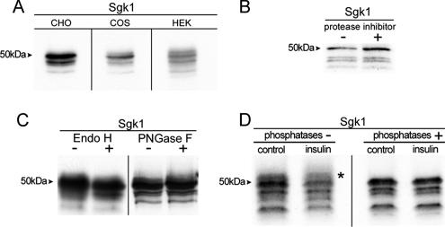
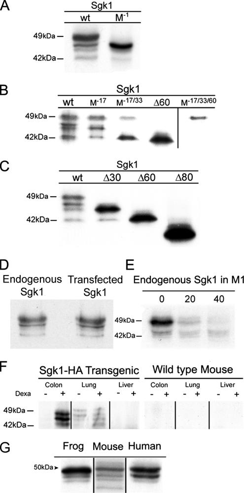
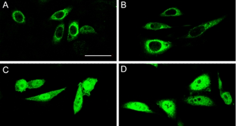
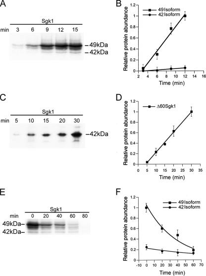
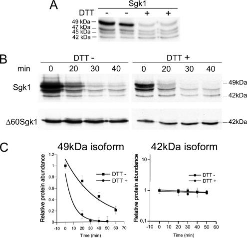
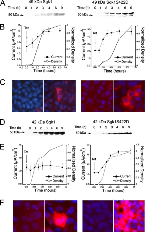
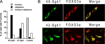
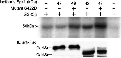
Serum- and glucocorticoid-induced kinase 1 is a ubiquitous kinase that regulates diverse processes such as ion transport and cell survival. We report that a single SGK1 mRNA produces isoforms with different N-termini owing to alternative translation initiation. The long isoforms, 49 and 47 kDa, are the most abundant, localize to the ER membrane, exhibit rapid turnover, their expression is decreased by ER stress, activate the epithelial sodium channel (ENaC) and translocate FoxO3a transcriptional factors from the nucleus to the cytoplasm. The short isoforms, 45 and 42 kDa, localize to the cytoplasm and nucleus, exhibit long half-life and phosphorylate glycogen synthase kinase-3beta. The data indicate that activation of Sgk1 in different cellular compartments is key to providing functional specificity to Sgk1 signaling pathways. We conclude that the distinct properties and functional specialization of Sgk1 given by the N-terminus confer versatility of function while maintaining the same core kinase domain.


Figures

Figure 1.
Figure 1.
Immunoprecipitation of Sgk1 from transfected CHO labeled with [35S]methionine/cysteine resolved on 7.5% SDS-PAGE gels. (A) Mouse Sgk1 cDNA transfected in various mammalian cell lines. (B) Cell lysates ± protease inhibitors. (C) IPs were treated with or without glycanases. (D) Control or cells pretreated with insulin were lysed and IP products were exposed to ± phosphatases. Arrowhead indicates molecular weights. Asterisk indicates a high-molecular-weight band removed by phosphatases.
Figure 2.
Figure 2.
Alternative initiation of translation produces Sgk1 isoforms in cells and mouse tissues and other vertebrates. (A) Immunoprecipitation (IP) of Sgk1 wild type and with deletion of the first Met (M−1). (B) IP of Sgk1 with mutations on Met17 (M−17), double (M−17/33), and triple mutant (M−17/33/60). (C) Similar experiments in cells transfected with Sgk1-truncated forms Δ33, Δ60, and Δ80. (D) Comparison of transfected and endogenous Sgk1 from M1 cells pretreated with 50 mM dexamethasone for 8 h. (E) Pulse-chase experiment of endogenous Sgk1 in M1 cells illustrating faster degradation of the 49 than the 42-kDa isoform. (F) IP of tissue homogenates from transgenic Sgk1-HA and wild-type mice ± pretreatment with dexamethasone using a polyclonal anti-HA followed by IB with monoclonal HA-HRP. (G) IP of frog, mouse, and human Sgk1 from transfected cell labeled with [35S]methionine/cysteine.
Figure 3.
Figure 3.
Subcellular localization of Sgk1 isoforms. Immunofluorescence of CHO cells transfected with the four Sgk1 isoforms: (A) 49, (B) 47, (C) 45 and (D) 42 kDa. Sgk1 proteins were detected with anti-HA monoclonal conjugated with fluorescein isothiocyanate on a confocal macroscope. Bar, 15 μm.
Figure 4.
Figure 4.
Rates of synthesis and degradation of 49- and 42-kDa Sgk1 isoforms. (A and B) Differential rate of synthesis of 49- and 42-kDa isoforms estimated by cumulative incorporation of [35S]methionine using Sgk1 full length. (C and D) Rate of synthesis of 42-kDa isoforms estimated with Δ60Sgk1 construct. (E and F) Differential rates of degradation of 49 and 42 isoforms estimated by pulse-chase experiments with [35S]methionine. Data points in graphs represent average of four independent experiments ± SD.
Figure 5.
Figure 5.
Effect of ER stress on expression of Sgk1 isoforms. (A) Steady state level of expression of Sgk1 isoforms in cells treated ± DTT. (B) Representative pulse-chase experiment of Sgk1 full-length and Δ60Sgk1 construct ± DTT. (C) Rate of degradation of 49- and 42-kDa isoforms ± DTT; n = 3.
Figure 6.
Figure 6.
Functional effects of 49- and 42-kDa Sgk1 isoforms on ENaC current. Clones of A6 cells expressing 49- or 42-kDa Sgk1 isoforms ± activating mutation S422D under control of tetracycline. (A and D) Time course of Sgk1 expression assessed by Western blotting at the indicated times. (B and E) Time course of amiloride-sensitive Isc after induction with tetracycline at time 0 (●) and quantification by densitometry of western blots at the corresponding time points (○). (C and F) Immunofluorescence of each clone grown of Transwells ± tetracycline with anti-HA antibody.
Figure 7.
Figure 7.
Effects of 49- and 42-kDa Sgk1 isoforms on Foxo3a translocation. (A) Cells transfected with FoxO3a-V5 alone or with Sgk1 49 or 42 isoforms were examined with anti-V5 antibody. Serum was removed 16 h before experiments. Quantification of FoxO3a localization in nucleus (hatched), cytoplasm (black), and nucleus+cytoplasm (white) in 100–150 cells. (B) Identification of Sgk1 isoforms (green) with anti-FLAG and FoxO3a (red) with anti-V5.
Figure 8.
Figure 8.
Representative autoradiography of immunoprecipitated GSK3β-HA after phosphorylation in cells cotransfected with empty vector or the Sgk1-FLAG isoforms indicated in the top panel. Immunoblot of expression levels of Sgk1 isoforms in the same experiments.

Similar articles

Cited by

References

    1. Alvarez de la Rosa D., Canessa C. M. Role of SGK in hormonal regulation of epithelial sodium channel in A6 cells. Am. J. Physiol. 2003;284:C404–C414. - PubMed
    1. Alvarez de la Rosa D., Coric T., Todorovic N., Shao D., Wang T., Canessa C. M. Distribution and regulation of expression of serum- and glucocorticoid-induced kinase-1 in the rat kidney. J. Physiol. 2003;551:455–466. - PMC - PubMed
    1. Arteaga M. F., Wang L., Ravid T., Hochstrasser M., Canessa C. M. An amphipathic helix targets serum and glucocorticoid-induced kinase 1 to the endoplasmic reticulum-associated ubiquitin-conjugation machinery. Proc. Natl. Acad. Sci. USA. 2006;103:11178–11183. - PMC - PubMed
    1. Arteaga M. F., Canessa C. M. Functional specificity of Sgk1 and Akt1 on ENaC activity. Am. J. Physiol. 2005;289:F90–F96. - PubMed
    1. Belova L., Sharma S., Brickley D. R., Nicolarsen J. R., Patterson C., Conzen S. D. Ubiquitin/proteasome degradation of serum and glucocorticoid-regulated kinase-1 (SGK-1) is mediated by the chaperone-dependent E3 ligase CHIP. Biochem. J. 2006 [Epub ahead of print] PMID: 16895519. - PMC - PubMed

Publication types

MeSH terms

LinkOut - more resources