

Regulation of cell diameter, For3p localization, and cell symmetry by fission yeast Rho-GAP Rga4p
- PMID: 17377067
- PMCID: PMC1877093
- DOI: 10.1091/mbc.e06-09-0883
Regulation of cell diameter, For3p localization, and cell symmetry by fission yeast Rho-GAP Rga4p
Abstract
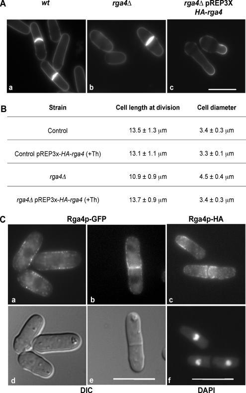
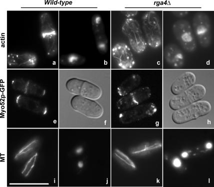
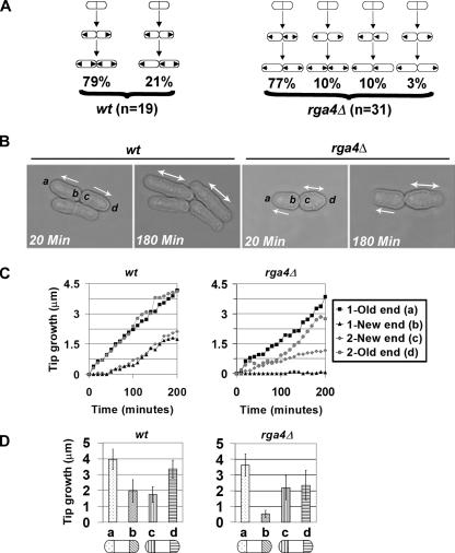
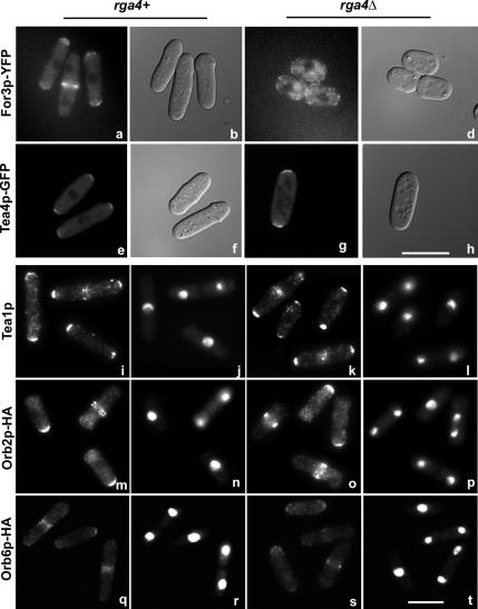
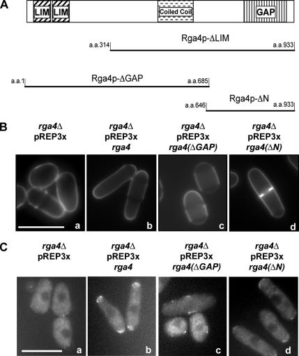
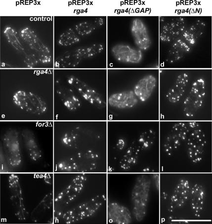
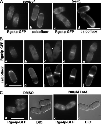
Control of cellular dimensions and cell symmetry are critical for development and differentiation. Here we provide evidence that the putative Rho-GAP Rga4p of Schizosaccharomyces pombe controls cellular dimensions. rga4 Delta cells are wider in diameter and shorter in length, whereas Rga4p overexpression leads to reduced diameter of the growing cell tip. Consistent with a negative role in cell growth control, Rga4p protein localizes to the cell sides in a "corset" pattern, and to the nongrowing cell tips. Additionally, rga4 Delta cells show an altered growth pattern similar to that observed in mutants of the formin homology protein For3p. Consistent with these observations, Rga4p is required for normal localization of For3p and for normal distribution of the actin cytoskeleton. We show that different domains of the Rga4p protein mediate diverse morphological functions. The C-terminal GAP domain mediates For3p localization to the cell tips and maintains cell diameter. Conversely, overexpression of the N-terminal LIM homology domain of Rga4p promotes actin cable formation in a For3p-dependent manner. Our studies indicate that Rga4p functionally interacts with For3p and has a novel function in the control of cell diameter and cell growth.
Figures
Similar articles
-
Dynamics of the formin for3p in actin cable assembly.Curr Biol. 2006 Jun 20;16(12):1161-70. doi: 10.1016/j.cub.2006.04.040. Curr Biol. 2006. PMID: 16782006
-
Regulation of the formin for3p by cdc42p and bud6p.Mol Biol Cell. 2007 Oct;18(10):4155-67. doi: 10.1091/mbc.e07-02-0094. Epub 2007 Aug 15. Mol Biol Cell. 2007. PMID: 17699595 Free PMC article.
-
Roles of the fission yeast formin for3p in cell polarity, actin cable formation and symmetric cell division.Curr Biol. 2001 Oct 30;11(21):1656-65. doi: 10.1016/s0960-9822(01)00525-5. Curr Biol. 2001. PMID: 11696322
-
Tea4p links microtubule plus ends with the formin for3p in the establishment of cell polarity.Dev Cell. 2005 Apr;8(4):479-91. doi: 10.1016/j.devcel.2005.02.008. Dev Cell. 2005. PMID: 15809031
-
Three's company: the fission yeast actin cytoskeleton.Trends Cell Biol. 2011 Mar;21(3):177-87. doi: 10.1016/j.tcb.2010.11.001. Epub 2010 Dec 7. Trends Cell Biol. 2011. PMID: 21145239 Free PMC article. Review.
Cited by
-
Explaining lengths and shapes of yeast by scaling arguments.PLoS One. 2009 Jul 10;4(7):e6205. doi: 10.1371/journal.pone.0006205. PLoS One. 2009. PMID: 19593452 Free PMC article.
-
Many roads to symmetry breaking: molecular mechanisms and theoretical models of yeast cell polarity.Mol Biol Cell. 2017 Feb 1;28(3):370-380. doi: 10.1091/mbc.E16-10-0739. Mol Biol Cell. 2017. PMID: 28137950 Free PMC article. Review.
-
Bot1p is required for mitochondrial translation, respiratory function, and normal cell morphology in the fission yeast Schizosaccharomyces pombe.Eukaryot Cell. 2008 Apr;7(4):619-29. doi: 10.1128/EC.00048-07. Epub 2008 Feb 1. Eukaryot Cell. 2008. PMID: 18245278 Free PMC article.
-
Roles of the novel coiled-coil protein Rng10 in septum formation during fission yeast cytokinesis.Mol Biol Cell. 2016 Aug 15;27(16):2528-41. doi: 10.1091/mbc.E16-03-0156. Epub 2016 Jul 6. Mol Biol Cell. 2016. PMID: 27385337 Free PMC article.
-
Cdc42 reactivation at growth sites is regulated by local cell-cycle-dependent loss of its GTPase-activating protein Rga4 in fission yeast.J Cell Sci. 2021 Oct 1;134(19):jcs259291. doi: 10.1242/jcs.259291. Epub 2021 Oct 11. J Cell Sci. 2021. PMID: 34523683 Free PMC article.
References
-
- Bardin A. J., Le Borgne R., Schweisguth F. Asymmetric localization and function of cell-fate determinants: a fly's view. Curr. Opin. Neurobiol. 2004;14(1):6–14. - PubMed
-
- Calonge T. M., Arellano M., Coll P. M., Perez P. Rga5p is a specific Rho1p GTPase-activating protein that regulates cell integrity in Schizosaccharomyces pombe. Mol. Microbiol. 2003;47(2):507–518. - PubMed
Publication types
MeSH terms
Substances
LinkOut - more resources
Full Text Sources
Molecular Biology Databases
Miscellaneous
