. 2007 May;11(5):204-10.
doi: 10.1016/j.tics.2007.03.001. Epub 2007 Mar 26.

The 'when' pathway of the right parietal lobe

Affiliations

The 'when' pathway of the right parietal lobe

Lorella Battelli et al. Trends Cogn Sci. 2007 May.

Abstract

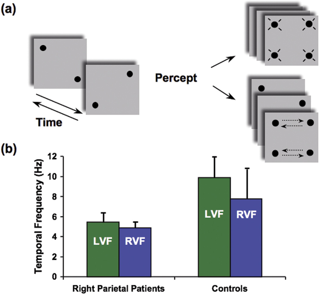
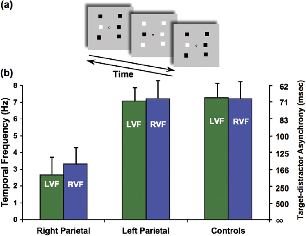
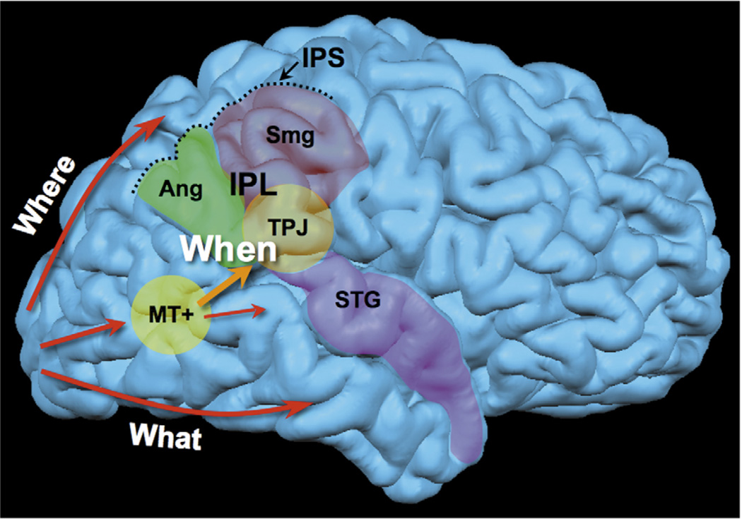
The order of events, whether two events are seen as simultaneous or successive, sets the stage for the moment-to-moment interpretation of the visual world. Evidence from patients who have lesions to the parietal lobes and transcranial magnetic stimulation studies in normal subjects suggest that the right inferior parietal lobe underlies this analysis of event timing. Judgment of temporal order, simultaneity and high-level motion are all compromised following right parietal lesions and degraded after transcranial magnetic stimulation over the right parietal but not elsewhere. The results suggest that the right parietal lobe serves as part of a when pathway for both visual fields. We propose that the disruption of this mechanism is the underlying cause of a wide range of seemingly unrelated tasks being impaired in right parietal patients.


Figures

Figure 1
Figure 1
Bilateral impairment in apparent motion. (a) A modified version of the apparent motion task (‘Ternus display’ [55]), using a quartet of dots. Two frames with two dots each are alternated at a variable frequency (left, arrows indicate alternation in time). When the interval between the frames is very short (at high frequency, more than 10 Hz), subjects can perceive only flickering dots and no motion is reported (top right). At appropriate time intervals (around a frequency of alternation of 7 or 8 Hz), subjects report motion (bottom right). The motion can be perceived either horizontally (as depicted in the panel; dashed arrows indicate motion and were not present in the actual display) or vertically. This apparent motion experiment examined whether motion was seen irrespective of its direction. (b) Patients who have right parietal lesions (average data from three patients) show a severe deficit in perceiving apparent motion both in the left visual field (LVF) and in the right visual field (RVF). Their psychophysical threshold is about half that of normal controls (average of six age-matched controls are reported). The patients cannot distinguish between four flashing and two moving dots unless the alternation rate is as low as 4 or 5 Hz, which corresponds to an average of ~200 ms interval between frames [13].
Figure 2
Figure 2
Phase discrimination task: bilateral deficit of identification of a visual event. (a) Subjects were asked to identify the odd target among six squares that alternated black and white at the same frequency as the target (middle item in the column left of fixation, top panel; arrows indicate alternation in time). The phase of the target square was shifted in time relative to the other five squares (when the target was black, the distractors were white). The target pops out easily at low alternation rates, but the task becomes more difficult at high alternation rates (for demonstrations of the task and patients’ performance, see http://visionlab.harvard.edu/Members/Lorella/movies/ movies.htm). (b) Normal controls and patients who had left parietal lesions could perform the task up to a speed of ~7 or 8 Hz, whereas patients who had right parietal lesions had a threshold speed of ~3 Hz for both the left visual field (LVF) and the right visual field (RVF). Asynchrony (msec) between the target and the distractors is reported on the right axis. [14].
Figure 3
Figure 3
The when pathway. The when pathway is represented in the brain. This pathway is lateralized in the right hemisphere. Information from the primary visual cortex (V1) travels along the dorsal pathway (spatial perception, determining where objects are) or the ventral pathway (object recognition, determining what objects are), according to the classical subdivision that has been proposed based on animal models [1]. A third pathway coming from V1 is dedicated to using time information to identify objects (e.g. determining when objects appeared or disappeared). Here, the temporoparietal junction (TPJ; considered the most common substrate of neglect [16]) is identified as a core anatomical locus, within the inferior parietal lobe (IPL); however, the when pathway is likely to include a bigger network of areas, including the right angular gyrus (Ang), the supramarginal gyrus (Smg) and the posterior superior temporal sulcus (included in the superior temporal gyrus, STG). All these areas are often involved in the cortical lesion of right parietal patients. The intraparietal sulcus (IPS) separates the IPL from the superior parietal lobe (not labeled). The middle temporal area MT+ is reported in yellow (also called the motion area, highly specialized in detecting and discriminating moving stimuli).

References

    1. Mishkin M, Ungerleider LG. Object vision and spatial vision: two cortical pathways. Trends Neurosci. 1983;6:414–417.
    1. Goodale MA, et al. Two distinct modes of control for object-directed action. Prog. Brain Res. 2004;144:131–144. - PubMed
    1. Janssen P, Shadlen MN. A representation of the hazard rate of elapsed time in macaque area LIP. Nat. Neurosci. 2005;8:234–241. - PubMed
    1. Nieder A, et al. Temporal and spatial enumeration processes in the primate parietal cortex. Science. 2006;313:1431–1435. - PubMed
    1. Leon MI, Shadlen MN. Representation of time by neurons in the posterior parietal cortex of the macaque. Neuron. 2003;38:317–327. - PubMed

Publication types