. 2007 May 11;282(19):14165-77.
doi: 10.1074/jbc.M701279200. Epub 2007 Mar 22.

Intracellular generation of sphingosine 1-phosphate in human lung endothelial cells: role of lipid phosphate phosphatase-1 and sphingosine kinase 1

Affiliations

Intracellular generation of sphingosine 1-phosphate in human lung endothelial cells: role of lipid phosphate phosphatase-1 and sphingosine kinase 1

Yutong Zhao et al. J Biol Chem. .

Abstract

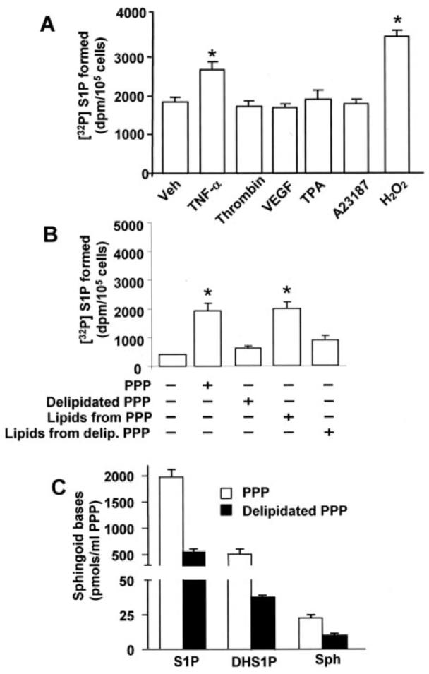
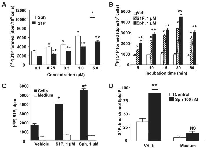
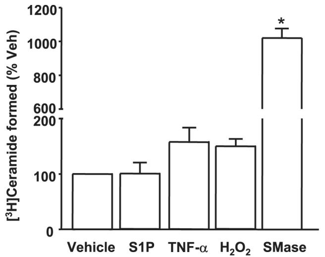
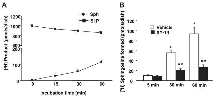
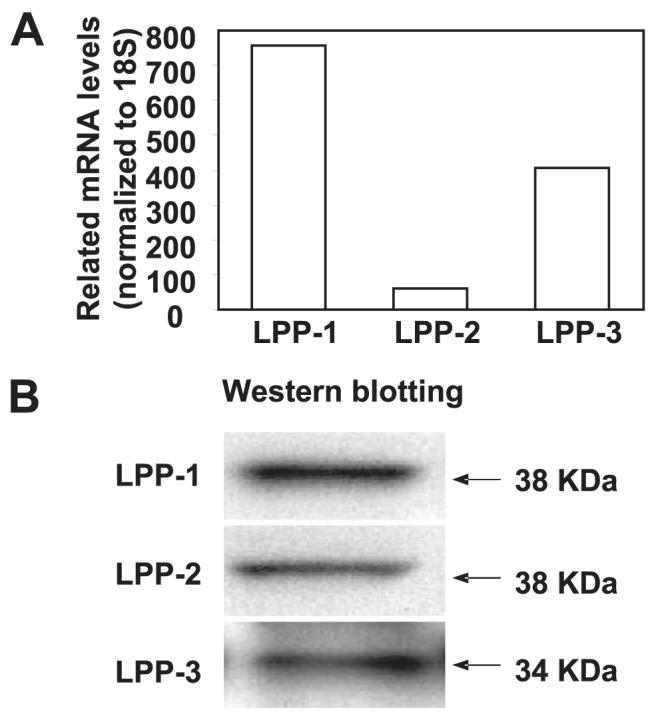
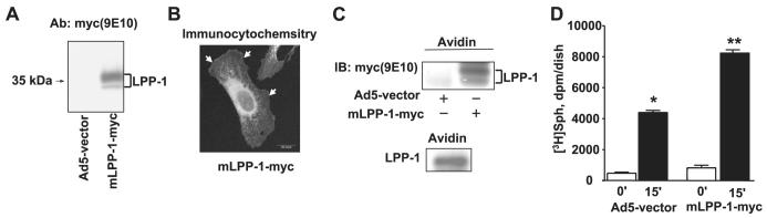
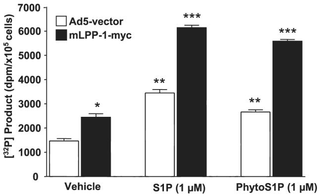
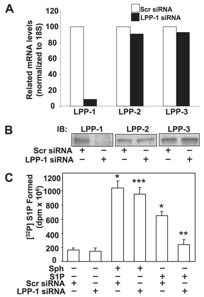
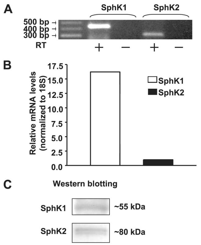
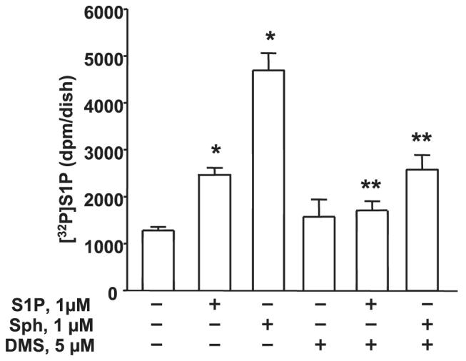
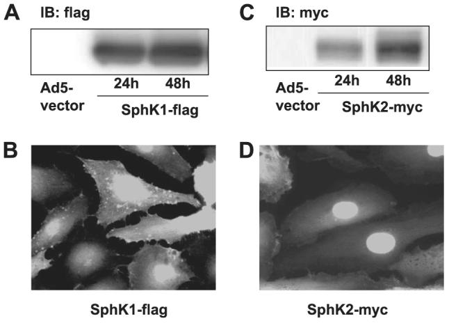
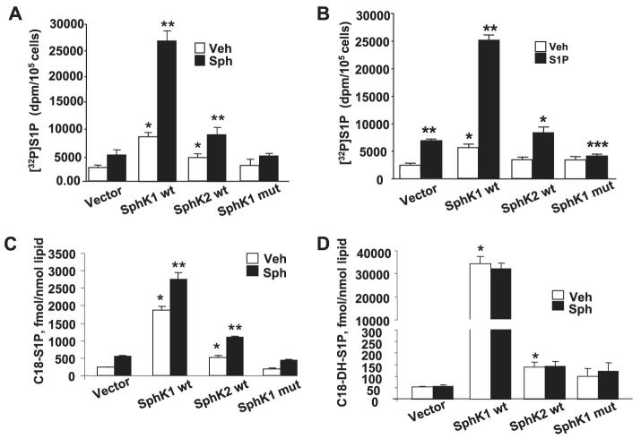
Sphingosine 1-phosphate (S1P) regulates diverse cellular functions through extracellular ligation to S1P receptors, and it also functions as an intracellular second messenger. Human pulmonary artery endothelial cells (HPAECs) effectively utilized exogenous S1P to generate intracellular S1P. We, therefore, examined the role of lipid phosphate phosphatase (LPP)-1 and sphingosine kinase1 (SphK1) in converting exogenous S1P to intracellular S1P. Exposure of (32)P-labeled HPAECs to S1P or sphingosine (Sph) increased the intracellular accumulation of [(32)P]S1P in a dose- and time-dependent manner. The S1P formed in the cells was not released into the medium. The exogenously added S1P did not stimulate the sphingomyelinase pathway; however, added [(3)H]S1P was hydrolyzed to [(3)H]Sph in HPAECs, and this was blocked by XY-14, an inhibitor of LPPs. HPAECs expressed LPP1-3, and overexpression of LPP-1 enhanced the hydrolysis of exogenous [(3)H]S1P to [(3)H]Sph and increased intracellular S1P production by 2-3-fold compared with vector control cells. Down-regulation of LPP-1 by siRNA decreased intracellular S1P production from extracellular S1P but had no effect on the phosphorylation of Sph to S1P. Knockdown of SphK1, but not SphK2, by siRNA attenuated the intracellular generation of S1P. Overexpression of wild type SphK1, but not SphK2 wild type, increased the accumulation of intracellular S1P after exposure to extracellular S1P. These studies provide the first direct evidence for a novel pathway of intracellular S1P generation. This involves the conversion of extracellular S1P to Sph by LPP-1, which facilitates Sph uptake, followed by the intracellular conversion of Sph to S1P by SphK1.


Figures

FIGURE 1
FIGURE 1. Agonist-induced intracellular generation of S1P in HPAECs
Panel A, HPAECs grown to ∼95% confluence in 35-mm dishes were labeled with [32P]orthophosphate (20 μCi/ml) in phosphate-free DMEM for 3 h. The radioactive medium was aspirated, and cells were rinsed in DMEM without serum and challenged with medium alone or medium containing TNF-α (20 ng/ml), thrombin (10 ng/ml), vascular endothelial growth factor (VEGF;20 ng/ml), phorbol ester (TPA,25nM), ionophore A23187 (1 μM), or H2O2 (250 μM). In panel B cells were treated with human PPP or lipids extracted from human PPP for 30 min. The reaction was terminated by the addition of 100 μl of 12 M HCl; lipids were extracted under acidic condition and separated by TLC for [32P]S1P generation. In panel C total lipids were extracted from PPP or prelipidated PPP, and sphingoid bases were quantified by LC-MS/MS as described under “Experimental Procedures.” Values are the means ± S.D. of six independent experiments. *, significantly different from cells exposed to medium alone (p < 0.05). Veh, vehicle.
FIGURE 2
FIGURE 2. Dose- and time-dependent formation of intracellular S1P from exogenous sphingosine or S1P
HPAECs grown to ∼95% confluence in 35-mm dishes were labeled with [32P]orthophosphate (20 μCi/ml) in phosphate-free DMEM for 3 h, the radioactive medium was aspirated, and cells were rinsed in DMEM without serum. In panel A, cells were challenged with medium alone or medium containing different concentrations of Sph or S1P in the presence of 0.1% BSA for 30 min; in panel B cells were challenged with medium alone or medium Sph (1 μM) or S1P (1 μM) in the presence of 0.1% BSA for 5, 10, 15, 30, and 60 min. The medium was removed, cell lipids were extracted, and intracellular [32P]S1P was quantified. In panel C, HPAECs labeled with [32P]orthophosphate (20 μCi/ml) for 3 h as described in panel A were exposed to either Sph (1 μM) or S1P (1 μM) for 30 min, and the lipids were extracted and quantified from the media and cells. The distribution of [32P]S1P in medium and cells was measured. Panel D shows LC-MS/MS quantification of S1P in media and HPAECs after exposure to Sph (100 nM) for 30 min. The values are the means ± S.D. of six independent experiments. *, significantly different compared with cells exposed to medium alone (p < 0.05); **, significantly different compared with cells exposed to medium alone (p < 0.01). NS, not significant; Veh, vehicle.
FIGURE 3
FIGURE 3. Agonist-induced generation of [3H]ceramide in HPAECs
HPAECs grown to ∼95% confluence in 35-mm dishes were labeled with L-[3H]serine (100 μCi/ml) in serine-free DMEM for 24 h. The radioactive medium was aspirated, and cells were rinsed and challenged with medium alone or medium containing S1P (1μM), TNF-α (20 ng/ml), H2O2 (250μM), or sphingomyelinase (SMase; 1 units/ml) for 30 min. Lipids were extracted, and accumulation of [3H]ceramide was calculated from the averages of two independent experiments in triplicate. Veh, vehicle.
FIGURE 4
FIGURE 4. Time course and effect of XY-14 on hydrolysis of [3H]S1P in HPAECs
In panel A, HPAECs (∼90% confluence) in 35-mm dishes were incubated with [3H]S1P (1 μM; specific radioactivity, 100 dpm/pmol) complexed with 0.1% BSA in BEGM medium (Clonetics) for up to 60 min. At each time point the medium was removed, and lipids were extracted and analyzed for [3H]Sph and [3H]S1P by TLC. Values are from two independent experiments in triplicate and expressed as dpm/dish. In panel B, HPAECs were pretreated with XY-14 (10 μM) before exposure to [3H]S1P (1 μM; specific activity 100 dpm/pmol) for 5, 30, and 60 min. At each time point, medium was removed, and the formation of [3H]Sph from [3H]S1P was quantified. Values are from three independent experiments in triplicate and are expressed as a percentage of total radioactivities (dpm) in the lipid extract.
FIGURE 5
FIGURE 5. Detection of LPPs by real-time RT-PCR and Western blotting in HPAECs
In panel A total RNA was extracted from HPAECs, and expression of LPPs was normalized to 18 S and quantified by real-time RT-PCR. Values are the average of three independent experiments. In panel B cell lysates (30 μg of protein) were subjected to SDS-PAGE and analyzed by Western blotting with anti-LPP-1, -LPP-2, and -LPP-3 antibodies. The figure shows a representative Western blot of three independent experiments.
FIGURE 6
FIGURE 6. Effect of overexpression of adenoviral construct of LPP-1 wt on hydrolysis of [3H]S1P in HPAECs
HPAECs (∼60% confluence in 35-mm dishes) were infected with adenoviral construct (10 m.o.i.) for the empty vector or vector containing cDNA for Myc-tagged mLPP-1 for 24 h. Panel A, cell lysates were subjected to SDS-PAGE and Western blotting with anti-LPP-1 antibody (Ab). Panel B, HPAECs grown on glass coverslips to ∼60% confluence were infected with cDNA for Myc-tagged mLPP-1 for 24 h, and cells were subjected to immunostaining with anti-Myc antibody (9E10) and examined by confocal fluorescent microscopy. Panel C, HPAECs (passage 6) grown on T-75 cm2 flasks were infected with adenoviral construct (10 m.o.i.) for the empty vector or vector containing cDNA for Myc-tagged mLPP-1 for 24 h. Surface labeling of cells with biotin was performed with the Cell Surface Protein Isolation kit (Pierce) as described under “Experimental Procedures.” Surface proteins were analyzed by Western blotting with anti-Myc (10E6) or anti-LPP-1 antibodies. Panel D, [3H]S1P (1 μM; specific activity 100 dpm/pmol) complexed with 0.1% BSA in BEGM was added to each dish, and hydrolysis was examined at the end of a 60-min incubation. Lipids were extracted and separated by TLC. Values for sphingosine production are the means ± S.D. of three independent experiments. *, significantly different compared with empty vector infected cells (p < 0.05). IB, immunoblot.
FIGURE 7
FIGURE 7. LPP-1 wt overexpression enhances intracellular accumulation of [lsqb]32P]S1P and [32P]phyto-S1P
HPAECs (∼60% confluence) were infected with adenoviral constructs (10 m.o.i.) that contained cDNA for the empty vector or Myc-tagged LPP-1 for 24 h. Cells were labeled with [32P]orthophosphate (20 μCi/ml) in phosphate-free DMEM for 3 h before exposure to either S1P (1 μM) or phyto-S1P (1 μM) for 30 min. The formation of 32P-labeled S1P or phyto-S1P was determined after separation by TLC. Values are expressed as the means ± S.D. of three independent experiments in triplicate. *, significantly different compared with cells infected with empty vector adenoviral construct (p < 0.05); **, significantly different compared with empty vector infected cells (p < 0.05); ***, significantly different compared with Myc-tagged LPP-1 wt-infected cells without S1P or phyto-S1P treatment (p < 0.01).
FIGURE 8
FIGURE 8. Knock down of LPP-1 by siRNA decreases intracellular generation of [32P]S1P from exogenous S1P but not from Sph
Panel A, total RNA was extracted from HPAECs transfected with scrambled siRNA or LPP-1 siRNA for 72 h, and transcription of the mRNA was determined by real-time RT-PCR. Panel B, cell lysates from scrambled and LPP-1 siRNA-transfected cells were analyzed by Western blotting (IB) for LPP-1, LPP-2, and LPP-3 protein expression using LPP-specific antibodies. Panel C, HPAECs were transfected with scrambled siRNA or LPP-1 siRNA for 72 h as described under “Experimental Procedures.” The transfected media was aspirated, and cells were labeled with [32P]orthophosphate (20 μCi/ml) in phosphate-free DMEM before exposure to exogenous S1P (1 μM) or Sph (1 μM) complexed with 0.1% BSA for 30 min. The accumulation of [32P]S1P was determined after separation by TLC. Values are the means ± S.D. of six independent experiments. *, significantly different compared with scrambled siRNA transfected cells (p < 0.05); **, significantly different compared with cells exposed to scrambled siRNA + S1P (p, 0.05); ***, not significantly different compared with cells transfected with scrambled siRNA + sphingosine (p > 0.05).
FIGURE 9
FIGURE 9. Detection of SphK1 and SphK2 by RT-PCR, real-time RT-PCR, and Western blotting in HPAECs
Panel A, total RNA was extracted from ∼95% confluent HPAECs and transcription of the genes encoding SphK1 and SphK2 was assessed by RT-PCR (- indicates the absence of reverse transcriptase, and + indicates the presence of reverse transcriptase during RT reaction) with primers indicated to SphKs. Panel B, one-step real-time RT-PCR was performed with total RNA from HPAECs. The relative abundance of target mRNA was calculated as 2 raised to the negative of its threshold cycle value multiplied by 106 normalized to the abundance of 18 S. Panel C, cell lysates (30 μg of protein) from HPAECs were subjected to SDS-PAGE and analyzed by Western blotting with anti-SphK1 and anti-SphK2 antibodies. Each Western blot is representative of three independent experiments.
FIGURE 10
FIGURE 10. Effect of DMS on intracellular generation of [32P]S1P
HPAECs (∼95% confluence) were labeled with [32P]orthophosphate (20 μCi/ml) in phosphate-free DMEM for 3 h before pretreatment with DMS (5 μM) for 1 h. The medium was aspirated, and cells were exposed to medium alone or medium containing either S1P (1 μM) or Sph (1 μM) complexed with 0.1% BSA for 30 min. Cellular lipids were extracted, and accumulation of [32P]S1P was measured after separation by TLC. Values are given as the means ± S.D. of three independent experiments in triplicate. *, significantly different compared with vehicle treatment (p < 0.05); **, significantly different compared either S1P or Sph exposed cells without DMS (p < 0.01).
FIGURE 11
FIGURE 11. Overexpression of adenoviral constructs of FLAG-tagged SphK and Myc-tagged SphK2 in HPAECs
Panels A and C, HPAECs (∼60% confluence in 35-mm dishes) were infected with empty vector or with cDNA for FLAG-tagged SphK1 or Myc-tagged SphK2 adenoviral constructs (10 m.o.i.) in complete BEGM for 24 and 48 h. At the indicated time points cell lysates were prepared and subjected to SDS-PAGE and Western blotting (IB) with anti-FLAG or anti-Myc (9E10) antibodies; Panels B and D, HPAECs grown on glass coverslips to ∼70% confluence were infected with cDNA for FLAG-tagged SphK1 or Myc-tagged SphK2 adenoviral constructs (10 m.o.i.) for 24 h. Cells were subjected to immunostaining with anti-FLAG or anti-Myc (9E10) antibody and examined by fluorescent microscopy. The Western blots and immunofluorescence images are representative of three independent experiments.
FIGURE 12
FIGURE 12. Effect of overexpression of SphK1 or SphK2 or SphK1 mutant on S1P formation in vitro and in vivo
HPAECs (∼70% confluence in 100-mm dishes) were infected with cDNA for FLAG-tagged SphK1, Myc-tagged SphK2 or FLAG-tagged SphK1 mutant (10 m.o.i.) for 24 h. Panel A, aliquots of 100 μg of protein were incubated with Sph (1 μM) complexed to 0.1% BSA and [γ-32P]ATP (10 μM, specific activity 1 × 10⁴ dpm/pmol) for 30 min at 37 °C. The formation of [32P]S1P was determined by TLC. Results are expressed as pmol of S1P formed/μg of protein/min. In panel B, cells were labeled with [32P]orthophosphate (20 μCi/ml) in phosphate-free DMEM for 3 h before challenge with S1P (1 μM) for 30 min. The intracellular accumulation of [32P]S1P was measured after separation by TLC. Values are the means ± S.D. of three independent experiments. *, significantly different from empty vector without S1P addition (p < 0.05); **, significantly different compared with empty vector (p < 0.01); ***, significantly different compared with cells infected with empty vector plus S1P (p < 0.05). In panels C and D cells were infected with SphK1 wt, SphK2 wt, or sphK1 mutant as described in panel A. Cells were challenged with DMEM or DMEM plus C18-sphingosine (1 μM) for 30 min, and intracellular accumulation of C18-S1P or C18-dihydro (DH)-S1P was measured by LC-MS/MS as described under “Experimental Procedures.” Values are the means ± S.D. of three independent experiments. *, significantly different from vector control in the absence or presence of sphingosine (p < 0.05); **, significantly different from vector control in the absence or presence of sphingosine (p < 0.01). Veh, vehicle.
FIGURE 13
FIGURE 13. Effect of down-regulation of SphK1, SphK2, SPP1, and SPL on intracellular generation of S1P in HPAECs
HPAECs (∼70% confluence in 35-mm dishes) were transfected with scrambled, SphK1, SphK2, SPP1, or SLP siRNA (100 nM) for 72 h. In panels A and B, total RNA was isolated, and mRNA expression of SphK1, SphK2, SPP1, and SPL, under different transfection, was evaluated by real-time RT-PCR and normalized to 18 S. Values are the averages of six independent experiments. The efficacy of SphK1 and SphK2 siRNA was also evaluated by Western blotting (IB, panel A). In panel C transfected cells were labeled with [32P]orthophosphate (20 μCi/ml) in phosphate-free DMEM for 3 h before exposure to either Sph (1 μM) or S1P (1 μM) for 30 min. The accumulation of intracellular [32P]S1P was quantified by TLC. Values given are the means ± S.D. of six independent experiments. *, significantly different compared with scrambled siRNA transfection alone (p < 0.05); **, significantly different compared with scrambled siRNA (p < 0.01); ***, significantly different compared with scrambled siRNA plus S1P or scrambled siRNA + Sph (p < 0.05). In panels D and E, cells were transfected with scrambled, SphK1, SphK2, SPP1, or SPL siRNA for 72 before exposure to medium or medium plus C18-sphingosine (1 μM) for 30 min. The accumulation of intracellular C18-S1P or C18- dihydro-S1P was quantified by LC-MS/MS as described under “Experimental Procedures.” *, significantly different compared with scrambled siRNA transfection (p < 0.05); **, significantly different compared with scrambled siRNA plus Sph (p < 0.01). Veh, vehicle.

References

    1. Pyne S, Pyne NJ. Biochem. J. 2000;349:385–402. - PMC - PubMed
    1. Spiegel S, Milstien S. Nat. Rev. Mol. Cell Biol. 2003;4:397–407. - PubMed
    1. Hla T. Pharmacol. Res. 2003;47:401–407. - PubMed
    1. Ozaki H, Hla T, Lee MJ. J. Atheroscler. Thromb. 2003;10:125–131. - PubMed
    1. Kluk MJ, Hla T. Biochim. Biophys. Acta. 2002;1582:72–80. - PubMed

Publication types

MeSH terms

LinkOut - more resources