. 2007 Jun 1;581(Pt 2):665-78.
doi: 10.1113/jphysiol.2007.128728. Epub 2007 Mar 22.

Postnatal androgen deprivation dissociates the development of smooth muscle innervation from functional neurotransmission in mouse vas deferens

Affiliations

Postnatal androgen deprivation dissociates the development of smooth muscle innervation from functional neurotransmission in mouse vas deferens

J A Brock et al. J Physiol. .

Abstract

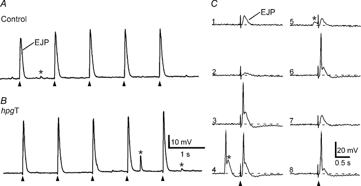
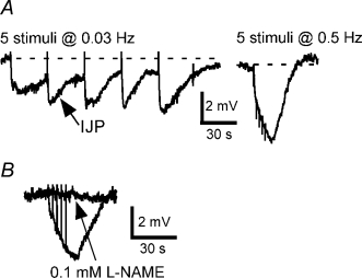
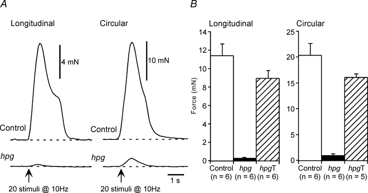
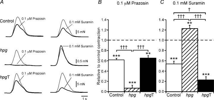
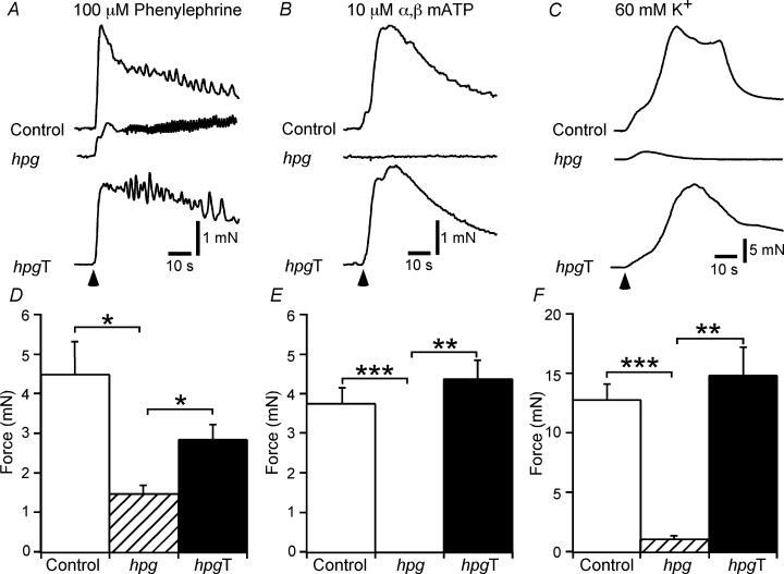
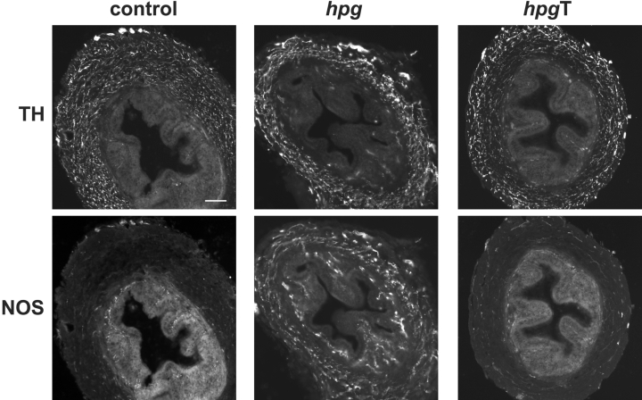
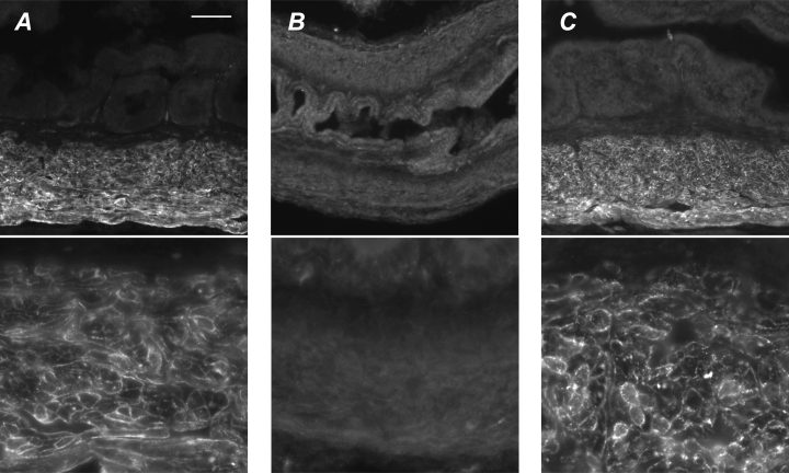
The pelvic autonomic nervous system is a target for circulating androgens in adults, with androgen exposure or deprivation affecting the structure and function of urogenital tract innervation. However, the critical period for androgen exposure to initially establish pelvic autonomic neuromuscular transmission has not been determined. We have examined the sympathetic innervation of the vas deferens in hypogonadal (hpg) mice that are deprived of androgens after birth but undergo normal prenatal sexual differentiation and remain androgen responsive throughout life. In vasa deferentia from hpg mice, purinergic excitatory junction potentials and contractions could not be elicited by electrical stimulation and P2X(1) purinoceptors could not be demonstrated by immunofluorescence. Moreover, a novel inhibitory nitrergic transmission developed. Administering testosterone to adult hpg mice restored purinergic excitatory transmission and P2X(1) purinoceptor immunofluorescence, and nitrergic inhibitory transmission was lost. Despite the deficit in excitatory neurotransmission in hpg mice, their vasa deferentia were innervated by numerous noradrenergic axons and pelvic ganglia appeared normal. In addition, noradrenergic contractions could be elicited by electrical stimulation. This study has revealed that postnatal androgen exposure has a profound effect on the development of excitatory transmission in vas deferens smooth muscle, primarily by a postjunctional action, but is not essential for development of the structural innervation of this organ. Our results also indicate that there is no postnatal critical period for androgen exposure to establish neuroeffector transmission and that postnatal androgen exposure can be delayed until adulthood, with little consequence for establishment of normal sympathetic neurotransmission.


Figures

Figure 1
Figure 1. Intracellularly recorded electrical activity in control and hpgT vasa deferentia
A and B, EJPs evoked by 5 stimuli at 1 Hz in a control (A) and an hpgT (B) vas deferens. C, representative traces from a cell in a control vas deferens in which EJPs evoked during trains at 1 Hz were more variable in amplitude (numbers indicate sequence of stimuli) and occasionally evoked muscle action potentials. Cells displaying this pattern of activity were also recorded in hpgT vas deferens. In both control and hpgT vasa deferentia, spontaneous EJPs were recorded (indicated by *). In trace 4 of panel C, a muscle action potential was triggered by a spontaneous EJP. In A, B and C, the resting membrane potentials were −76, −79 and −61 mV, respectively.
Figure 2
Figure 2. Intracellularly recorded electrical activity recorded in hpg vasa deferentia
A, inhibitory junction potentials (IJPs) evoked by 5 stimuli at 0.03 and 0.5 Hz. B, overlaid traces showing responses to 5 stimuli at 0.5 Hz before and during application of l-NAME (0.1 mm) in another tissue. In A and B, the resting membrane potentials were −58 and −52 mV, respectively.
Figure 3
Figure 3. Neurally evoked mechanical responses of control, hpg and hpgT vasa deferentia
A, representative traces showing contractions of the longitudinal and circular smooth muscle of control and hpg vasa deferentia to 20 stimuli at 10 Hz. B, bar graphs showing the contraction amplitude data for the longitudinal and circular smooth muscle of vasa deferentia from control, hpg and hpgT mice.
Figure 4
Figure 4. Neurally evoked contractions of control and hpgT vasa deferentia were reduced by either prazosin or suramin but those of hpg vasa deferentia were only reduced by prazosin
A, overlaid traces showing contractions of the circular smooth muscle of control (upper traces), hpg (middle traces) and hpgT (lower traces) vasa deferentia evoked by 20 stimuli at 10 Hz under control conditions (thin line) and in the presence of either prazosin (0.1 μm) or suramin (0.1 mm) (thick line). B and C, bar graphs showing the relative change in contraction of control, hpg and hpgT vasa deferentia produced by prazosin (B) and suramin (C). Comparisons with control recordings in the same tissues were made with paired t tests (**P < 0.01, ***P < 0.001). Comparisons between the groups of tissues were made with one-way ANOVA followed by Games–Howell tests (†P < 0.05, †††P < 0.001).
Figure 5
Figure 5. Mechanical responses of control, hpg and hpgT vasa deferentia to phenylephrine, α,β-methylene ATP and 60 mm K+
A–C, representative traces showing contractions of the circular smooth muscle of control, hpg and hpgT vasa deferentia to phenylephrine (100 μm, A), α,β-methylene ATP (10 μm, B) and 60 mm K+ (C). D–F, bar graphs showing the peak amplitude of contractions of control, hpg and hpgT vasa deferentia to phenylephrine, α,β-methylene ATP and 60 mm K+. Comparisons between the groups of tissues were made with one-way ANOVA followed by Games–Howell tests (*P < 0.05, **P < 0.01, ***P < 0.001).
Figure 6
Figure 6. Autonomic innervation of vasa deferentia in adult control, hpg and hpgT mice
Transverse sections of vasa deferentia show noradrenergic (TH-immunoreactive) and nitrergic cholinergic (NOS-immunoreactive) nerves in the muscle and mucosal layers. TH axons are prevalent in the muscle of all three animal groups. In control and hpgT mice, NOS axons provide a sparse supply to muscle and innervate the subepithelial region. In hpg mice the NOS innervation of the muscle and the mucosa appears substantially increased. Calibration bar represents 100 μm (control, hpgT) or 50 μm (hpg).
Figure 7
Figure 7. Chemical characteristics of vas deferens autonomic innervation in adult control and hpg mice
A and B, numerous axons in the muscle show catecholamine histofluorescence (glyoxylic acid method) in the control (A) and hpg (B) vas deferens. Panels C–G show vasa from hpg mice. Panels A, B, D and F show vasa sectioned longitudinally and panels C, E and G are transverse sections. C, TH-immunoreactive axons coexpress dopamine-β-hydroxylase (DBH). D, TH-immunoreactive axons coexpress the synaptic protein, synapsin (SAP). E, NOS-immunoreactive axons coexpress vasoactive intestinal peptide (VIP). F, vesicular acetylcholine transporter (VAChT)-immunoreactive axons are prevalent in the muscle and mucosa. G, sensory fibres immunolabelled for calcitonin gene-related peptide (CGRP) are rare in the muscle and mucosa, but nerve bundles can be seen in the serosa. Calibration bar represents 50 μm in all micrographs except C (20 μm).
Figure 8
Figure 8. Pelvic ganglia from adult male control and hpg mice
Double-staining fluorescence immunohistochemical analysis of pelvic ganglion whole mounts from adult male hpg and control mice showing separate populations of neurons immunostained for tyrosine hydroxylase (TH, noradrenergic) and nitric oxide synthase (NOS, cholinergic). Neither TH- or NOS-immunostained neurons are less prevalent in ganglia from hpg mice. Calibration bar represents 500 μm.
Figure 9
Figure 9. P2X1 immunoreactivity in vasa deferentia
A, P2X1 immunoreactivity in smooth muscle of control vasa shows bright punctae in the plasma membrane of all smooth muscle cells but no labelling in other tissues. Occasional bright spots near the epithelium are due to autofluorescence of red blood cells. B, in hpg mice there is very pale or no P2X1 immunoreactivity associated with vas deferens smooth muscle. C, following testosterone treatment of hpg mice bright P2X1 immunoreactivity is restored to smooth muscle cells, which show bright punctate labelling in all smooth muscle cells. Calibration bar in A represents 100 μm in top row and 25 μm in bottom row.

Similar articles

Cited by

References

    1. Becker JB, Arnold AP, Berkley KJ, Blaustein JD, Eckel LA, Hampson E, Herman JP, Marts S, Sadee W, Steiner M, Taylor J, Young E. Strategies and methods for research on sex differences in brain and behavior. Endocrinology. 2005;146:1650–1673. - PubMed
    1. Bianco JJ, Handelsman DJ, Pedersen JS, Risbridger GP. Direct response of the murine prostate gland and seminal vesicles to estradiol. Endocrinology. 2002;143:4922–4933. - PubMed
    1. Brock JA, Cunnane TC, Starke K, Wardell CF. α2adrenoceptor-mediated autoinhibition of sympathetic transmitter release in guinea-pig vas deferens studied by intracellular and focal extracellualr recording of junction potentials and currents. Naunyn Schmiedebergs Arch Pharmacol. 1990;342:45–52. - PubMed
    1. Bustamante D, Lara H, Belmar J. Changes of norepinephrine levels, tyrosine hydroxylase and dopamine-β-hydroxylase activities after castration and testosterone treatment in vas deferens of adult rats. Biol Reprod. 1989;40:541–548. - PubMed
    1. Calixto JB, Rae GA. Influence of castration of the neonatal rat on the pharmacological reactivity of the isolated vas deferens. Biol Reprod. 1981;25:481–486. - PubMed

Publication types

MeSH terms

LinkOut - more resources