Neovascularization of ischemic myocardium by newly isolated tannins prevents cardiomyocyte apoptosis and improves cardiac function
- PMID: 17380192
- PMCID: PMC1829195
- DOI: 10.2119/2006–00039.Gu
Neovascularization of ischemic myocardium by newly isolated tannins prevents cardiomyocyte apoptosis and improves cardiac function
Retraction in
-
Notice of retraction.Mol Med. 2009 Sep-Oct;15(9-10):359. doi: 10.2119/molmed.2009.00039.Retraction. Mol Med. 2009. PMID: 19750127 Free PMC article. No abstract available.
Abstract
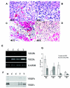
During remodeling progress post myocardial infarction, the contribution of neoangiogenesis to the infarct-bed capillary is insufficient to support the greater demands of the hypertrophied but viable myocardium resulting in further ischemic injury to the viable cardiomyocytes at risk. Here we reported the bio-assay-guided identification and isolation of angiogenic tannins (angio-T) from Geum japonicum that induced rapid revascularization of infarcted myocardium and promoted survival potential of the viable cardiomyocytes at risk after myocardial infarction. Our results demonstrated that angio-T displayed potent dual effects on up-regulating expression of angiogenic factors, which would contribute to the early revascularization and protection of the cardiomyocytes against further ischemic injury, and inducing antiapoptotic protein expression, which inhibited apoptotic death of cardiomyocytes in the infarcted hearts and limited infarct size. Echocardiographic studies demonstrated that angio-T-induced therapeutic effects on acute infarcted myocardium were accompanied by significant functional improvement by 2 days after infarction. This improvement was sustained for 14 days. These therapeutic properties of angio-T to induce early reconstitution of a blood supply network, prevent apoptotic death of cardiomyocytes at risk, and improve heart function post infarction appear entirely novel and may provide a new dimension for therapeutic angiogenesis medicine for the treatment of ischemic heart diseases.
Figures


Similar articles
-
Neovascularization of ischemic myocardium by human bone-marrow-derived angioblasts prevents cardiomyocyte apoptosis, reduces remodeling and improves cardiac function.Nat Med. 2001 Apr;7(4):430-6. doi: 10.1038/86498. Nat Med. 2001. PMID: 11283669
-
Repair of infarcted myocardium by an extract of Geum japonicum with dual effects on angiogenesis and myogenesis.Clin Chem. 2006 Aug;52(8):1460-8. doi: 10.1373/clinchem.2006.068247. Epub 2006 Jun 22. Clin Chem. 2006. PMID: 16873297
-
New directions in strategies using cell therapy for heart disease.J Mol Med (Berl). 2003 May;81(5):288-96. doi: 10.1007/s00109-003-0432-0. Epub 2003 Apr 16. J Mol Med (Berl). 2003. PMID: 12698252 Review.
-
Role of nitric oxide in the vasorelaxant and hypotensive effects of extracts and purified tannins from Geum japonicum.J Ethnopharmacol. 2007 Jan 3;109(1):128-33. doi: 10.1016/j.jep.2006.07.015. Epub 2006 Jul 21. J Ethnopharmacol. 2007. PMID: 16939706
-
Myocardial neovascularization by adult bone marrow-derived angioblasts: strategies for improvement of cardiomyocyte function.Ann Hematol. 2002;81 Suppl 2:S21-5. Ann Hematol. 2002. PMID: 12611063 Review.
Cited by
-
Current Status of Primary, Secondary, and Tertiary Prevention of Coronary Artery Disease.Int J Angiol. 2021 Aug 25;30(3):177-186. doi: 10.1055/s-0041-1731273. eCollection 2021 Sep. Int J Angiol. 2021. PMID: 34776817 Free PMC article. Review.
-
The Antiapoptosis Effect of Geum japonicum Thunb. var. chinense Extracts on Cerebral Ischemia Reperfusion Injury via PI3K/Akt Pathway.Evid Based Complement Alternat Med. 2018 Nov 14;2018:7290170. doi: 10.1155/2018/7290170. eCollection 2018. Evid Based Complement Alternat Med. 2018. PMID: 30538763 Free PMC article.
-
Isolation, characterization, and transplantation of cardiac endothelial cells.Biomed Res Int. 2013;2013:359412. doi: 10.1155/2013/359412. Epub 2013 Oct 27. Biomed Res Int. 2013. PMID: 24282814 Free PMC article.
References
-
- Pu L, Sniderman A, Brassard R, et al. Enhanced revascularization of the ischemic limb by angiogenic therapy. Circulation. 1993;88:208–15. - PubMed
-
- Risau W. Mechanisms of angiogenesis. Nature. 1997;386:671–4. - PubMed
-
- Folkman J. Clinical Applications of Research on Angiogenesis. N Engl J Med. 1995;333:1757–63. - PubMed
-
- Arras M, Mollnau H, Strasser R, et al. The delivery of angiogenic factors to the heart by microsphere therapy. Nat Biotech. 1998;16:159–62. - PubMed
Publication types
MeSH terms
Substances
LinkOut - more resources
Full Text Sources
Other Literature Sources
Medical
