. 2007 Apr;117(4):953-60.
doi: 10.1172/JCI30537. Epub 2007 Mar 22.

Thrombin-initiated platelet activation in vivo is vWF independent during thrombus formation in a laser injury model

Affiliations

Thrombin-initiated platelet activation in vivo is vWF independent during thrombus formation in a laser injury model

Christophe Dubois et al. J Clin Invest. 2007 Apr.

Abstract

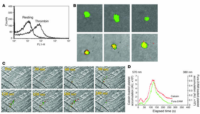
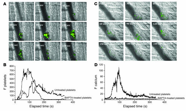
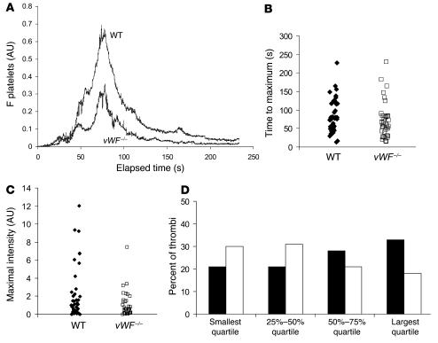
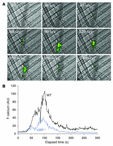
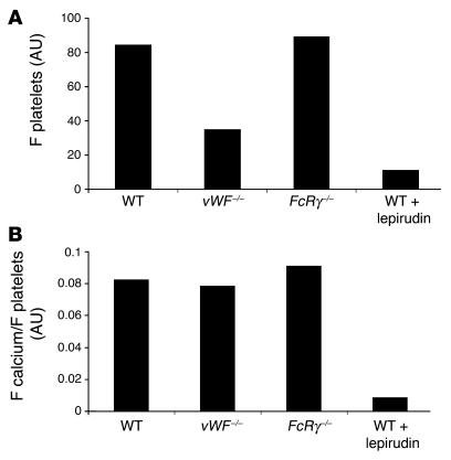
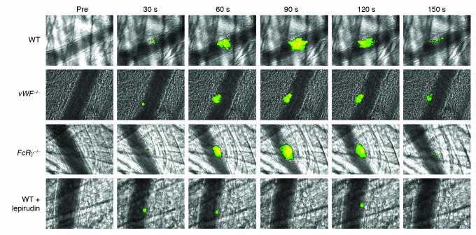
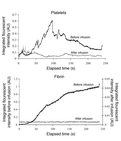
Adhesion of platelets to an injured vessel wall and platelet activation are critical events in the formation of a thrombus. Of the agonists involved in platelet activation, thrombin, collagen, and vWF are known to induce in vitro calcium mobilization in platelets. Using a calcium-sensitive fluorochrome and digital multichannel intravital microscopy to image unstimulated and stimulated platelets, calcium mobilization was monitored as a reporter of platelet activation (as distinct from platelet accumulation) during thrombus formation in live mice. In the absence of vWF, platelet activation was normal, but platelet adherence and aggregation were attenuated during thrombus formation following laser-induced injury in the cremaster muscle microcirculation. In WT mice treated with lepirudin, platelet activation was blocked, and platelet adherence and aggregation were inhibited. The kinetics of platelet activation and platelet accumulation were similar in FcRgamma(-/-) mice lacking glycoprotein VI (GPVI), GPVI-depleted mice, and WT mice. Our results indicate that the tissue factor-mediated pathway of thrombin generation, but not the collagen-induced GPVI-mediated pathway, is the major pathway leading to platelet activation after laser-induced injury under the conditions employed. In the tissue factor-mediated pathway, vWF plays a role in platelet accumulation during thrombus formation but is not required for platelet activation in vivo.


Figures

Figure 1
Figure 1. Properties of fura-2–loaded platelets in vitro and in vivo.
(A) P-selectin expression, determined by FACS analysis, on unstimulated or thrombin-activated mouse platelets containing fura-2/AM. (B) Calcium mobilization in unstimulated (top row) or thrombin-activated fura-2–loaded mouse platelets (bottom row) was determined by fluorescence microscopy in vitro. Emission at 510 nm after excitation at 380 nm is represented in green, whereas emission at 510 nm after excitation at 340 nm is represented in red. Merge is presented in yellow. (C) Platelets loaded with red-orange calcein AM or with fura-2/AM were injected into the circulation of a mouse, and vessel wall injury induced. Images of thrombus formation indicate platelets containing calcein (red) and platelets containing fura-2/AM (green). (D) The median integrated fluorescence intensity of red-orange calcein–labeled platelets or fura-2–labeled platelets in 18 thrombi from 3 mice is plotted as a function of time.
Figure 2
Figure 2. In vivo imaging of calcium mobilization during platelet activation during thrombus formation in WT mice.
Platelets isolated from WT mice were loaded with fura-2/AM. Platelets (250 × 106 to 300 × 106 cells) were infused into the circulation of a WT mouse. Following laser-induced vessel wall injury, thrombus formation was observed and images recorded over time. (A) Representative composite images of fluorescence and bright-field data depicting thrombus formation show fura-2–loaded platelet accumulation (green, yellow) and calcium mobilization (yellow). Pre, before injury. (B) Platelet accumulation during thrombus formation represented by the median integrated fluorescence intensity (y axis) of fura-2–labeled platelets excited at 380 nm. Platelets were studied in the absence (upper curve) or presence (lower curve) of BAPTA-AM. F platelets, median integrated fluorescence of platelets, with excitation at 380 nm and emission at 510 nm. (C) Representative composite images of fluorescence and bright-field data depicting thrombus formation show fura-2–loaded platelet accumulation (green, yellow) and calcium mobilization (yellow) in the presence of BAPTA-AM. (D) Calcium mobilization during thrombus formation depicted by the median integrated fluorescence intensity (y axis) of fura-2–labeled platelets excited at 340 nm. Platelets were studied in the absence (upper curve) or presence (lower curve) of BAPTA-AM. These data represent the median of experiments performed in 18 thrombi from 3 mice. F calcein, median integrated fluorescence of the signal corresponding to calcein-labeled platelets.
Figure 3
Figure 3. Platelet accumulation during thrombus formation after vessel wall injury in WT and vWF–/– mice.
Platelets were labeled with anti-mouse CD41 Fab fragments conjugated to Alexa Fluor 647. (A) The median integrated platelet fluorescence (y axis) for 43 thrombi in 4 WT mice and for 39 thrombi in 4 vWF–/– mice is presented versus time after vessel wall injury. (B) The distribution of the time to reach maximal size for each thrombus in WT mice and in vWF–/– mice. No significant difference was observed by the Wilcoxon rank sum test. (C) The distribution of the integrated platelet fluorescence for each thrombus in WT and vWF–/– mice at maximal size. WT thrombi were significantly larger than vWF–/– thrombi by the Wilcoxon rank sum test (P < 0.001). (D) The quartile distribution of the maximal integrated platelet fluorescence for each thrombus in WT and vWF–/– mice. Forty-three thrombi in WT mice and 39 thrombi in vWF–/– mice were ranked, and the percentage of thrombi of each genotype was determined independently in each quartile of the rank order. Black bars, WT mice; white bars, vWF–/– mice.
Figure 4
Figure 4. In vivo imaging of calcium mobilization during platelet activation during thrombus formation in vWF–/– mice.
(A) Platelets isolated from vWF–/– mice were loaded with fura-2/AM. Platelets (250 × 106 to 300 × 106 cells) were infused into the circulation of a vWF–/– mouse. Following laser-induced vessel wall injury, thrombus formation was observed and images recorded over time. (A) Representative composite images of fluorescence and bright-field data depicting thrombus formation show fura-2–loaded platelet accumulation (green, yellow) and calcium mobilization (yellow) in a vWF–/– mouse. (B) Calcium mobilization during thrombus formation represented by the median integrated fluorescence intensity (y axis) of fura-2–labeled platelets excited at 340 nm. Black: WT mice; blue: vWF–/– mice.
Figure 5
Figure 5. Calcium mobilization per platelet in WT mice, vWF–/– mice, FcRγ–/– mice, and WT mice treated with lepirudin.
In order to calculate calcium mobilization per platelet, the ratios of the signals corresponding to platelet accumulation and calcium mobilization (as assessed by the median integrated fluorescence intensity of each) were compared. (A) Platelet accumulation. (B) Calcium mobilization per platelet, presented as a ratio. FcRγ–/–, FcRγ–/– mice lacking GPVI; WT + lepirudin, WT mice treated with lepirudin.
Figure 6
Figure 6. Role of thrombin, vWF, and GPVI in platelet accumulation and calcium mobilization.
WT, vWF–/–, or FcRγ–/– platelets loaded with fura-2/AM were infused into a recipient mouse of the same genotype. In some experiments, the WT recipient mouse was treated with lepirudin. Thrombus formation was performed as described in Figure 2 and fura-2–loaded platelets imaged for platelet accumulation and calcium mobilization. Representative images were obtained before injury (pre) and 30, 60, 90, 120, and 150 seconds after injury.
Figure 7
Figure 7. Effect of lepirudin on platelet accumulation and fibrin deposition in WT mice.
Thrombus formation was induced as described in Figure 1, and platelets and fibrin detected using Fab fragments of antibodies against CD41 and fibrin-specific antibodies, respectively. The median integrated fluorescence over time is presented for 30 thrombi generated in 3 WT mice. After generating a series of 10 thrombi per mouse, lepirudin (8 U/g mouse) was infused and 9–10 additional thrombi generated per mouse. A total of 28 thrombi before infusion and 29 thrombi after infusion were studied in 3 mice. Black, before infusion; gray, after infusion.

References

    1. Ruggeri Z.M. Platelets in atherothrombosis. Nat. Med. 2002;8:1227–1234. - PubMed
    1. Gibbins J.M. Platelet adhesion signalling and the regulation of thrombus formation. J. Cell Sci. 2004;117:3415–3425. - PubMed
    1. Furie B., Furie B.C. Thrombus formation in vivo. J. Clin. Invest. 2005;115:3355–3362. doi: 10.1172/JCI26987. - DOI - PMC - PubMed
    1. Jackson S.P., Nesbitt W.S., Kulkarni S. Signaling events underlying thrombus formation. J. Thromb. Haemost. 2003;1:1602–1612. - PubMed
    1. Savage B., Saldivar E., Ruggeri Z.M. Initiation of platelet adhesion by arrest onto fibrinogen or translocation on von Willebrand factor. Cell. 1996;84:289–297. - PubMed

Publication types