. 2007 Apr;117(4):1049-57.
doi: 10.1172/JCI30235. Epub 2007 Mar 22.

Mesenchymal cells regulate the response of acute lymphoblastic leukemia cells to asparaginase

Affiliations

Mesenchymal cells regulate the response of acute lymphoblastic leukemia cells to asparaginase

Shotaro Iwamoto et al. J Clin Invest. 2007 Apr.

Abstract

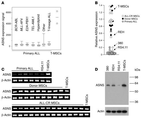
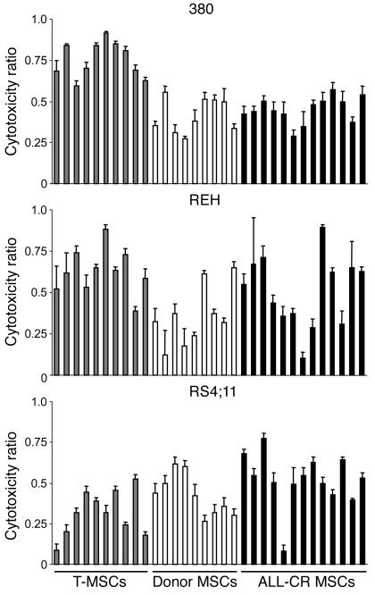
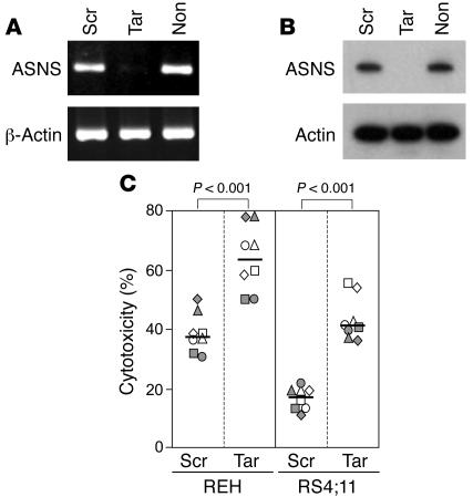
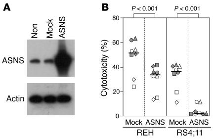
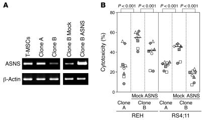
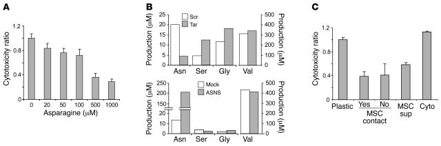
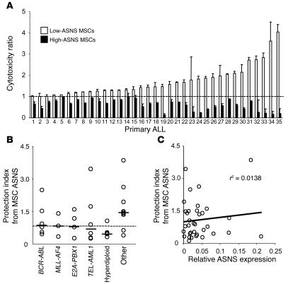
Because of their low asparagine synthetase (ASNS) expression and asparagine biosynthesis, acute lymphoblastic leukemia (ALL) cells are exquisitely sensitive to asparagine depletion. Consequently, asparaginase is a major component of ALL therapy, but the mechanisms regulating the susceptibility of leukemic cells to this agent are unclear. In 288 children with ALL, cellular ASNS expression was more likely to be high in T-lineage ALL and low in B-lineage ALL with TEL-AML1 or hyperdiploidy. However, ASNS expression levels in bone marrow-derived mesenchymal cells (MSCs), which form the microenvironment where leukemic cells grow, were on average 20 times higher than those in ALL cells. MSCs protected ALL cells from asparaginase cytotoxicity in coculture experiments. This protective effect correlated with levels of ASNS expression: downregulation by RNA interference decreased the capacity of MSCs to protect ALL cells from asparaginase, whereas enforced ASNS expression conferred enhanced protection. Asparagine secretion by MSCs was directly related to their ASNS expression levels, suggesting a mechanism - increased concentrations of asparagine in the leukemic cell microenvironment - for the protective effects we observed. These results provide what we believe to be a new basis for understanding asparaginase resistance in ALL and indicate that MSC niches in the bone marrow can form a safe haven for leukemic cells.


Figures

Figure 1
Figure 1. Expression of ASNS in ALL cells and bone marrow–derived MSCs.
(A) Expression of ASNS mRNA in 288 samples of primary ALL cells compared with that of TERT-immortalized MSCs (T-MSCs; mean of 2 measurements denoted by circles) by Affymetrix GeneChip arrays. Boxes denote interquartile range; whiskers denote range; lines denote median. *Cases lacking known genetic abnormalities. (B) Expression of ASNS mRNA in ALL cell lines, primary ALL samples and primary MSCs as measured by real-time RT-PCR. Primary MSCs were obtained from the bone marrow of 9 healthy donors (Donor MSCs) and 13 patients with ALL undergoing therapy and in complete remission (ALL-CR MSCs). Shown is ASNS expression relative to that of the TBP gene. (C) ASNS mRNA expression in primary ALL cell samples, the RS4;11 ALL cell line, and primary MSCs by semiquantitative RT-PCR; signals obtained with TERT-immortalized MSCs are also shown. β-Actin was used as a control. (D) Western blot analysis of ASNS protein expression in ALL cell lines and TERT-immortalized MSCs. After probing with the anti-ASNS antibody, the membrane was stripped and reprobed with an anti-actin antibody.
Figure 2
Figure 2. MSCs protect ALL cells from asparaginase cytotoxicity.
The ALL cell lines 380, REH, and RS4;11 were cultured with and without MSCs for 48 hours in the presence of the concentration of asparaginase that causes 50% killing in 2 days of culture, as previously determined for each cell line in experiments without MSCs (1.0 IU/ml for 380 and REH; 0.001 IU/ml for RS4;11). Values are mean ± SD of cell killing obtained in cultures with MSCs relative to that obtained in parallel cultures without MSCs. Ten experiments were performed for each ALL cell line with TERT-immortalized MSCs; MSCs from 9 healthy donors and 13 patients with ALL in complete remission were also tested for each ALL cell line. Each experiment consisted of 4 measurements.
Figure 3
Figure 3. Downregulation of ASNS expression in MSCs by RNA interference decreases their capacity to protect ALL cells from asparaginase cytotoxicity.
(A) Expression of ASNS mRNA in TERT-immortalized MSCs transduced with an ASNS-targeted siRNA construct (Tar) compared with that of cells transduced with a scrambled control construct (Scr) and that of nontransduced MSCs (Non) by semiquantitative RT-PCR. β-Actin was used as a control. (B) Expression of ASNS protein in the same cells by Western blotting. After probing with the anti-ASNS antibody, the membrane was stripped and reprobed with an anti-actin antibody. (C) Cytotoxicity of asparaginase against the ALL cell lines REH and RS4;11 was measured in coculture with MSCs transduced with the target siRNA construct or the scrambled control construct. Cytotoxicity was measured after 48 hours of exposure to 1 IU/ml and 0.001 IU/ml asparaginase for REH and RS4;11, respectively. Each symbol indicates the results of 1 experiment (mean of 2 measurements). Bars correspond to the median of 8 experiments.
Figure 4
Figure 4. Upregulation of ASNS expression in MSCs increases their capacity to protect ALL cells from asparaginase cytotoxicity.
(A) Expression of ASNS protein in TERT-immortalized MSCs transduced with an ASNS retroviral vector (ASNS) compared to that of cells transduced with an empty control vector (Mock) and that of nontransduced MSCs by Western blotting. After probing with the anti-ASNS antibody, the membrane was stripped and reprobed with an anti-actin antibody. (B) Cytotoxicity of asparaginase on the ALL cell lines REH and RS4;11 was measured in coculture with MSCs transduced with either ASNS or empty vector. Cytotoxicity was measured after 48 hours of exposure to 1 IU/ml and 0.001 IU/ml asparaginase for REH and RS4;11, respectively. Each symbol indicates the results of 1 experiment (mean of 2 measurements). Bars correspond to the median of 8 experiments.
Figure 5
Figure 5. MSC clones with different levels of endogenous ASNS expression have different capacities to protect ALL cells from asparaginase cytotoxicity.
(A) Different levels of expression of ASNS mRNA in TERT-immortalized unsorted MSCs and in 2 MSC clones (clones A and B) by semiquantitative RT-PCR. Expression of ASNS transcripts in clone B after retroviral transduction of ASNS or empty vector is also shown. β-Actin was used as a control. (B) Cytotoxicity of asparaginase against the ALL cell lines REH and RS4;11 was measured in coculture with MSCs with different levels of ASNS expression. Cytotoxicity was measured after 48 hours of exposure to 1 IU/ml and 0.001 IU/ml asparaginase for REH and RS4;11, respectively. Each symbol indicates the results of 1 experiment (mean of 2 measurements). Bars correspond to the median of 8 experiments.
Figure 6
Figure 6. The protective effects of MSCs against asparaginase cytotoxicity are mediated by asparagine biosynthesis.
(A) RS4;11 ALL cells were cultured for 48 hours in the presence of asparaginase (0.001 IU/ml). The effect of adding increasing concentrations of asparagine to the tissue culture medium are shown. Values are mean and SD of 4 measurements. (B) Concentration of asparagine in asparagine-free, FCS-free tissue culture medium (MEM) was measured after 24 hours of culture with MSCs expressing different levels of ASNS. Compared are MSCs with downregulated ASNS by siRNA and MSCs transduced with a scrambled sequence as well as MSCs with upregulated ASNS by retroviral transduction and MSCs transduced with an empty vector. Levels of control amino acids serine, glycine, and valine are also shown. (C) RS4;11 cells were cultured either in the absence of MSCs (Plastic), in direct contact with MSCs, in Transwell inserts suspended over MSCs (no MSC contact), in MSC-conditioned medium (collected after 48 hours of culture; MSC Sup) or with a mixture of MSC-derived cytokines (IL-1α, IL-1β, IL-3, IL-6, IL-7, IL-11, stem cell factor, and Fms-like tyrosine kinase 3 ligand; Cyto). Cytotoxicity was measured after 48 hours of exposure to 0.001 IU/ml asparaginase. Values are mean and SD of 4 measurements.
Figure 7
Figure 7. Expression of ASNS in MSCs determines the susceptibility of primary ALL cells to asparaginase.
(A) Primary ALL cells, obtained from the diagnostic bone marrow of 35 children with ALL, were exposed for 48 hours to asparaginase (1.0 IU/ml) in the presence of MSCs with ASNS expression downregulated by siRNA or upregulated by ASNS transduction. Results were compared to those of parallel cultures using MSCs transduced with a scrambled siRNA construct or empty vector, respectively. Values are mean ± SD cell killing obtained in cultures with each MSC type relative to that of its corresponding control (indicated by the dashed line). Differences between the test and control cultures were significant (P < 0.05, Student’s t test) in all samples except 1–6 and 23 in cultures with MSCs underexpressing ASNS and 5 and 14–16 in cultures with MSCs overexpressing ASNS. (B) The mean cytotoxicity ratio obtained in cultures with MSCs overexpressing ASNS were subtracted from those with MSCs underexpressing ASNS to derive a protection index. Figure shows the relation between this protection index and genetic subtype in the 35 ALL samples. P = 0.068, hyperdiploid cases; *Cases lacking known genetic abnormalities, P = 0.023; Wilcoxon rank-sum test. (C) Relationship between protection index and ASNS transcript expression in ALL cells by real-time PCR. r2 by regression analysis is shown.

References

    1. Oettgen H.F., et al. Inhibition of leukemias in man by L-asparaginase. Cancer Res. 1967;27:2619–2631. - PubMed
    1. Tallal L., et al. E. coli L-asparaginase in the treatment of leukemia and solid tumors in 131 children. Cancer. 1970;25:306–320. - PubMed
    1. Pratt C.B., Simone J.V., Zee P., Aur R.J., Johnson W.W. Comparison of daily versus weekly L-asparaginase for the treatment of childhood acute leukemia. J. Pediatr. 1970;77:474–483. - PubMed
    1. Jaffe N., et al. L-asparaginase in the treatment of neoplastic diseases in children. Cancer Res. 1971;31:942–949. - PubMed
    1. Sutow W.W., et al. L-asparaginase therapy in children with advanced leukemia. The Southwest Cancer Chemotherapy Study Group. Cancer. 1971;28:819–824. - PubMed

Publication types

MeSH terms