. 2007 Apr;117(4):1078-87.
doi: 10.1172/JCI30087. Epub 2007 Mar 22.

Intersectin links WNK kinases to endocytosis of ROMK1

Affiliations

Intersectin links WNK kinases to endocytosis of ROMK1

Guocheng He et al. J Clin Invest. 2007 Apr.

Abstract

With-no-lysine (WNK) kinases are a novel family of protein kinases characterized by an atypical placement of the catalytic lysine. Mutations of 2 family members, WNK1 and WNK4, cause pseudohypoaldosteronism type 2 (PHA2), an autosomal-dominant disease characterized by hypertension and hyperkalemia. WNK1 and WNK4 stimulate clathrin-dependent endocytosis of renal outer medullar potassium 1 (ROMK1), and PHA2-causing mutations of WNK4 increase the endocytosis. How WNKs stimulate endocytosis of ROMK1 and how mutations of WNK4 increase the endocytosis are unknown. Intersectin (ITSN) is a multimodular endocytic scaffold protein. Here we show that WNK1 and WNK4 interacted with ITSN and that the interactions were crucial for stimulation of endocytosis of ROMK1 by WNKs. The stimulation of endocytosis of ROMK1 by WNK1 and WNK4 required specific proline-rich motifs of WNKs, but did not require their kinase activity. WNK4 interacted with ROMK1 as well as with ITSN. Disease-causing WNK4 mutations enhanced interactions of WNK4 with ITSN and ROMK1, leading to increased endocytosis of ROMK1. These results provide a molecular mechanism for stimulation of endocytosis of ROMK1 by WNK kinases.


Figures

Figure 1
Figure 1. Effects of long WNK1 on ROMK1.
(A) Long WNK1 decreases surface abundance of GFP-ROMK1 via a dynamin-dependent mechanism. Surface abundance was measured by biotinylation assays. Equal expression of the total ROMK1 in transfected cells was confirmed by Western blot analysis. Expression of Myc-tagged full-length long WNK1 and dynamin-2 (WT or dominant-negative; DN) was examined by an anti-myc antibody. The molecular size of proteins is shown. (B) Knockdown of endogenous long WNK1 by siRNA (top) increased surface abundance of ROMK1 (bottom). HEK cells were transfected with GFP-ROMK1 alone. Control oligo, control oligonucleotides. (C) Validation of the integrity of biotinylation assay. Cells were transfected with GFP-ROMK1 and incubated with or without biotin before lysis. Biotinylated proteins were precipitated by streptavidin beads. HC, heavy chain. (D) Effects of long WNK1 on whole-cell and single ROMK1 channel activity. Top left: Current-voltage relationship of whole-cell ROMK1 currents. Top right: Long WNK1 decreased whole-cell ROMK1 currents. Bottom: Long WNK1 had no effects on single-channel conductance (at –100 mV) or open probability (Po) of ROMK1. Open probability was measured based on approximately 15 minutes recording from patches containing 1 active channel. *P < 0.05 vs ROMK alone. C, closed state; O, open state.
Figure 2
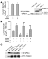
Figure 2. ITSN is important for long WNK1 regulation of ROMK1.
(A) Domain structure of ITSN1s. It contains 2 EH domains, a central coiled-coil (CC) region, and 5 consecutive SH3 domains. The EH domains anchor ITSN to clathrin coat. The SH3 domains recruit dynamin, synaptojanin, and other endocytic proteins. (B) Amino acids 1–491 of long WNK1 interacted specifically with the ITSN SH3 C domain. GST fusion proteins were used to pull down myc-tagged WNK11–491 from transfected cell lysates. Western blot analysis was performed with anti-GST and anti-myc antibodies, respectively. Amphi, amphiphysin; Endo, endophilin. (C) Evidence for knockdown of endogenous ITSN1. Endogenous ITSN1s was detected by an anti-ITSN antibody. Western blot analysis of lysates from mock-transfected (control) cells and cells transfected with HA-tagged ITSN1s probed by anti-ITSN and anti-HA antibodies is shown on the right. (D) Knockdown of ITSN1 increased surface abundance of ROMK1. (E) Knockdown of ITSN1 increased baseline whole-cell ROMK1 current density and prevented the decrease caused by WNK11–491. Cells were transfected with ROMK1 plus indicated constructs. (F) Overexpression of dominant-negative ITSN1 increased baseline whole-cell ROMK1 current density and prevented the decrease caused by WNK11–491. *P < 0.05 versus ROMK1 alone.
Figure 3
Figure 3. Specific proline-rich motifs of long WNK1 are critical for regulation of ROMK1 via ITSN.
(A) The region of amino acids 1–119 of rat long WNK1 contains 3 PXXP motifs conserved among mouse, rat, and human proteins; the triple WNK11–491 mutant P94A/P103A/P114A is denoted as PXXA3. The catalytic K233 and conserved D368 of the WNK1 kinase domain are shown. (B) Effects of various N-terminal long WNK1 fragments on ROMK1 current density. Cells were transfected with ROMK1 plus indicated constructs. FL, full-length. *P < 0.05 versus ROMK1 alone. (C) Effects of various long WNK1 constructs on ROMK1 current density. The concentration that causes a maximal inhibition of ROMK1 current (3 μg DNA per transfection per 6-well dish) for wild-type WNK11–491 and each mutant were used. Equal levels of protein expression for each construct were confirmed by Western blot analysis using an anti-myc antibody. Modified statistics adjusted for multiple comparisons was used (see Methods). P < 0.01 versus ROMK1 alone. #P < 0.01 versus ROMK1 plus WT WNK11–491. NS, not significantly different versus ROMK1 alone. (D) Pulldown of various long WNK1 constructs by GST fusion protein of ITSN SH3C. Cells were transfected with myc-tagged WNK11–119, WNK11–491, or the triple WNK11–491 mutant P94A/P103A/P114A. GST-ITSN-SH3C was used to pull down lysates from transfected cells. Shown are Western blots probed by anti-GST and anti-myc antibodies, respectively. Equal expression of long WNK1 constructs (WNK1 input) was confirmed.
Figure 4
Figure 4. Role of WNK1 kinase domain in regulation of ROMK1 and interaction with ITSN.
(A) Role of WNK1120–220 in WNK11–119 inhibition of ROMK1. *P < 0.05. (BD) Effects of K233 and/or D368 mutations of WNK11–491 on WNK1 kinase activity (B), on ROMK1 current density (C), and on the interaction with ITSN SH3C (D). WNK11–491 carrying K233D and D368K single mutations are denoted K233D and D368, respectively. Double WNK11–491 mutant K233D/D368K is denoted KDDK. (B) Wild-type and mutant WNK11–491 were immunoprecipitated from lysates of transfected cells and used to phosphorylate purified kinase-dead GST-OSR1. Kinase-dead OSR1 was used here to prevent its autophosphorylation (37, 38). Autophosphorylation of WNK11–491 is denoted as WNK1. (C) Cells were transfected with ROMK1 plus wild-type WNK11–491 or one of the mutants. *P < 0.05 versus ROMK1 alone. NS, not significantly different versus ROMK1 alone. (D) GST-ITSN-SH3C was used to pull down wild-type WNK11–491 or mutants as described in Figure 3D.
Figure 5
Figure 5. WNK4 decreases ROMK1 surface abundance via ITSN.
(A) Domains of WNK4 involved in regulation of ROMK1 current density. Cells were transfected with ROMK1 plus indicated constructs. A, autoinhibitory domain; C, coiled-coil domain. (B) WNK41–584 decreased ROMK1 surface abundance. (C) WNK41–584 interacted with ITSN SH3A, SH3B, and SH3C. GST fusion proteins were used to pull down myc-tagged WNK41–584 from lysates of transfected cells. (D) WNK41–444 did not interact with ITSN. GST fusion proteins were used to pull down myc-tagged WNK41–444 from lysates of transfected cells. ROMK1 C, C terminus of ROMK1. (E) A dominant-negative ITSN1 containing SH3A–SH3E increased baseline ROMK1 currents and prevented the decrease caused by WNK41–584. (F) Knockdown of ITSN1 increased baseline ROMK1 currents and prevented the decrease caused by WNK41–584. *P < 0.05 versus ROMK1 alone.
Figure 6
Figure 6. Representative experiments showing the effects of PHA2 mutation on WNK4 regulation of ROMK1 and interaction with ITSN.
(A) Amino acids 545–561 of WNK4 contain 3 PXXP motifs and disease-causing mutations. Double mutations of P548A/P555A disrupted all 3 PXXP motifs (denoted PA2). (B) Triple PXXP mutations prevented the decrease of surface ROMK1 caused by WNK41–584. (C) ITSN did not interact with triple PXXP mutations of WNK41–584. GST fusion proteins were used to pull down myc-tagged WT WNK41–584 or WNK41–584 with the triple PXXP mutation from lysates of transfected cells. (D) Disease-causing mutations increased the ability of WNK4 to decrease surface ROMK1 levels. (E) Effects of PXXP motifs and disease-causing mutations of WNK4 on ROMK1 currents. Cells were transfected with ROMK1 plus indicated constructs. *P < 0.05 versus ROMK1 alone. #P < 0.05 versus ROMK1 plus WNK41–584. (F) SH3A–SH3E interacted with WT WNK41–584 and the E559K mutant. H6-tagged WNK41–584 protein was used to pull down HA-tagged ITSN SH3A–SH3E from lysates of transfected cells. The amount of ITSN SH3 shown for supernatant (S) and pellet (P) represent 2% and 50% of their totals, respectively. To compare the efficacy of ITSN SH3 domain pulldown by WT WNK41–584 and E559K, the intensity of ITSN SH3 band in pellet was quantified and normalized to that in supernatant (WNK41–584, 0.32; WNK41–584 plus E559K, 0.83). This is in spite of that the intensity of E559K mutant (used for pulldown) was 65% that of WT (mean ± SEM, WNK41–584, 0.38 ± 0.15; WNK41–584 plus E559K, 0.81 ± 0.21; P < 0.05; n = 4 per group).
Figure 7
Figure 7. Role of direct interaction with ROMK1 in the regulation by WNK4.
(A) WNK41–584 interacts with the C terminus of ROMK1 but not with the C terminus of IRK1. H6-tagged ROMK1 and IRK1 C termini were used to pull down myc-tagged WNK11–584 from transfected cell lysates. Western blot analysis of proteins was performed using anti-H6 and anti-myc antibody. WNK41–584 had no effects on IRK1 current density. (B) Effects of various combinations of WNK1 and WNK4 constructs on ROMK1 currents. P < 0.01 versus ROMK1 alone. NS, not significantly different versus ROMK1 plus WNK41–444. Modified statistics adjusted for multiple comparisons were used (see Methods). (C) Interaction between disease-causing WNK4 mutants and full-length ROMK1. Detergent-solubilized GFP-ROMK1 was incubated with WNK4 fusion proteins. ROMK1 was present in all 5 conditions. Without WNK4, nickel beads alone (Ni-NTA beads) did not pull down ROMK1.
Figure 8
Figure 8. Localization of ITSN1 in rat kidney.
(A) Cortical section stained by anti-ITSN antibody. Fluorescent image was merged with differential interference contrast image. White arrow and arrowhead indicate a CCT and a DCT, respectively. G, glomerulus; P, proximal tubule. Scale bar is shown. (B) Cortical section stained as in panel A, but without the anti-ITSN antibody. (C) Cortical section double-stained by antibodies against ITSN (red) and NCX1 (green). (D) Cortical section double-stained by antibodies against ITSN (red) and AQP2 (green). Scale bars: 50 μm (A and B); 10 μm (C and D).

References

    1. Xu B., et al. WNK1, a novel mammalian serine/threonine protein kinase lacking the catalytic lysine in subdomain II. J. Biol. Chem. 2000;275:16795–16801. - PubMed
    1. Verissimo F., Jordan P. WNK kinases, a novel protein kinase subfamily in multi–cellular organisms. Oncogene. 2001;20:5562–5569. - PubMed
    1. Wilson F.H., et al. Human hypertension caused by mutations in WNK kinases. Science. 2001;293:1107–1112. - PubMed
    1. Gordon R.D. Syndrome of hypertension and hyperkalemia with normal glomerular filtration rate. Hypertension. 1986;8:93–102. - PubMed
    1. Delaloy C., et al. Multiple promoters in the WNK1 gene: one controls expression of a kidney–specific kinase–defective isoform. Mol. Cell. Biol. 2003;23:9208–9221. - PMC - PubMed

Publication types

MeSH terms

Substances